
Product Overview
EnkiLife APC labeling kit contains multiple components for labeling antibody proteins, such as the activated lyophilized APC fluorescent dye, labeling buffer, and other chemical reagents, enabling easy and reliable antibody labeling for use as fluorescent probes.
Product Features
- Activated dye with extended arm:A specially designed long flexible arm minimizes spatial hindrance between antibodies and the fluorescent dye. This protects antibody activity and enhances labeling efficiency.
- Site-specific conjugation:Optimized site-specific conjugation technology ensures uniform antibody labeling and improves efficiency.
- Rapid process:The simple procedure allows for efficient labeling completion in as fast as 3 - 4 hours.
- High dye brightness: The selected high-performance fluorescent dye boasts high quantum yield and brightness, ensuring high detection sensitivity.
- Dye stability:The chosen high-performance fluorescent dye is cross-linked and stable, resistant to degradation, and suitable for various subsequent applications, such as fixed sample detection.
- User-friendly operation:Each dye reagent is pre-optimized for corresponding antibody amounts. No complex calculations or ultrafiltration steps are required if the antibody sample meets the labeling conditions. The solid dye form ensures batch stability. Simply mix the reagents according to the steps to achieve good results.
- Flexibility: In the large-scale kit, the dye is pre-portioned for consistent batch performance, facilitating multiple labeling uses.
- Comprehensive reagents: The kit includes a labeling preservation solution, covering the entire process from labeling to preservation. No additional reagents are needed, making the process worry-free and convenient.
Dye Introduction
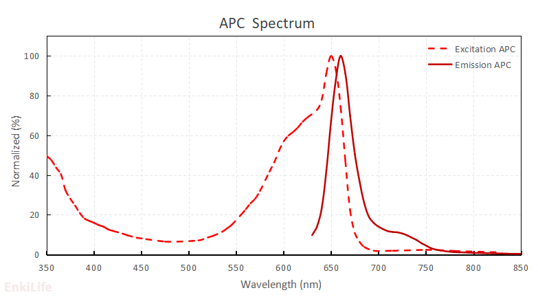
Allophycocyanin (APC) is a phycobiliprotein isolated from Spirulina (a type of blue-green algae), with a molecular weight of approximately 104 kDa. It is composed of α and β subunits aggregated into an (αβ)₃ structure and possesses a very high quantum yield. It can be excited at 594 nm or 633 nm, with a maximum absorption peak at ~650 nm and a maximum fluorescence emission peak at ~660 nm. APC exhibits very high fluorescence brightness and is one of the most commonly used fluorescent dyes in flow cytometry.
Activated Dye Properties
Basic Characteristics
Synonyms:APC, Cl-APC,cross-linked APC,Allophycocyanin
Molecular Weight: ~ 104 kDa
Common Excitation Sources: 630~640 nm
Maximum Excitation Wavelength:650 nm
Maximum Emission Wavelength: 662 nm
Molar Extinction Coefficient (L/(mol・cm)): 700000
Purity:A650/A280 ≥ 5.0
Cross Link Ratio: ≥ 1.2
Solubility:Highly soluble in aqueous solutions
Appearance:Blue solid
Kit Specifications
| Product Components | Component Content in Different Specifications | Storage Temperature | ||
|---|---|---|---|---|
| 40μg Antibody | 200μg Antibody | 1mg Antibody | ||
| Activated APC | 1 tube | 1 tube | 5 tubes | -20°C after opening, protected from light |
| Labeling Buffer L | 2 mL | 2 mL | 2 mL | 4 °C, away from light |
| Antibody Modification Reagents | 60 μL | 60 μL | 60 μL | -20 °C, away from light |
| Ultrafiltration Tube * ,50K MWCO | 1 set ** | 1 set ** | 1 set ** | RT |
| Blocking reagent | 300 μg | 300 μg | 300 μg | -20 °C, away from light |
| DMSO (dissolve blocking reagent) | 50 μL | 50 μL | 50 μL | -20 °C, away from light |
| Labeled protein storage solution | 200 μL | 1 mL | 5 mL | 2-8 °C |
| Recommended amount of labeled antibody | Each tube dyes for labeling 20-40μg antibody Recommended: 20μg antibody | Each tube dyes for labeling 50-200μg antibody Recommended: 100μg antibody | Each tube dyes for labeling 50-200μg antibody Recommended: 100μg antibody | 2-8 °C |
* * Ultrafiltration tube molecular weight cutoff: 50KDa
** 1 set = 1 ultrafiltration tube and collection tube
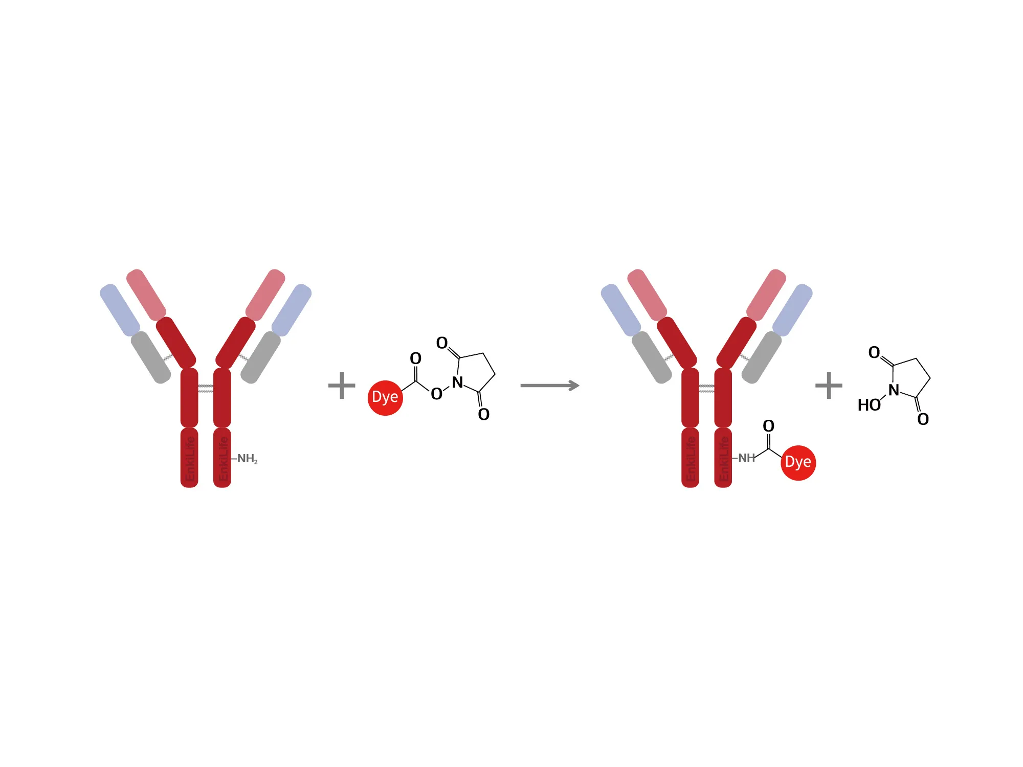
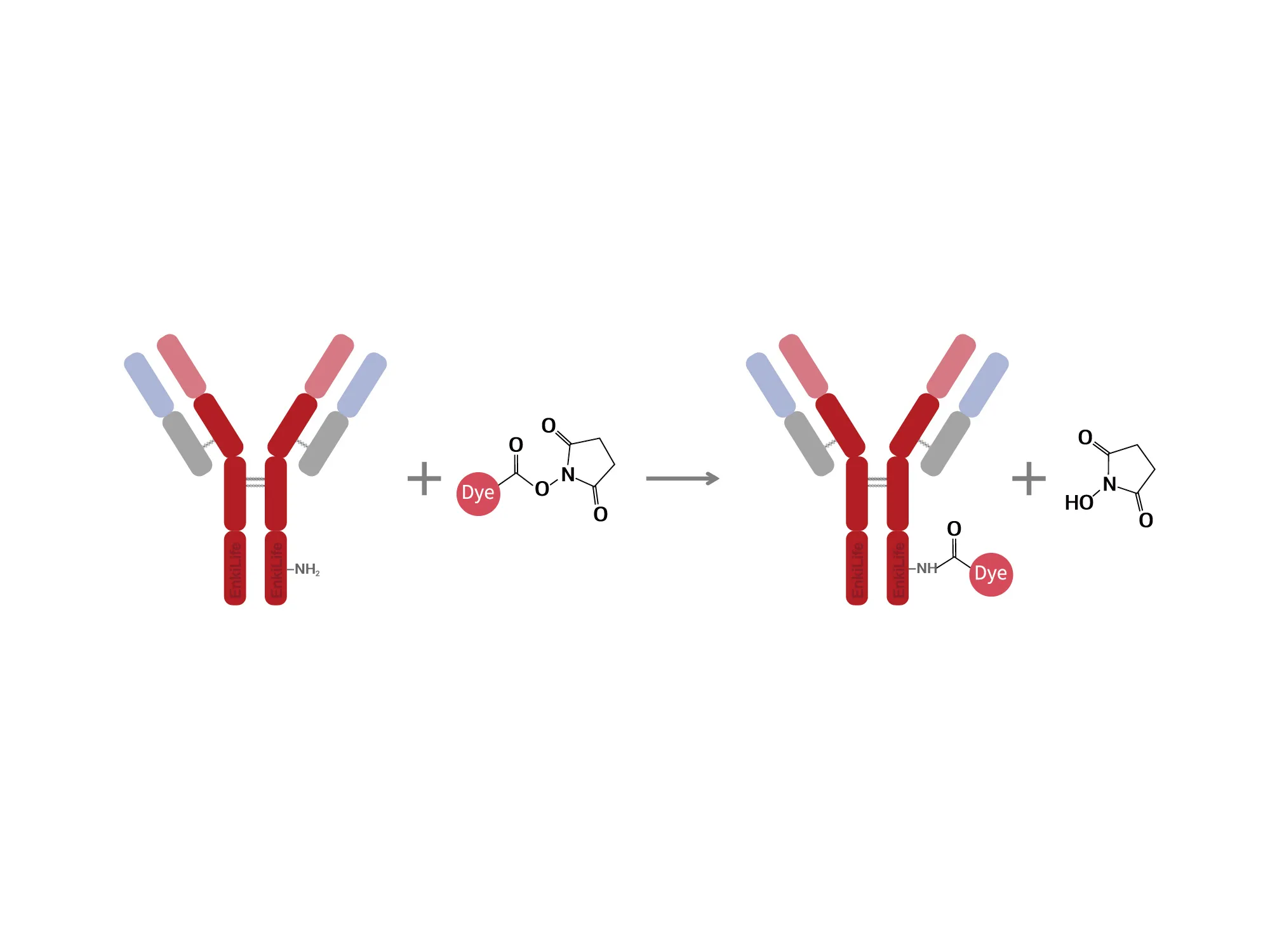
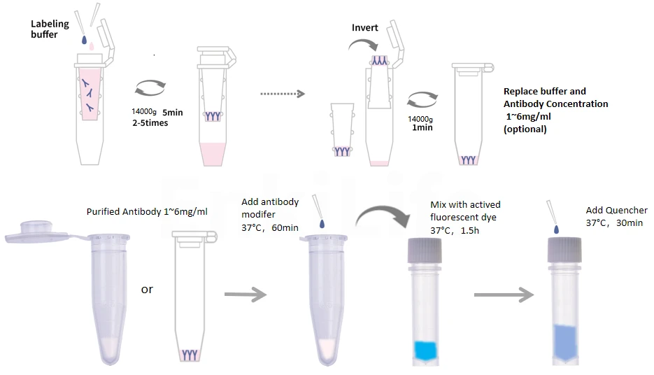
Labeling Principle
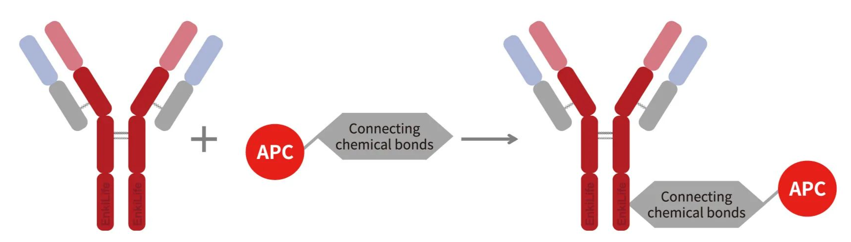
This kit utilizes the cysteine characteristics of the antibody and the free amino groups on the dye to covalently couple the antibody molecule with the dye using a directed docking coupling technique.
Standard Labeling Procedure
- STEP 1:Ultrafiltration buffer exchange and concentration (optional)
- STEP 2:Antibody activation
- STEP 3:Labeling reaction
- STEP 4:Quench/Blocking and preservation
Data of APC labeling kit application
Flow Cytometry Analysis
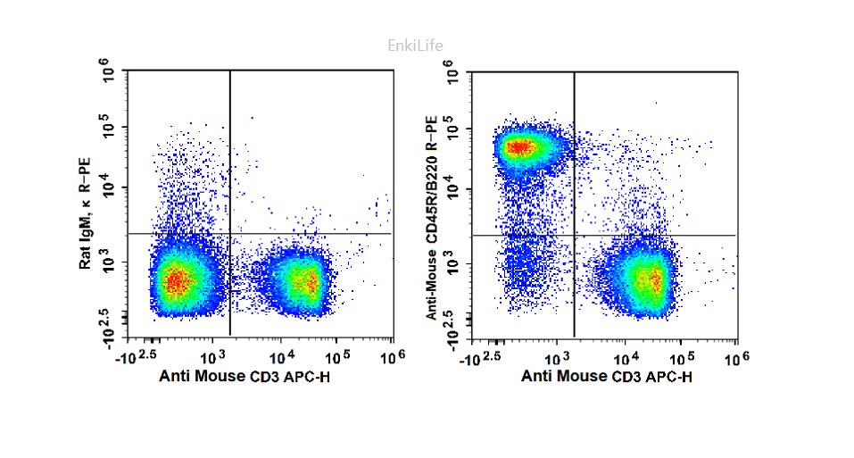
FC experiment of mouse lymphocyte using FC antibodies preparaed with two kinds of labeling kits
- Sample: mouse spleen lymphocyte cells from C57BL/6 mice
- Antibody: PE anti-mouse CD45R/CD220 antibody and APC anti-mouse CD3 antibody preparaed with two kinds of labeling kits(RE80005p and RE80006p)
- Isotype control: Rat IgM,kappa
- Instrument: Agilent ACEA NovoCyte™3130
Conclusion: The antibody staining showed clear signals with a high signal-to-noise ratio.The two FC antibody preparaed with two kinds of labeling kits is consistent with expected results. RE80006p kit is suitable for labeling anti-mouse CD3 antibodies.
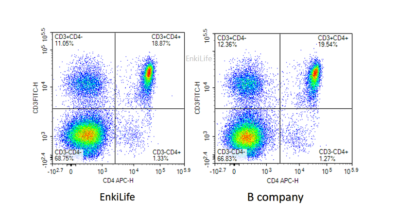
Flow Cytometry Analysis
Using the APC(RE80006p) and FITC labeling kit(RE80001p), label two antibodies. At the same time, use the corresponding internationally brand FC antibodies. After preparaing mouse spleen cell suspension, red blood cell lysis, 10^6 cells per test, perform flow cytometry detection.
- Antibody:
B company:0.05ug APC-anti Mouse CD4(GK1.5) and 0.2ug FITC-anti mouse CD3 from B company
EnkiLife:0.05ug APC-anti Mouse CD4(GK1.5) and 0.2ug FITC-anti mouse CD3 preparaed with two kinds of labeling kits from EnkiLife
Conclusion: The antibody preparaed with APC labeling kit and FITC labeling kit staining showed clear signals with a high signal-to-noise ratio and is similar with the internationally brand antibody.