Cell Lysis for Western Blotting: Choosing a Method and Preparing Protein Lysates
A practical guide to releasing, preserving, separating, and detecting proteins from cultured cells and tissues.
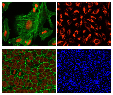
Cell fluorescence staining utilizes fluorescent probes to specifically bind to specific components within cells (such as DNA, proteins, and organelles) or react under specific physiological conditions, emitting characteristic fluorescence when irradiated by excitation light. With the aid of platforms including fluorescence microscopes, laser confocal microscopes, and flow cytometers, it enables cell morphology observation, target localization, expression quantification, and cell state analysis. Its core process covers two links: fluorescence labeling and signal detection. Probes first accumulate in target structures via specific binding or physiological response, then emit fluorescence at characteristic wavelengths under excitation light, and finally complete visualization and quantification through microscopic imaging or flow cytometry detection.
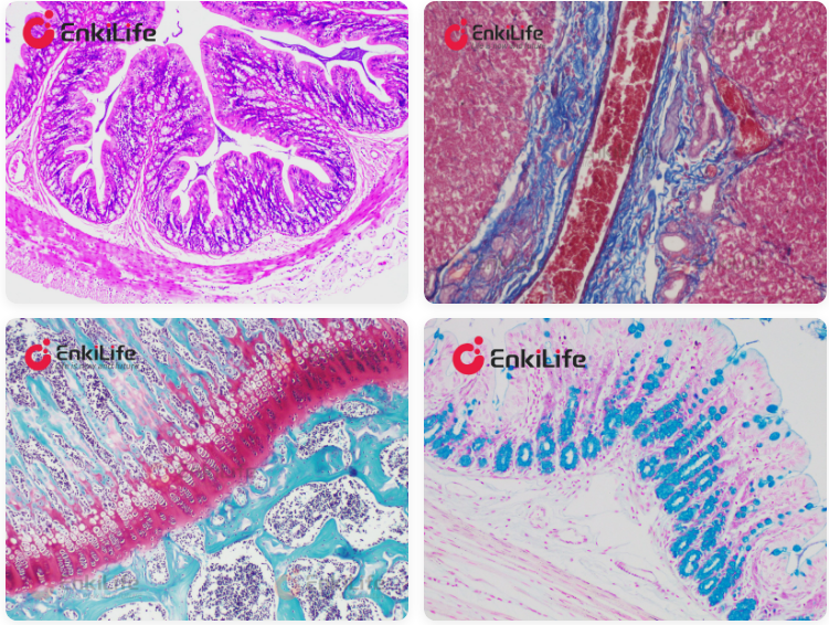
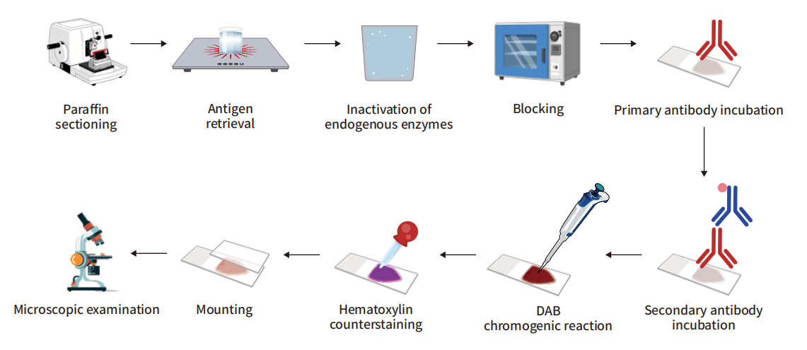
The core principle of pathological staining is to exploit differences in chemical affinity between tissue or cellular components and specific dyes, rendering them in different colors through chemical reactions or physical adsorption, so that cell structures, tissue types, or specific substances can be clearly distinguished under the microscope.

Tyramide Signal Amplification (TSA) is an enzyme‑catalyzed in situ detection method that overcomes the sensitivity and multiplexing limitations of conventional immunofluorescence. The core mechanism relies on the high catalytic activity of horseradish peroxidase (HRP) to achieve specific, high‑density labeling of target proteins and nucleic acids.

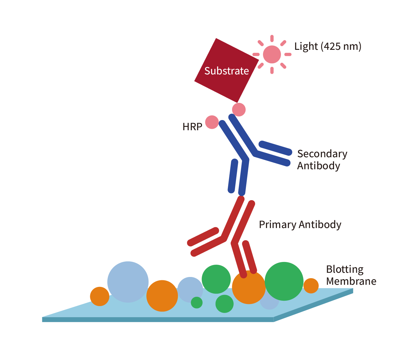
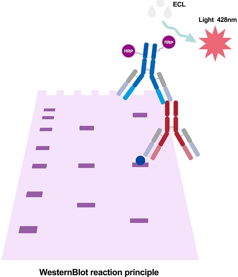
Western blotting is a widely used immunological method for detecting specific proteins in complex mixtures. It combines the separation power of gel electrophoresis with the specificity of antibodies. The process begins with protein extraction from cells or tissues, followed by separation by SDS-PAGE based on molecular weight. Proteins are then transferred to a membrane (typically nitrocellulose or PVDF) and detected using specific antibodies. The primary antibody binds to the target protein, while the secondary antibody, conjugated to a reporter enzyme, enables visualization through chemiluminescent, fluorescent, or colorimetric detection.