Cell Fluorescent Staining Technical Manual
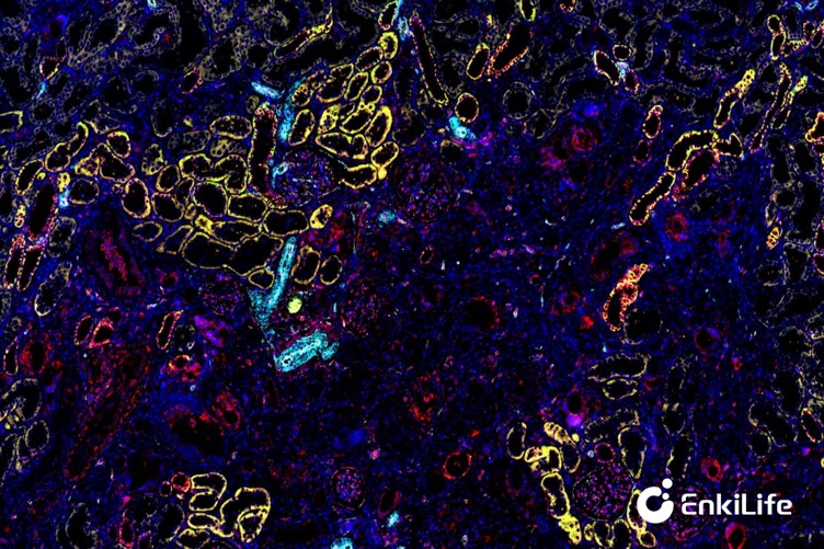
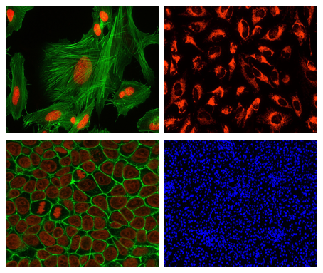
Cell fluorescence staining utilizes fluorescent probes to specifically bind to specific components within cells (such as DNA, proteins, and organelles) or react under specific physiological conditions, emitting characteristic fluorescence when irradiated by excitation light. With the aid of platforms including fluorescence microscopes, laser confocal microscopes, and flow cytometers, it enables cell morphology observation, target localization, expression quantification, and cell state analysis. Its core process covers two links: fluorescence labeling and signal detection. Probes first accumulate in target structures via specific binding or physiological response, then emit fluorescence at characteristic wavelengths under excitation light, and finally complete visualization and quantification through microscopic imaging or flow cytometry detection.