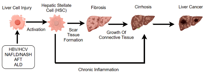
Hepatocellular carcinoma is the most common type of primary liver cancer worldwide (accounting for 70%-90% of primary liver cancer) and one of the leading causes of cancer-related deaths. The development of liver cancer after hepatocyte injury is a multi-stage, multi-factor-driven complex pathological process involving molecular genetic changes and microenvironmental abnormalities, and the core logic is a vicious circle of "continuous injury→ abnormal repair→ cell malignancy→ tumor progression". This process usually takes years to decades, and the mechanism of damage varies slightly between different triggers (e.g., hepatitis B virus, alcohol, non-alcoholic fatty liver disease, etc.), but there are commonalities in the final pathways[1].
Viral infection (HBV/HCV, the main trigger);
Alcoholic liver damage;
Non-alcoholic fatty liver disease (NAFLD/NASH);
Chemical carcinogens (e.g., aflatoxin B1, AFB1)[2].
After hepatocyte necrosis/apoptosis, the liver releases "hepatocyte growth factor (HGF)" to stimulate the proliferation (regeneration) of residual hepatocytes, but proliferation that is too fast increases the probability of DNA replication errors (equivalent to "more replication, higher risk of mutation");
Hepatic stellate cells (HSCs) are activated by inflammatory factors and converted into "myofibroblasts", secreting a large amount of collagen to form liver fibrosis - fibrosis not only leads to structural deformation of the liver, but also forms a "hypoxic microenvironment" (collagen fibers compress blood vessels and reduce blood flow), and hypoxia activates "hypoxia-inducible factor (HIF-1α)", which subsequently promotes tumor angiogenesis (paving the way for cancer).
Inflammatory cells (macrophages, T cells) continue to infiltrate, and the released inflammatory factors (such as IL-6) promote hepatocyte survival and proliferation through the "JAK-STAT3 pathway", even if the cells already have DNA damage, they are "forcibly retained" rather than eliminated by apoptosis (which is the core mechanism of "inflammatory cancer")[3,4].
p53: The most common inactivated tumor suppressor gene (p53 mutations are present in about 50% of HCCs), which cannot prevent damaged cell division after inactivation and cannot induce abnormal apoptosis, resulting in "indefinite survival" of mutant cells;
p16INK4a: Inactivated by methylation or deletion, unable to inhibit "cyclin-dependent kinase (CDK4/6)", resulting in cell cycle loss (direct entry from G1 phase to S phase, skipping DNA repair checkpoints);
PTEN: phosphatase gene, which cannot inhibit the "PI3K-Akt pathway" after inactivation, and continued activation of this pathway will promote cell proliferation, inhibit apoptosis, and enhance cell invasion.
Ras family (K-Ras, H-Ras): Continuous activation of the "Ras-MAPK pathway" after mutation, which is the core signal of cell proliferation, will "force" hepatocytes to divide indefinitely;
β-catenin: a key molecule of the Wnt signaling pathway, accumulates in cells after mutation, binds transcription factors after entering the nucleus, promotes the expression of proliferation-related genes such as "cyclin D1 and c-Myc", and inhibits cell differentiation (keeping cells in an "immature" proliferation state);
MET: The receptor gene for HGF, which is continuously activated after amplification or mutation, promotes hepatocyte proliferation and migration (paving the way for subsequent metastasis)[5,6].
Angiogenesis initiation: Hypoxia-inducible factor (HIF-1α) activated by the hypoxic microenvironment promotes the expression of vascular endothelial growth factor (VEGF), which attracts endothelial cells to migrate to tumor tissue and form "abnormal tumor blood vessels" - a key marker of cancer, as tumor cells need blood vessels to provide nutrients to grow from "millimeter-sized nodules" to "centimeter-scale tumors";
Immune escape: Tumor cells evade immune system clearance in several ways:
Expressing the "PD-L1" molecule, which binds to PD-1 of immune cells (T cells) and inhibits T cell activity;
Secrete "transforming growth factor β (TGF-β)" to induce regulatory T cell (Treg) aggregation and inhibit immune response;
Reduce the expression of "major histocompatibility complex (MHC)", so that immune cells cannot recognize tumor cells[7].

Diagram of the mechanisms of hepatocellular carcinoma
| Target | Catalog# | Product Name | Reactivity | Application |
|---|---|---|---|---|
| Antibodies related to the uncontrolled proliferation pathway of hepatic stellate cells: | ||||
| KRAS | APRab13128 | K-Ras Rabbit Polyclonal Antibody | Human,Mouse,Rat | WB,IHC-P,IF-P,IF-F,ICC/IF,ELISA |
| B-Raf | APRab07417 | BACE Rabbit Polyclonal Antibody | Human,Mouse,Rat | IHC-P,IF-P,IF-F,ICC/IF,ELISA |
| MEK1 | AMRe04007 | MEK1 Rabbit Monoclonal Antibody | Human,Mouse,Rat | WB,IHC-F,IHC-P,ICC/IF,FC |
| MEK1 | AMRe13797 | MEK1 (15N17) Rabbit Monoclonal Antibody | Human,Mouse,Rat | WB,IHC-P,ICC/IF,FC,IF-P |
| MEK2 | AMRe03082 | MEK2 Rabbit Monoclonal Antibody | Human,Mouse | WB,IHC-F,IHC-P,ICC/IF,IP |
| ERK1 | AMRe03741 | ERK1/2 Rabbit Monoclonal antibody | Human,Mouse,Rat | WB,ICC/IF,IP |
| mTOR | AMRe02286 | Phospho-mTOR (Ser2448) Rabbit Monoclonal Antibody | Human,Mouse | WB,IHC-P |
| AKT1 | AMRe06740 | AKT1 (5O1) Rabbit Monoclonal Antibody Rabbit Monoclonal Antibody | Human,Mouse | WB,IHC-P,ICC/IF,FC,IP,IF-P |
| β-catenin | AMRe03762 | beta Catenin Rabbit Monoclonal Antibody | Human,Mouse,Rat | WB,ICC/IF |
| β-catenin | AMRe03746 | beta Catenin Rabbit Monoclonal Antibody | Human,Mouse,Rat,Hamster | WB,IHC-P,IP |
| GPC3 | AMRe11520 | Glypican 3 (18Q6) Rabbit Monoclonal Antibody | Human | WB |
| c-Myc | AMRe05879 | Phospho-c-Myc (S62) (9Z2) Rabbit Monoclonal Antibody | Human,Mouse,Rat | WB,IHC-P,ICC/IF,FC,IP,IF-P |
| c-Myc | AMRe05880 | Phospho-c-Myc (T58) (1A2) Rabbit Monoclonal Antibody | Human,Mouse,Rat | WB,ICC/IF,FC |
| Antibodies related to the tumour microenvironment and angiogenesis pathways: | ||||
| P53 | AMRe03901 | Phospho-p53 (Ser392) Rabbit Monoclonal Antibody | Human, Mouse, Rat | WB,IHC-F,IHC-P,IP |
| P53 | AMRe02388 | p53 Rabbit Monoclonal antibody | Mouse | WB,ICC/IF,IP |
| PTEN | AMRe16636 | PTEN (16Q18) Rabbit Monoclonal Antibody | Human,Mouse,Rat | WB,IHC-P,FC,IP,IF-P |
| p16INK4A | AMRe01811 | CDKN2A/p16INK4a Rabbit Monoclonal Antibody | Human,Mouse | WB,ICC/IF |
| VEGFA | AMRe02757 | VEGFA Rabbit Monoclonal Antibody | Human,Mouse,Rat | WB |
| VEGFA1 | AMRe19767 | VEGF Receptor 1 (16I17) Rabbit Monoclonal Antibody | Human,Mouse,Rat | WB,IHC-P,IP,IF-P |
| VEGFR2 | APRab04679 | Flk-1 (phospho Tyr1214) Rabbit Polyclonal Antibody | Human,Mouse,Rat | WB,IHC-P,IF-P,IF-F,ICC/IF,ELISA |
| VEGFR2 | APRab04678 | Flk-1 (phospho Tyr1175) Rabbit Polyclonal Antibody | Human,Mouse,Rat | WB,IHC-P,IF-P,IF-F,ICC/IF,ELISA |
| VEGFR3 | APRab11039 | Flt-4 Rabbit Polyclonal Antibody | Human,Mouse,Rat | WB,IHC-P,IF-P,IF-F,ICC/IF,ELISA |
| CA9 | AMRe07799 | CA9 (14N17) Rabbit Monoclonal Antibody | Human,Mouse,Rat | WB,IHC-P,IP,IF-P |
| TERT | AMRe18798 | TERT (9Y18) Rabbit Monoclonal Antibody | Human | WB,IP |
| YAP1 | AMRe06050 | Phospho-YAP1 (S127) (14M14) Rabbit Monoclonal Antibody | Human,Mouse,Rat | WB,IHC-P,IHC-F |
| YAP1 | AMRe02781 | YAP1 Rabbit Monoclonal Antibody | Human,Mouse | WB,ICC/IF |
| FGF-21 | AMRe10930 | FGF21 (7E19) Rabbit Monoclonal Antibody | Human,Mouse,Rat | WB,IHC-P,IF-P |
| Antibodies related to inflammation and immune regulation pathways: | ||||
| TNFα | AMM19084 | TNF α(Q34)Mouse Monoclonal Antibody | Human,Mouse,Rat | WB,IHC-P,IF-P,IF-F,ICC/IF |
| IL-6 | APRab03851 | IL-6 Rabbit Polyclonal Antibody | Human | WB,IHC-P,ELISA |
| IL-10 | AMRe12483 | IL10 (8U9) Rabbit Monoclonal Antibody | Human | WB,ICC/IF,FC |
| TGF-β1 | AMM00661 | TGF beta 1 (8F6) Mouse Monoclonal Antibody | Human,Mouse,Rat | WB,IHC-P |
| STAT3 | AMRe06021 | Phospho-STAT3 (Y705) (13H8) Rabbit Monoclonal Antibody | Human,Mouse,Rat | WB,IHC-P,ICC/IF,FC,IP,IF-P |
| STAT3 | AMRe18352 | STAT3 (11W6) Rabbit Monoclonal Antibody | Human,Mouse,Rat | WB,IHC-P,ICC/IF,FC,IF-P |
| Galectin-9 | APRab11278 | Galectin-9 Rabbit Polyclonal Antibody | Human,Mouse,Rat | WB,IHC-P,IF-P,IF-F,ICC/IF,ELISA |
| NLRP3 | AMRe01571 | NLRP3 Rabbit Monoclonal Antibody | Human,Mouse,Rat | WB |
| NLRP3 | AMRe14399 | NALP3 (8Q17) Rabbit Monoclonal Antibody | Human,Mouse,Rat | WB,FC,IP |
| PD-L1 | AMRe15922 | PD-L1 (CD274) (5R18) Rabbit Monoclonal Antibody | Human | WB,IHC-P,ICC/IF,FC,IP,IF-P |
| PD-1 | PD-L2 Rabbit Polyclonal Antibody | Human,Mouse,Rat | WB,IHC-P,IF-P,IF-F,ICC/IF,ELISA | |
| CTLA-4 | AMRe09507 | CTLA4 (CD152) (14H2) Rabbit Monoclonal Antibody | Human,Mouse | WB,IHC-P,FC,IP,IF-P |
| Other relevant antibodies: | ||||
| α-Fetoprotein | AMRe06665 | AFP (1J18) Rabbit Monoclonal Antibody | Human | WB,IHC-P,IP,IF-P |
| Albumin | AMRe17769 | Serum Albumin (14W10) Rabbit Monoclonal Antibody | Human | WB |
| Arginase-1 | AMRe07109 | ARG1 (7H3) Rabbit Monoclonal Antibody | Human | WB,IHC-P,IP,IF-P |
| GLUL | AMM82509 | GLUL Mouse Monoclonal Antibody | Human,Mouse | WB,IHC,ICC,FC,ELISA |
| S100A6 | AMRe03193 | S100 alpha6 Rabbit Monoclonal Antibody | Human,Mouse | WB,ICC/IF,IP |
| Vimentin | AMRe03745 | Vimentin Rabbit Monoclonal Antibody | Human,Mouse,Rat,Hamster | WB,IHC-F,IHC-P,ICC/IF |
| Vimentin | AMM80578 | Vimentin Mouse Monoclonal Antibody | Human,Mouse,Monkey | WB,IHC,FC,ELISA |
| KRT7 | AMM08857 | CK7(12D7)Mouse Monoclonal Antibody | Human,Mouse,Rat | IF-P,IF-F,ICC/IF,WB,IHC-P,IP |
| E-Cadherin | AMRe01411 | E-Cadherin Rabbit Monoclonal Antibody | Human | WB,IHC-F,IHC-P,ICC/IF,IP |
| HSP70 | AMRe21556 | Hsp70 Rabbit Monoclonal antibody | Human,Mouse,Rat | WB,IHC,IF,IP,ELISA |
| HSP70 | AMRe03777 | Hsp70 1B Rabbit Monoclonal antibody | Human,Mouse,Rat | WB,IHC-P,IP |
| RASSF1 | AMRe16920 | RASSF1 (19I9) Rabbit Monoclonal Antibody | Human | WB,IHC-P,IF-P |
| Target | Catalog# | Product Name | Reactivity | Detection Range | Sensitivity |
|---|---|---|---|---|---|
| VEGFA | EM10657 | Mouse VEGF-A (Vascular Endothelial Cell Growth Factor A) ELISA Kit | Mouse | 31.25-2000pg/mL | 18.75pg/mL |
| α-Fetoprotein | EH10304 | Human αFP (Alpha-Fetoprotein) ELISA Kit | Human | 1.56-100ng/mL | 0.94ng/mL |
| TNFα | EH10021 | Human TNF-α (Tumor Necrosis Factor Alpha) ELISA Kit | Human | 7.81-500pg/mL | 4.69pg/mL |
| TNFα | EM27661S | High Sensitivity Mouse TNF-α (Tumor Necrosis Factor Alpha) ELISA Kit | Mouse | 1.56-100pg/mL | 0.93pg/mL |
| IL-1β | EM27654S | Mouse IL-1β (Interleukin 1 Beta) ELISA Kit | Mouse | 3.13-200pg/mL | 1.87pg/mL |
| IL-6 | EM21023S | High Sensitivity Mouse IL-6 (Interleukin 6) ELISA Kit | Mouse | 0.781-50pg/mL | 0.47pg/mL |
| IL-6 | EH10020 | Human IL-6 (Interleukin 6) ELISA Kit | Human | 1.56-100pg/mL | 0.94pg/mL |
Related Products
[1].Ilyas SI, Wang J, El-Khoueiry AB. Liver Cancer Immunity. Hepatology. 2021 Jan;73 Suppl 1(Suppl 1):86-103. [PMID: 32516437].
[2].Sun Y, Li H, Chen Q, Luo Q, Song G. The distribution of liver cancer stem cells correlates with the mechanical heterogeneity of liver cancer tissue. Histochem Cell Biol. 2021 Jul;156(1):47-58. [PMID: 33710418].
[3].Bakrania A, Joshi N, Zhao X, Zheng G, Bhat M. Artificial intelligence in liver cancers: Decoding the impact of machine learning models in clinical diagnosis of primary liver cancers and liver cancer metastases. Pharmacol Res. 2023 Mar;189:106706. Epub 2023 Feb 20. [PMID: 36813095].
[4].Wu M, Wang H, Wu X, Zeng H, Miao M, Song Y. Research Progress of Liver Cancer Recurrence Based on Energy Metabolism of Liver Cancer Stem Cells. J Hepatocell Carcinoma. 2025 Mar 3;12:467-480. [PMID: 40061164].
[5].Chao X, Qian H, Wang S, Fulte S, Ding WX. Autophagy and liver cancer. Clin Mol Hepatol. 2020 Oct;26(4):606-617. [PMID: 33053934].
[6].Feng M, Pan Y, Kong R, Shu S. Therapy of Primary Liver Cancer. Innovation (Camb). 2020 Aug 28;1(2):100032. [PMID: 32914142].
[7].Yang Y, Yu S, Lv C, Tian Y. NETosis in tumour microenvironment of liver: From primary to metastatic hepatic carcinoma. Ageing Res Rev. 2024 Jun;97:102297. [PMID: 38599524].
 | Flora Flora is a technical support expert at EnkiLife, familiar with immunology and neuroscience, dedicated to providing customers with high-quality product combinations and technical support to help achieve research in neurodegenerative diseases and other neuroscience areas. |
© 2025 EnkiLife Hepatocellular carcinoma Research Materials | Providing Professional Antibodies and ELISA Kits
