
Summary
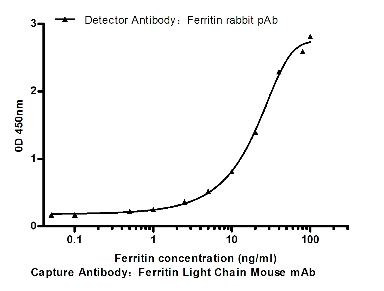
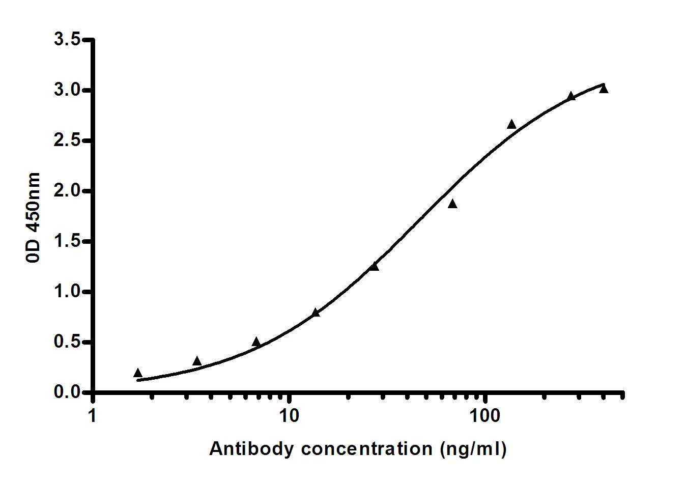
Performance
Immunogen
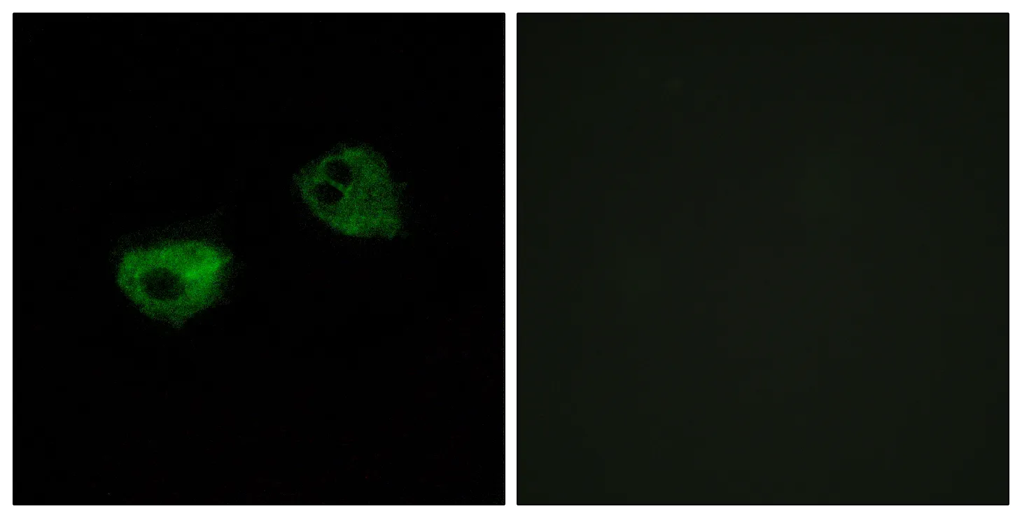
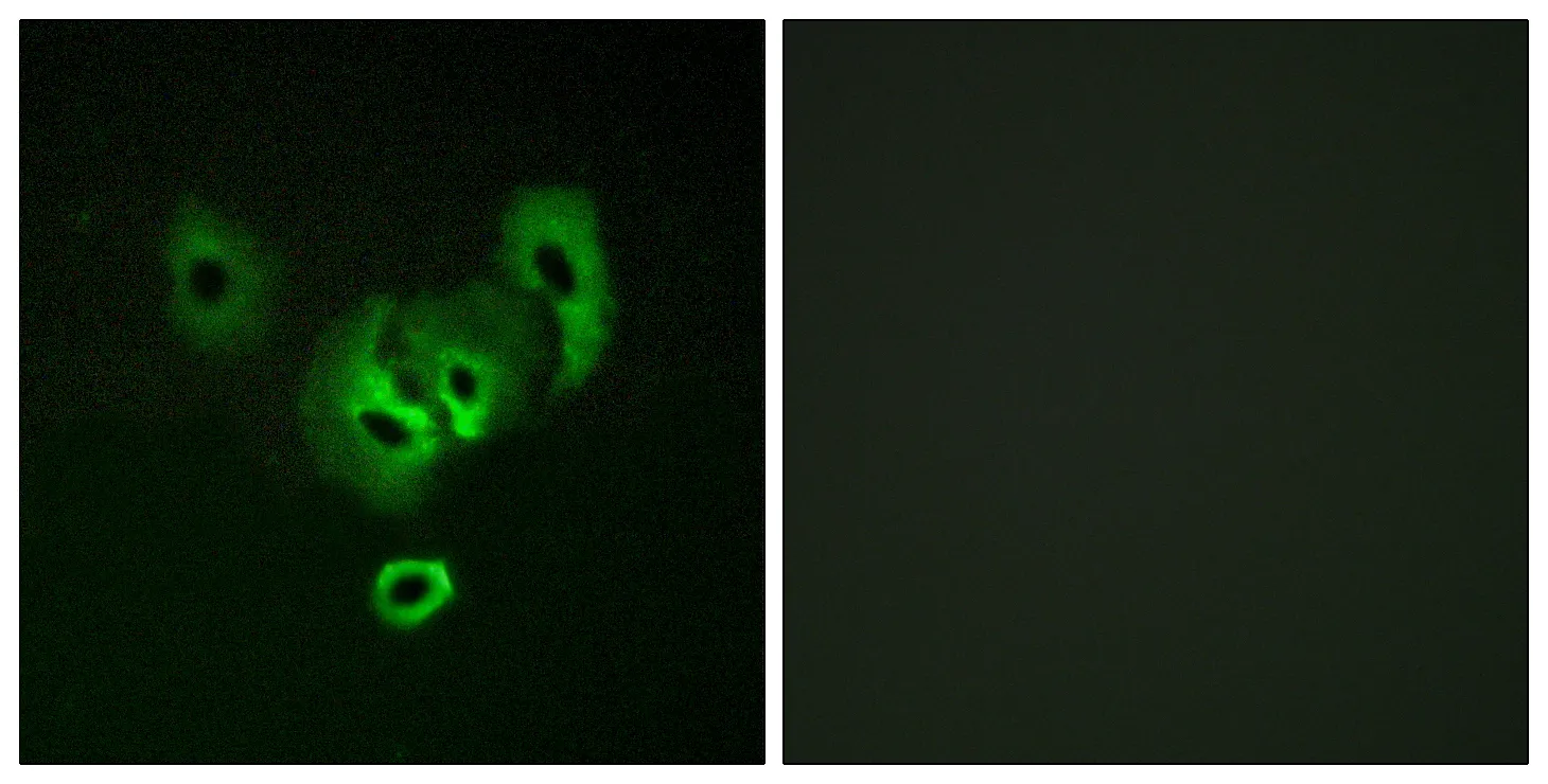
Application
Background
The A-kinase anchor proteins (AKAPs) are a group of structurally diverse proteins, which have the common function of binding to the regulatory subunit of protein kinase A (PKA) and confining the holoenzyme to discrete locations within the cell. This gene encodes a member of the AKAP family. The encoded protein binds to the RII-beta regulatory subunit of PKA, and also to protein kinase C and the phosphatase calcineurin. It is predominantly expressed in cerebral cortex and may anchor the PKA protein at postsynaptic densities (PSD) and be involved in the regulation of postsynaptic events. It is also expressed in T lymphocytes and may function to inhibit interleukin-2 transcription by disrupting calcineurin-dependent dephosphorylation of NFAT. [provided by RefSeq, Jul 2008],domain:RII-alpha binding site, predicted to form an amphipathic helix, could participate in protein-protein interactions with a complementary surface on the R-subunit dimer.,function:May anchor the PKA protein to cytoskeletal and/or organelle-associated proteins, targeting the signal carried by cAMP to specific intracellular effectors. Association with to the beta2-adrenergic receptor (beta2-AR) not only regulates beta2-AR signaling pathway, but also the activation by PKA by switching off the beta2-AR signaling cascade.,miscellaneous:The N-terminal region, which is highly basic, is required for interaction with calmodulin.,similarity:Contains 1 AKAP domain.,subcellular location:Associated with particulate fractions.,subunit:Binding protein for dimer of the RII-beta regulatory subunit of cAMP-dependent protein kinase (PKA) and also for the protein kinase C (PKC) and the phosphatase calcineurin (PP2B). Each enzyme is inhibited when bound to the anchoring protein. Also binds the beta2-adrenergic receptor.,tissue specificity:Predominantly in the cerebral cortex and the postsynaptic densities of the forebrain, and to a lesser extent in adrenal medulla, lung and anterior pituitary.,
Research Area