

Summary
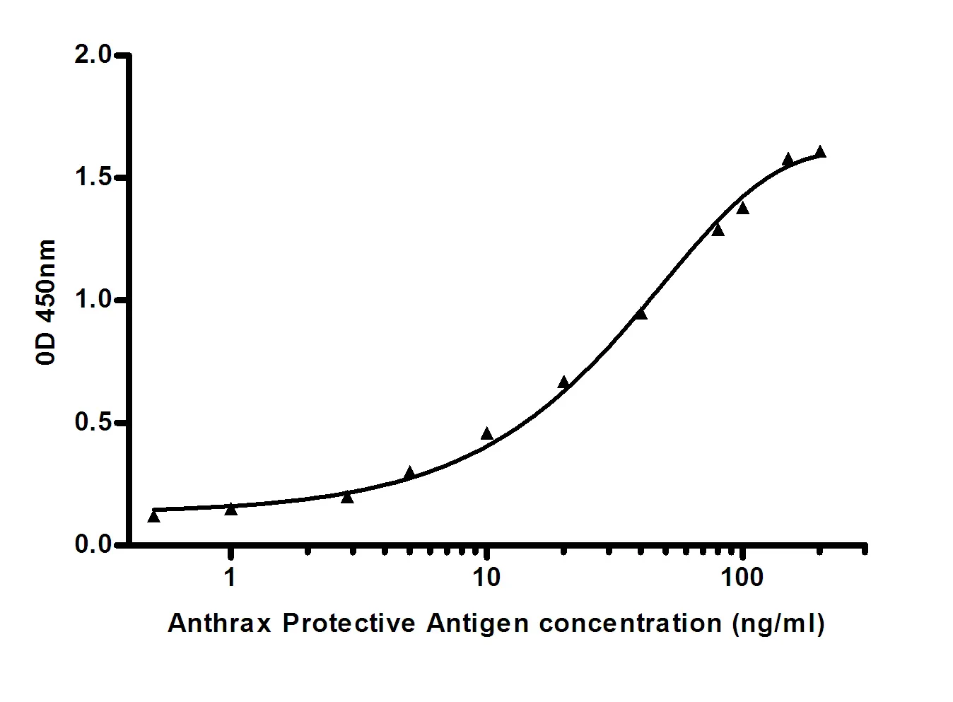
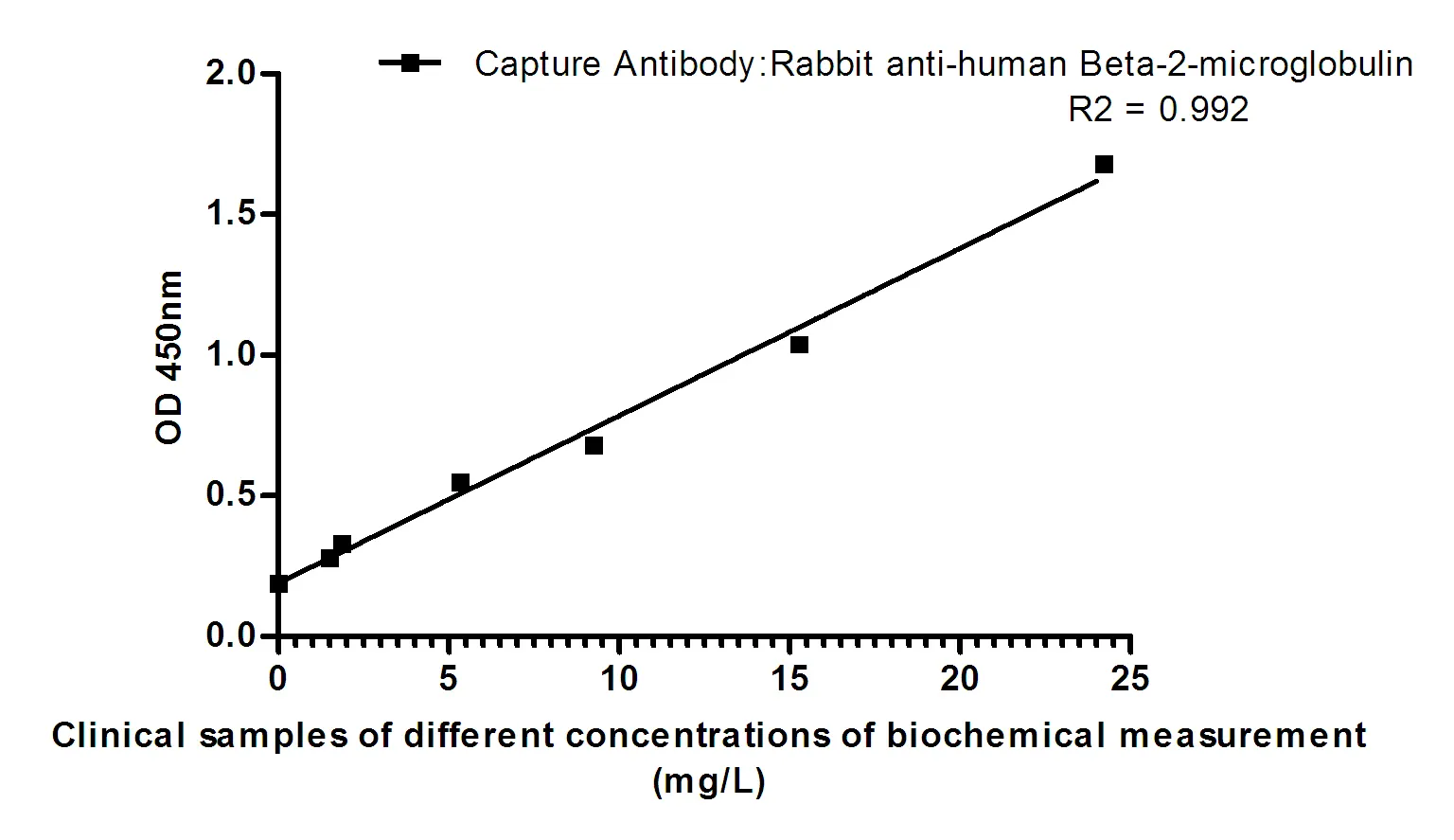
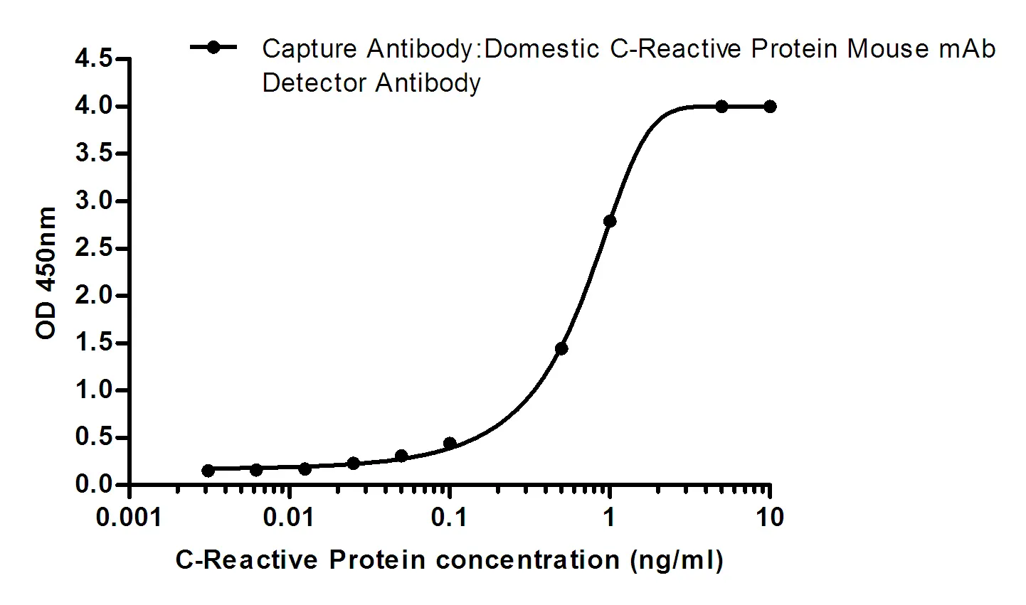
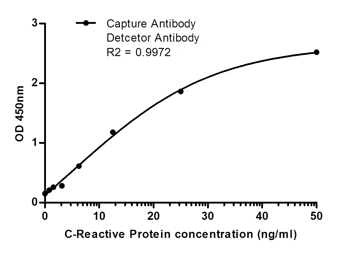
Performance
Immunogen
Application
Background
The product of this gene belongs to the integrin alpha chain family. Integrins are heterodimeric integral membrane proteins composed of an alpha chain and a beta chain. This gene encodes the integrin alpha 5 chain. Alpha chain 5 undergoes post-translational cleavage in the extracellular domain to yield disulfide-linked light and heavy chains that join with beta 1 to form a fibronectin receptor. In addition to adhesion, integrins are known to participate in cell-surface mediated signalling. Integrin alpha 5 is a heterodimer that associates noncovalently with CD29/integrin beta 1 subunit to form the alpha-5-beta-1 very late antigen (VLA-5) complex. VLA-5 is a fibronectin receptor that is expressed on thymocytes, T-cells, monocytes and platelets. It is also found on very early B-cells and activated B-cells. VLA-5-mediated binding to fibronectin sends a costimulatory signal to T-cells and enhances Fc-gamma-R- and complement receptor-mediated phago-cytosis. It is also involved in monocyte migration into extracellular tissues.
Research Area
PI3K-Akt signaling pathway