
Product Overview
The EnkiLife Cyanine7 Labeling Kit contains multiple components for labeling antibody proteins, such as high-performance fluorescent dye, purification ultrafiltration tubes, elution labeling buffer, and labeled protein storage solution. It enables easy and reliable labeling of antibodies, proteins, and other molecules for use as fluorescent probes.
Product Features
- Fast: Labeling reaction takes only about 30 minutes, balancing both labeling efficiency and effectiveness.
- Simple: Each dye reagent is pre-optimized for the corresponding antibody amount, eliminating tedious calculations. Follow the steps to achieve excellent results.
- Flexible: Dyes in large sizes kits can be aliquoted with consistent batch performance, convenient for multiple uses.
- Excellent Labeling: Optimized labeling buffer enables relatively fixed labeling sites, enhancing the homogeneity of labeled antibodies.
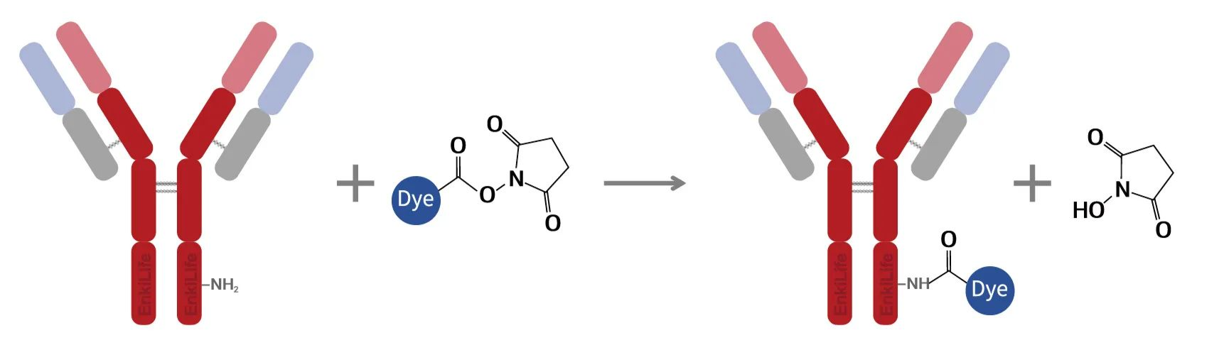
- Amine-Specific Labeling: The reactive group on Cyanine7 efficiently binds to primary amine groups on antibodies or other purified proteins, achieving covalent labeling.
- Protein Labeling: Primarily designed for IgG antibodies, but can also label proteins of similar size and properties to IgG.
Dye Introduction
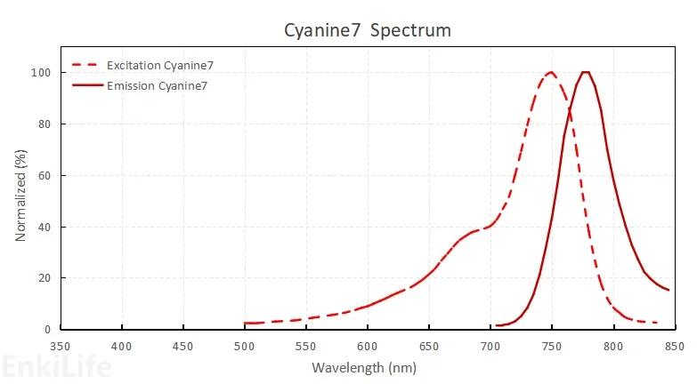
Cyanine7 is a near-infrared (NIR) fluorescent dye. Its emitted fluorescence is invisible to the naked eye but can be detected by most fluorescence instruments. It is a very commonly used dye for small animal imaging, frequently employed to label nucleic acids, proteins, peptides, nanoparticles, and polymers. Due to its longer emission wavelength compared to Cyanine5.5, it generally offers higher sensitivity and lower background in small animal imaging experiments. It can also be used in tandem with PE dyes for flow cytometry.
Dye Properties
Basic Characteristics
Similar Dyes:Alexa Fluor® 750, DyLight® 750, IRDye® 750
Common Excitation Sources: 680 or 685 nm
Maximum Excitation Wavelength: 750nm
Maximum Emission Wavelength: 773nm
Molar Extinction Coefficient: 200000L/(mol·cm)
Quantum Yield:0.3
Appearance:Dark green solid
Kit Specifications
| Product Components | Component Content in Different Specifications | Storage Temperature | ||
|---|---|---|---|---|
| 40μg Antibody | 200μg Antibody | 2mg Antibody | ||
| Activated Dye | 1 tube | 5 tubes | 5 tubes | -20°C after opening, protected from light |
| 50KDa Ultrafiltration Tube* | 1 set** | 1 set** | 1 set** | RT |
| Labeling Buffer S | 10 mL | 10 mL | 10 mL | 2~8°C |
| 1×PBS (pH 7.4) | 10 mL | 10 mL | 10 mL | 2~8°C |
| DMF | 100 μL | 100 μL | 100 μL | 2~8°C, protected from light |
| Labeled Protein Storage Solution | 200 μL | 1 mL | 5 mL | 2~8°C |
| Recommended Labeling Amount | Each tube dyes for labeling 20-40μg antibody Recommended: 20μg antibody | Each tube dyes for labeling 20-40μg antibody Recommended: 20μg antibody | Each tube dyes for labeling 100-400μg antibody Recommended: 200μg antibody | |
* Ultrafiltration tube molecular weight cutoff: 50KDa
** 1 set = 1 ultrafiltration tube and collection tube
Labeling Principle
Under specific chemical reaction conditions, the fluorescent dye activation group specifically reacts with primary amine groups on antibody proteins, forming stable amide bonds, thereby achieving labeling conjugation with antibody proteins.
Standard Labeling Procedure
- 1.Ultrafiltration for buffer exchange and concentration
- 2.Labeling reaction
- 3.Purification
- 4.DOL calculation, collection and storage