

Summary
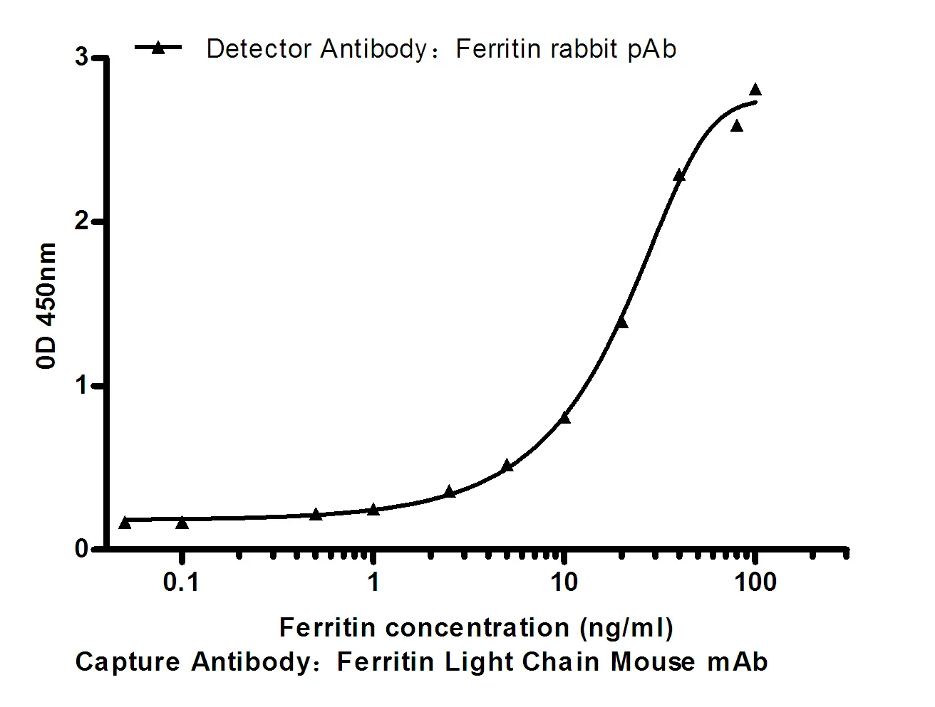
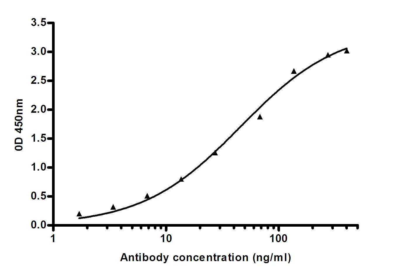
Performance
Immunogen
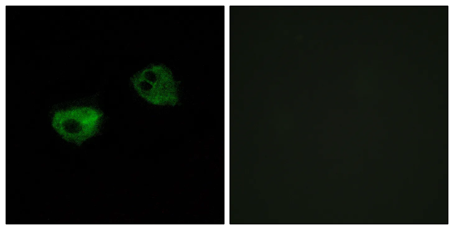
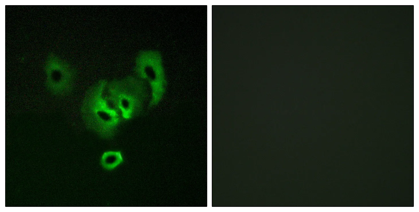
Application
Background
This gene encodes a member of the A-kinase anchor protein family. A-kinase anchor proteins bind to the regulatory subunits of protein kinase A (PKA) and confine the holoenzyme to discrete locations within the cell. The encoded protein is localized to mitochondria and interacts with both the type I and type II regulatory subunits of PKA. Polymorphisms in this gene may be associated with increased risk of arrhythmias and sudden cardiac death. [provided by RefSeq, May 2012],domain:RII-alpha binding site, predicted to form an amphipathic helix, could participate in protein-protein interactions with a complementary surface on the R-subunit dimer.,function:Differentially targeted protein that binds to type I and II regulatory subunits of protein kinase A and anchors them to the mitochondria or the plasma membrane. Although the physiological relevance between PKA and AKAPS with mitochondria is not fully understood, one idea is that BAD, a proapoptotic member, is phosphorylated and inactivated by mitochondria-anchored PKA. It cannot be excluded too that it may facilitate PKA as well as G protein signal transduction, by acting as an adapter for assembling multiprotein complexes. With its RGS domain, it could lead to the interaction to G-alpha proteins, providing a link between the signaling machinery and the downstream kinase.,similarity:Contains 2 RGS domains.,subcellular location:Predominantly mitochondrial but also membrane associated and cytoplasmic.,
Research Area