

Summary
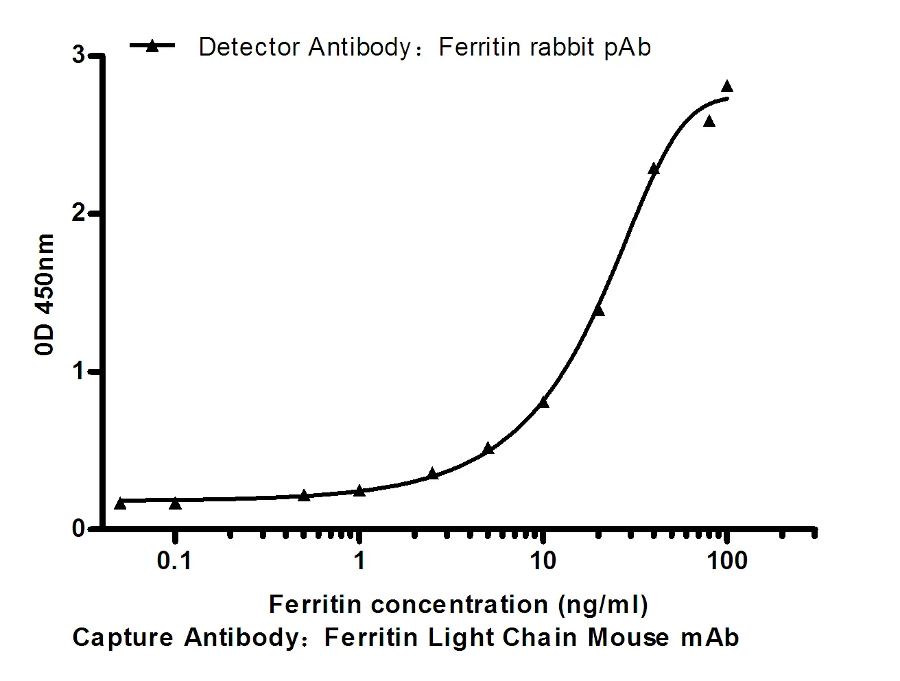
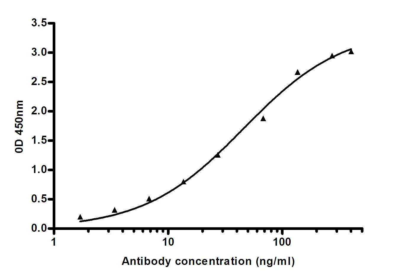
Performance
Immunogen
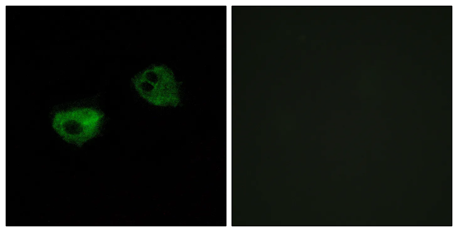
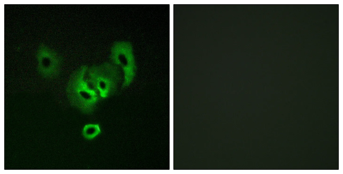
Application
Background
The giant protein titin, together with its associated proteins, interconnects the major structure of sarcomeres, the M bands and Z discs. The C-terminal end of the titin string extends into the M line, where it binds tightly to M-band constituents of apparent molecular masses of 190 kD (myomesin 1) and 165 kD (myomesin 2). This protein, myomesin 1, like myomesin 2, titin, and other myofibrillar proteins contains structural modules with strong homology to either fibronectin type III (motif I) or immunoglobulin C2 (motif II) domains. Myomesin 1 and myomesin 2 each have a unique N-terminal region followed by 12 modules of motif I or motif II, in the arrangement II-II-I-I-I-I-I-II-II-II-II-II. The two proteins share 50% sequence identity in this repeat-containing region. The head structure formed by these 2 proteins on one end of the titin string extends into the center of the M band. The integrating structurefunction:Major component of the vertebrate myofibrillar M band. Binds myosin, titin, and light meromyosin. This binding is dose dependent.,similarity:Contains 5 fibronectin type-III domains.,similarity:Contains 5 Ig-like C2-type (immunoglobulin-like) domains.,subunit:Interacts with TTN/titin (By similarity). Interacts with PNKD.,
Research Area