A Comprehensive Overview of Lateral Flow Chromatographic Assay Strips
Lateral Flow Chromatographic Assay (LFCA) originated from paper chromatography developed by Martin and Synge in 1943, and was elaborated in detail by Consden, Gordon, and Martin in 1944. After 1945, research in this field surged. ELISA technology was developed in 1971. Armkel LLC began applying for a series of LFT patents starting in 1988.
Lateral flow chromatography technology offers many advantages, such as low cost, ease of operation, rapid results, potential for on-site testing, and visually interpretable results. Based on these advantages, LFCA has achieved rapid development in both basic and applied research fields since its emergence. In 1980, the first LFCA strip for detecting human chorionic gonadotropin (hCG) was developed for early pregnancy diagnosis. Since then, LFCA has been widely used to detect various molecules, including tumor markers, microorganisms, mycotoxins, heavy metals, and pesticides.
1. Antibody-Antigen Binding Based LFCA
Antigens and antibodies are essentially proteins. As long as it is a protein, there is a corresponding antibody or antigen that can pair with it. Therefore, whether detecting an antigen or an antibody, the principle is essentially the same; only the immobilized materials are swapped.
When antibodies are used as biorecognition elements, LFCA is also called Immunochromatographic Rapid Test Technology. It is a solid-phase membrane immunoassay method developed in the 1980s that combines labeled immunoassay technology with chromatographic separation technology. Based on the different binding modes of antigens and antibodies during the immunochromatographic reaction, it is mainly divided into two types: the double-antibody sandwich immunochromatographic assay and the competitive immunochromatographic assay.
1.1 Double-Antibody Sandwich Immunochromatographic Assay (Sandwich Assay)
The double-antibody sandwich immunochromatographic assay, also known as the sandwich assay, is typically used for larger analytes as they often have multiple binding sites. When the sample migrates during the analysis process, it first encounters a specific antibody, usually labeled with colloidal gold. The antibody binds to the target protein in the sample, and they migrate together until reaching the test line (T line). The test line contains immobilized antibodies that can specifically bind the target protein; these antibodies bind to the conjugated molecules (e.g., colloidal gold-labeled antibodies) that are bound to the migrating analyte. Then, due to the concentration of colloidal gold, the test line shows a visual change, confirming the presence of the target molecule. Most sandwich assays also have a control line (C line) that appears regardless of the presence of the target analyte, ensuring the normal function of the lateral flow pad. This rapid, low-cost sandwich detection method is commonly used in home pregnancy tests to detect human chorionic gonadotropin (hCG) in the urine of pregnant women.
1.2 Competitive Immunochromatographic Assay
The competitive assay is typically used for smaller analytes because smaller analytes have fewer binding sites. The sample first encounters an antibody against the target analyte, labeled with a visual marker (colored particles). The test line contains the target analyte immobilized on the surface. When the target analyte is not present in the sample, the unbound antibodies will bind to these fixed analyte molecules, meaning the visual marker will be displayed. Conversely, when the target analyte is present in the sample, it binds to the antibodies, preventing them from binding to the fixed analytes on the test line, so no visual marker is displayed. This differs from the sandwich assay, as the absence of a band indicates the presence of the analyte.
There are two modes of the competitive method: one for detecting antigens, and another for detecting specific antibodies when interfering substances in the antigen material are difficult to remove or when sufficient purified antigen is not readily available. Taking colloidal gold labeling as an example:
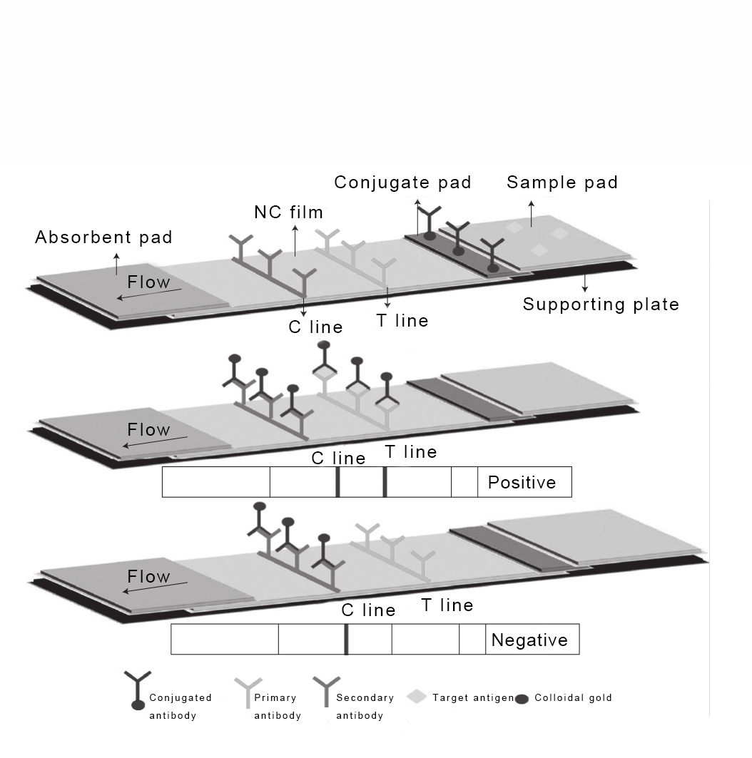
In the first mode (detecting antigen), a certain amount of target antigen is labeled with colloidal gold and fixed on the conjugate pad. The T line and C line are sprayed with an antibody specifically binding the target antigen (primary antibody) and an "anti-antibody" (commonly known as secondary antibody) against species-specific IgG, respectively. For a negative sample, the gold-labeled antigen migrates laterally with the sample, binds to the primary antibody at the T line, and binds to the secondary antibody at the C line, resulting in both T and C lines showing red color. For a positive sample, the unlabeled target antigen in the sample competes with the gold-labeled target antigen on the conjugate pad for binding to the primary antibody at the T line. The more target antigen in the sample, the less gold-labeled antigen binds to the T line, resulting in lighter color. The excess gold-labeled antigen binds to the secondary antibody at the C line, so only the C line shows red.
Schematic Diagram of Competitive LFCA Principle (Mode 1)
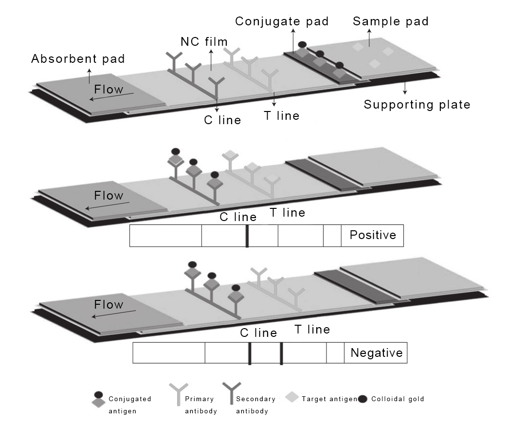
In the second mode (detecting antibody), the antibody is labeled with colloidal gold on the conjugate pad. The T line and C line contain an antigen-carrier molecule (usually Bovine Serum Albumin, BSA) conjugate and an "anti-antibody" (secondary antibody) against species-specific IgG, respectively. The target antibody in the sample and the gold-labeled antibody on the conjugate pad compete for binding to the antigen-carrier conjugate on the T line. For a negative sample, the gold-labeled antibody on the conjugate pad reacts and binds to the immobilized materials on the T and C lines, causing both lines to show red. For a positive sample, the target antibody in the sample preferentially binds specifically to the antigen-carrier conjugate on the T line. The more target antibody in the sample, the less gold-labeled antibody binds to the T line, resulting in lighter color. The excess gold-labeled antibody binds to the secondary antibody at the C line, so only the C line shows red.
Schematic Diagram of Competitive LFCA Principle (Mode 2)
2. Nucleic Acid-Based LFCA
The high specificity and strong affinity between avidin and biotin, allowing them to bind various molecules, form the Avidin-Biotin System (ABS), which facilitates combining nucleic acids with LFCA. Similar to ABS, systems like biotin-anti-biotin antibody, biotin-streptavidin, fluorescein-anti-fluorescein antibody, digoxigenin-anti-digoxigenin antibody also serve as bridging links. Based on this, complementary nucleotide sequences (DNA-DNA, DNA-RNA, RNA-RNA, etc.) form non-covalent bonds through Watson-Crick base pairing, enabling the nucleic acid hybridization process to form stable homologous or heterologous double-stranded molecules. Thus, nucleic acids can also be used as biorecognition elements in LFCA. The detection results and analysis are as follows: Samples showing bands on both T and C lines are positive; samples where the C line is colored but the T line is not are negative; if the C line does not develop color, the result is invalid. Based on whether antibodies are required in the process, nucleic acid-based LFCA is divided into two types: antibody-dependent (also known as Nucleic Acid Lateral Flow Immunoassay, NALFIA) and antibody-independent (also known as Nucleic Acid Lateral Test Strips, NALTS).
2.1 NALFIA Technology
Antibody-dependent LFCA utilizes nucleic acid-antibody interactions and specific labeling of double-stranded DNA, hence it is called NALFIA. NALFIA is essentially a combined application of Polymerase Chain Reaction (PCR) technology and ELISA technology.
Taking colloidal gold NALFIA as an example: First, a pair of corresponding PCR primers are designed for the specific gene fragment of the detection target, and labeled with two different markers (e.g., biotin labeling one primer, and fluorescein labeling the other). PCR amplification produces double-labeled double-stranded amplicons. A test strip is manufactured by fixing a complex of a substance that specifically binds one of the amplicon markers (e.g., streptavidin or avidin) and colloidal gold on the conjugate pad. The T line is sprayed with a labeled antibody specific to the other marker of the amplicon (e.g., anti-fluorescein antibody). The C line is sprayed with a substance that can specifically bind the complex on the conjugate pad (e.g., biotin). The manufactured colloidal gold test strip is used to detect the PCR amplicons.
For a positive sample, the double-labeled amplicon migrates laterally from the sample pad to the conjugate pad due to capillary action. Its biotin binds specifically to streptavidin or avidin on the conjugate pad. The complex then migrates to the T line, where the fluorescein on the double-labeled amplicon binds specifically to the anti-fluorescein antibody on the T line. Excess streptavidin or avidin from the conjugate pad binds specifically to the biotin on the C line. Because the streptavidin or avidin on the conjugate pad is labeled with colloidal gold, both T and C lines appear red simultaneously. For a negative sample, the streptavidin or avidin on the conjugate pad binds specifically to the biotin on the C line, so only the C line appears red. If the C line does not develop color, the result is invalid.
2.2 NALTS Technology
Antibody-independent LFCA is called NALTS because it does not rely on antibodies. Unlike NALFIA, the sample tested in NALTS is single-stranded nucleotides rather than double-stranded nucleotides, and its key step is nucleic acid hybridization. Aptamers are short, artificially synthesized single-stranded oligonucleotides (RNA or DNA) obtained through in vitro selection (SELEX). They can specifically recognize and bind target nucleic acids, are easy to synthesize, and do not lose activity during labeling. Therefore, aptamers are often used in NALTS to replace antibodies. Taking colloidal gold NALTS as an example, the conjugate pad is fixed with a certain amount of a complex of a capture probe labeled with colloidal gold and a marker (e.g., biotin). The T line is sprayed with a binding probe or its conjugate with BSA. The C line is sprayed with a marker that specifically binds the marker on the conjugate pad (e.g., streptavidin).
Supplement on what nucleic acid aptamers are: Aptamers are single-stranded DNA obtained through the Systematic Evolution of Ligands by Exponential Enrichment (SELEX) process. They have stable secondary structures, typical ones including stems, bulges, hairpins, pseudoknots, triplexes, and G-quadruplex structures. Aptamers fold into three-dimensional structures through conformational changes and bind stably to targets via shape complementarity, stacking interactions between aromatic compounds, base stacking, electrostatic interactions, and hydrogen bonding. To date, aptamers have been found for nearly 300 targets, including ions (e.g., Hg²⁺), mycotoxins, and molecules. Currently, antibodies are commonly used for rapid detection, but their production and purification require strict control, and they are limited by operating environment, pH, temperature, etc., making rapid and accurate recognition of small molecules with similar structures challenging. In contrast, aptamers are easy to modify and label, offer high sensitivity, good specificity, low cost, and strong affinity, garnering increasing attention in fields like environmental monitoring, medical diagnosis, and food safety.
For a positive sample, the single-stranded amplicon hybridizes with the capture nucleic acid probe on the conjugate pad. The complex then hybridizes with the binding probe on the T line. Streptavidin on the C line specifically binds the excess biotin on the conjugate pad. Because the capture probe and biotin on the conjugate pad are labeled with colloidal gold, both T and C lines show red bands simultaneously. For a negative sample, the streptavidin on the C line specifically binds the biotin (or streptavidin-binding molecule) on the conjugate pad, so only the C line shows a red band. If the C line does not develop color, the result is invalid.
Due to different detection principles, the detection sensitivity and time differ between NALFIA and NALTS. Because the hybridization process takes more time than the affinity reaction between markers, NALTS requires a longer detection time than NALFIA. Sensitivity, however, depends on more conditions and cannot be generalized. For example, Ochratoxin A (OTA) is a secondary metabolite of the Aspergillus and Penicillium families. It is a small molecule suitable for competitive LFCA. Both aptamer-based and antibody-based LFCAs can be used for OTA detection. The detection limit based on NALTS can reach 0.18 ng/mL, while based on NALFIA it is 0.5 ng/mL.
3. Quantitative Testing
Most LFCAs operate on a purely qualitative basis. However, the intensity of the test line can be measured to determine the amount of analyte in the sample. Some companies use handheld diagnostic devices called lateral flow readers to provide fully quantitative analytical results. By utilizing unique light wavelengths for illumination combined with CMOS or CCD detection technology, signal-rich images of the actual test line can be generated. Using image processing algorithms designed for specific test types and media, the line intensity can be correlated with analyte concentration. Detekt Biomedical L.L.C. manufactures one such handheld lateral flow device platform. Other non-optical technologies are also capable of reporting quantitative analytical results. One example is the Magnetic Immunoassay (MIA) in the form of LFCA, which can also obtain quantitative results. Reducing variations in sample fluid capillary pumping is another method to move from qualitative to quantitative results.
4. Lateral Flow Chromatographic Analysis Technology Based on Different Detection Methods
4.1 Optical Detection-Based Lateral Flow Chromatographic Biosensors
4.1.1 Fluorescent Lateral Flow Chromatographic Assay Technology
Fluorescent aptamer-based lateral flow chromatographic assay technology offers advantages such as good selectivity, high sensitivity, convenient operation, and speed. Fluorescent dyes have some inherent problems, such as short lifespan and poor stability, which limit their application in on-site analysis. In a report on detecting Aflatoxin B1 (AFB1) using fluorescently labeled aptamer lateral flow chromatography, the target competes with the aptamer complementary strand on the detection line. When AFB1 is present, the aptamer first binds to AFB1 to form a complex. The signal intensity of the T line is negatively correlated with the target content. Excess free Cy5-modified DNA probe migrates to the C line, is captured by the capture probe, and the fluorescent signal is observed. In the absence of the target, the Cy5-DNA hybridizes with the DNA complementary strand on the detection line.

Schematic Diagram of Fluorescent Lateral Flow Chromatographic Assay Technology for Detecting OTA
4.1.2 Molecular Hairpin/Fluorescent Microsphere-Based Lateral Flow Chromatographic Assay Technology
For quantitative detection based on molecular hairpin/fluorescent microsphere lateral flow chromatography, the "loop" of a DNA molecular hairpin probe labeled with fluorescent microspheres is coated on the conjugate pad. The detection line is coated with a base sequence complementary to the "stem" sequence of the DNA molecular hairpin. A molecular hairpin containing an AFB1 aptamer is conjugated to fluorescent microspheres, and the formed labeled probe is sprayed onto the conjugate pad of the lateral flow strip. As shown in the figure below, when the sample contains AFB1, AFB1 first binds to the AFB1 aptamer in the labeled probe on the conjugate pad. Simultaneously, the double strand of the DNA molecular hairpin "stem" in the labeled probe is opened. The complex formed by AFB1 and the labeled probe migrates to the detection line, where it is captured by the nucleotide sequence on the detection line, resulting in a bright line reaction.
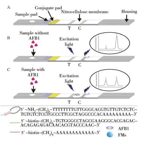
Schematic Diagram of Molecular Hairpin/Fluorescent Microsphere-Based Lateral Flow Chromatographic Assay Technology for Detecting AFB1
4.1.3 Quantum Dot Lateral Flow Chromatographic Technology
In recent years, Quantum Dots (QDs) have advantages as fluorescent markers, such as narrow full width at half maximum (FWHM), long fluorescence lifetime, and tunable maximum emission peaks. Common efficient fluorescent dye donor QDs include CdTe and CdSe/ZnS. Richarte et al. used CdSe/ZnS QDs as the labeling signal to develop a lateral flow chromatographic technology for the simultaneous detection of three toxins, DON, ZEN, and T2, in grains. This detection method offers high sensitivity, cost-effectiveness, and speed. Wang et al. designed a lateral flow chromatographic assay for rapid detection of OTA, using OTA and a DNA probe (DNA1) fixed on the detection line to simultaneously compete for the QD-labeled aptamer. In the absence of OTA, the QD-labeled aptamer binds to DNA1, and excess free labeled aptamer binds to the poly T probe (DNA2) fixed on the quality control line. Conversely, when OTA is present, the signal value of the detection line decreases, and the signal value of the quality control line increases, with an LOD reaching 4.7 nmol/L. This is consistent with the fluorescent polarization immunoassay method but is more time-saving.

Schematic Diagram of the Reaction Principle Based on Quantum Dot Aptamer Lateral Flow Chromatographic Technology
4.2 Colorimetric Detection-Based Lateral Flow Chromatographic Biosensors
In lateral flow chromatography, colloidal gold lateral flow chromatography is the most commonly used technology for rapid on-site detection without any instruments. Colloidal gold is an inert metal with advantages such as good stability, good biocompatibility, and low cost. Gold nanoparticles appear red, making them visible to the naked eye, a trend in rapid detection that has accelerated research on colloidal gold in the detection field. Aptamers modified with thiol groups form stable composite structures with colloidal gold via Au-S bonds. The T line contains a nucleic acid fragment modified with biotin that is complementary to the target aptamer. The C line contains a nucleic acid fragment modified with biotin, consisting of thymine or adenine, which can bind to the target aptamer. When there is no target in the solution, migration to the T line will cause a color reaction with the aptamer's complementary strand. Excess aptamer solution migrates forward and reacts with the C line nucleic acid fragment, showing red. When the solution contains the target, migration to the T line results in reduced or no reaction, showing light red or no color, while excess free aptamer binds to the C line nucleic acid fragment, showing red. Wu et al. proposed an aptamer-based colloidal gold lateral flow chromatographic technology for recognizing ZEN. A thiolated ZEN aptamer was conjugated to colloidal gold as the signal probe for lateral flow chromatography. The detection line on the nitrocellulose membrane surface was fixed with the complementary strand of the aptamer's recognition sequence, and the quality control line was fixed with the complementary strand of the aptamer's non-recognition sequence. Good linearity was achieved within the detection range of 5-200 ng/mL, with a visual detection limit of 20 ng/mL. However, this detection method also has some problems. First, the sample matrix effect, meaning actual samples always contain other impurity molecules that may produce non-specific signals during detection. Second, the reproducibility of nano-gold; complex nanostructures, shapes, and porosity are difficult to prepare accurately. Third, existing immunochromatographic test strips can only be used for qualitative or semi-quantitative detection.

Schematic Diagram of the Structure and Principle of Aptamer-Based Lateral Flow Chromatographic Technology
There are many types of nanomaterials, such as AuNPs, CBNs, MNPs, QDs, and UCNPs. A lateral flow chromatographic assay using silver core and gold shell (AgAu) as markers was used for sensitive and rapid detection of AFB1 in food. Compared with gold nanoparticles, Ag-Au nanoparticles enhanced the sensitivity, reproducibility, and stability of the assay. As the concentration of AFB1 increased, the color intensity of the nanocomposite on the detection line increased, with a detection limit for AFB1 of 0.1 ng/mL. Kolosova et al. reported a colloidal gold lateral flow chromatographic assay for the simultaneous detection of Zearalenone (ZEN) and Deoxynivalenol (DON). Currently, the advantages of colorimetric lateral flow chromatographic technology are sensitivity, simplicity, speed, and the absence of multiple labeling and separation steps. However, this technology is not yet mature, and there are not many reported articles. It has great potential for development in the field of on-site rapid analysis and detection in food safety, drug research and development, and medical diagnosis.
Advantages and Limitations of Various Nanomaterials Used in Rapid Detection
4.3 Surface Enhanced Raman Scattering (SERS) Lateral Flow Chromatographic Assay Technology
Surface Enhanced Raman Scattering is a technique that enhances the intensity of Raman scattering. It offers advantages such as high sensitivity and the provision of fingerprint spectra. Metal nanomaterials can significantly enhance Raman scattering intensity, so NP technology based on Raman scattering has always been a research hotspot. The excitation light and scattered Raman signal can be adjusted to the near-infrared region, enabling highly sensitive quantitative analysis in the laboratory. The application of SERS in lateral flow chromatographic detection is based on Raman scattering enhanced by nanoparticles, significantly improving the sensitivity of the experiment.
Li et al. designed and constructed a SERS sensor for rapid detection of AFB1 based on aptamer recognition with magnetic separation assistance. They modified carboxylated magnetic beads with a biotin-labeled AFB1 aptamer as the detection probe. They synthesized signal molecules (4-MBA) embedded in gold-silver core-shell nanorods and modified them with thiolated complementary DNA to assemble SERS probes as capture probes. The gold-silver core-shell nanorods and magnetic beads were assembled via DNA base complementarity to construct an AFB1 detection sensor. When AFB1 is present, it is recognized by the aptamer, causing the separation of the magnetic beads and the gold-silver core-shell nanorods. The SERS signal subsequently weakens, thus enabling rapid detection of AFB1 through changes in SERS intensity. The linear range of this sensor is 1 pg/mL - 1000 pg/mL, with a detection limit of 0.4 pg/mL. As a novel identification method, it has great potential for accurate, objective, and direct rapid detection of targets and is expected to become a powerful tool for identification.
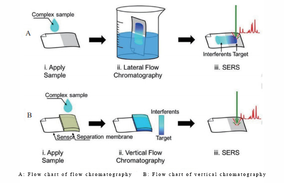
Operational Flow Chart of SERS-Based Lateral Flow Chromatographic Assay Technology
4.4 Smartphone-Based Aptamer Lateral Flow Chromatographic Assay Technology
Smartphone-based aptamer detection devices use converted nanoparticle-modified aptamer complementary strands fixed on three detection lines of the same lateral flow strip as detection probes. This allows for simultaneous specific and sensitive rapid detection of multiple different targets (e.g., OTA, mercury ions, and Salmonella), thus providing an economical, efficient, and rapid means for multi-target detection. Under optimal conditions, the detection limits for mercury ions, OTA, and Salmonella were 5 ng/mL, 3 ng/mL, and 85 CFU/mL, respectively. Jin et al. developed an imaging-based lateral flow chromatographic analyzer that captures the signals of the T and C lines into a single image, which is then processed by mobile phone software. This analyzer has been successfully used for the quantitative analysis of Creatine Kinase - brain (CK-MB), with an LOD value of 2 ng/mL and small variability. Mobile phones have advantages such as ease of use, good portability, ubiquity, and strong data transmission capabilities, making them suitable as Point-of-Care Testing (POCT) readers. Therefore, smartphone-based aptamer lateral flow chromatographic technology is expected to be widely used in rapid detection fields such as food safety, toxin detection, medical diagnosis, and environmental monitoring.
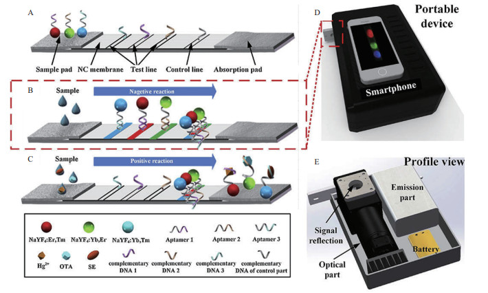
Schematic Diagram of LFAs for Simultaneous Detection of Multiple Targets and Smartphone-Based Portable Device
5. Influencing Factors of LFCA
5.1 Antibody
The sensitivity and specificity of LFCA are based on the selection of immunoreagents. As summarized above, LFCA has extremely high requirements for the type and quality of antibodies. The selected antibodies must have high affinity, sensitivity, and specificity. Often, LFCA requires a matched pair of antibodies (labeled antibody and T line antibody), increasing the requirements for antibodies and the difficulty of developing LFCA. To address this issue, in 2016, Song Chunmei et al. first invented a semi-quantitative immunofluorescence test strip technology for foodborne pathogens using only one antibody, with a detection time of 5-10 minutes for E. coli O157:H7 in real samples.
5.2 NC Membrane
Another key factor affecting the detection limit of LFCA is the choice of Nitrocellulose (NC) membrane. Membranes with different pore sizes have different effects on the detection limit and detection time. Laura and Majdinasab et al. used Hi-Flow 180 and Millipore NC membranes, with detection limits of 1.5 ng/mL and 0.25 ng/mL, and detection times of 20 minutes and 15 minutes, respectively.
5.3 Labeling Material
The labeling material serves both the "visualization" of LFCA detection results and the "antibody labeling" function. It is crucial for the sensitivity, detection limit, and other performance aspects of the method. Gold nanoparticles have advantages such as high stability, good antibody compatibility, and clear color development, making them the most commonly used antibody labeling material for LFCA. In recent years, research on new labeling materials such as carbon nanomaterials, quantum dots, upconversion fluorescent materials, and superparamagnetic nanoparticles has made breakthrough progress, significantly improving detection performance. In 2015, Wang Chunying et al. used multicolor quantum dots to label antibodies for simultaneous dual-target detection of alpha-fetoprotein and carcinoembryonic antigen, achieving detection limits of 3 ng/mL and 2 ng/mL, respectively, with results obtainable within 15 minutes. Their sensitivity was higher than the ELISA method.
6. Research Progress and Applications of LFCA
In LFCA, the label is one of the key factors affecting its sensitivity. Labeling antibodies or antigens allows the relative content of the corresponding antigen or antibody in the reaction system to be displayed through the enhancement and amplification effect of the label.
6.1 Labeling Materials for LFCA
Based on the labeling principle, they can be divided into two major categories: one is enzyme-based labeling methods; the other is self-colored labeling methods. For immunochromatographic strips that do not require instrument assistance, self-colored labeling is primary. To increase the sensitivity of test strips, people have been continuously searching for new types of labels, including nanoparticles (gold nanoparticles, carbon nanoparticles), luminescent nanoparticles (quantum dots, fluorescence quenching materials, upconversion fluorescent materials), superparamagnetic nanoparticles, liposomes, and enzymes. Currently, the most widely used is colloidal gold labeling.
6.1.1 Nanoparticles
Nanoparticles are used as tracers in LFCA due to their unique optical properties (including fluorescence or color changes caused by assembly and aggregation). Currently, colloidal gold, silver and carbon nanoparticles, and carbon nanotubes have been widely used in developing LFCA.
6.1.1.1 Colloidal Gold
Colloidal gold is easy to prepare, low cost, forms a visible color quickly, is very stable in both liquid and dry states, and does not fade easily, making it widely used in many fields. However, the shape, size, uniformity, and stability of colloidal gold are key factors affecting the success of colloidal gold test strips. The color and size of colloidal gold are closely related to the amount of trisodium citrate used to reduce the gold. Simultaneously, colloidal gold labeling is affected by many factors, such as the pH of the ligand and colloidal gold, and the ratio of colloidal gold to ligand protein.
6.1.1.2 Carbon Nanotubes and Carbon Nanoparticles
Carbon nanoparticles are used in Lateral Flow Immunochromatographic Assay (LFICA) analysis due to their large surface area and good photoelectric properties. Their color is darker than colloidal gold, making them very suitable as labeling materials, and they are low cost, easy to prepare, and have stable conjugation. However, the labeling effect of carbon nanoparticles can be affected by conditions such as reaction time and temperature. The widespread application of carbon nanoparticles could also have adverse effects on the environment and human health.
6.1.1.3 Other Nanomaterials
Other nanomaterials used in LFCA include Fe₃O₄@Si nanoparticles, silicon atom nanoparticles, latex microspheres, platinum nanoparticles, etc. Among them, latex microspheres are widely used due to their wide availability and low price. They can be coupled to many substances through simple adsorption and covalent linkages generated by amino, carbonyl, and thiol groups. They are easy to operate, stable, reliable, and highly sensitive. However, carbodiimide/N-hydroxysuccinimide is needed to activate the reactive groups in the latex microspheres to effectively label and bind antibodies or antigens.
6.1.2 Fluorescent Nanoparticles
Many recent studies have shown that using fluorescent nanoparticles achieves lower detection limits than using colorimetric labeling materials. Fluorescent nanoparticles have low background signal, less interference, high sensitivity, and the fluorescent dyes are inexpensive. They can directly couple biospecific antibodies without surface modification. However, their fluorescent signal is unstable and prone to quenching, and the accuracy of semi-quantitative analysis is relatively poor.
6.1.2.1 Quantum Dots
Quantum dots, with their narrow emission spectra, wide excitation range, and high fluorescence quantum yield, possess excellent brightness, size-tunable fluorescence emission, large absorption coefficients, good stability, and high signal-to-noise ratio. They have been widely used to improve the detection sensitivity of LFCA. However, quantum dot labeling can induce the production of oxygen free radicals, thus having certain toxicity to bioactive substances, although this toxicity can be reduced by coupling to protein molecules or coating with a low-toxicity substance. The combination of quantum dots and certain proteins may lead to fluorescence weakening or quenching of the quantum dots; for example, copper/zinc-superoxide dismutase has a significant quenching effect on CdSe quantum dots. Additionally, if quantum dot labeling is used for test strip preparation, a UV light source is needed to observe the color change during use, making the operating procedure slightly more complex compared to other types of test strips where results can be judged by the naked eye.
6.1.2.2 Fluorescence Quenching Materials
Fluorescence quenching materials (organic fluorescent dyes, fluorescent proteins, colloidal gold, quantum dots, etc.) have been used to detect small analytes, and studies have found a positive correlation between signal intensity and analyte concentration. These materials have advantages such as low background signal, high sensitivity, and the ability to label multiple biorecognition elements.
6.1.2.3 Lanthanide Elements
Lanthanide chelates that have been used to label LFCA to improve its detection limit include Eu³⁺, Tb³⁺, Sm³⁺, and Dy³⁺. Their principle as labeling materials is based on fluorescence reactions. They are chosen as labeling materials for LFCA because their nanoparticle size is quite close to that of colloidal gold and they eliminate background fluorescence. Compared to conventional fluorescent labels and quantum dots, they have unique and advantageous fluorescence characteristics, such as long fluorescence lifetime, narrow emission spectrum, large Stokes shift, and outstanding photostability.
6.1.2.4 Upconversion Phosphors
Upconversion phosphors are submicron-sized ceramic particles containing lanthanide elements. Their special composition and structure give them excellent optical properties; they emit visible light when excited by infrared light. Compared to other fluorescent labels, their main advantages include high sensitivity, long shelf life, permanent recording (no fading), low matrix interference, and low cost.
6.1.2.5 Other Fluorescent Materials
Researchers often use fluorescent signals to enhance detection signals and improve the sensitivity of LFCA. Fluorescent microspheres (FMs) have a stable configuration and high fluorescence intensity, making them colorful and safe labeling materials. Due to the lack of sensitivity in colloidal gold applications, FMs are mainly used to detect several compounds, such as Aflatoxin M1 and Tumor Necrosis Factor α.
6.1.3 Superparamagnetic Nanoparticles
Superparamagnetic nanoparticles can be completely captured by devices due to their stable magnetic signal, potentially increasing detection sensitivity by 10-100 times and shortening detection time. However, they can generate background noise. Since the rapid detection technology established using magnetic particle labeling requires an external magnetic field, it is more troublesome compared to colloidal gold labeling. Additionally, magnetic particles have high surface free energy and strong magnetic dipole moment interactions, making them prone to aggregate with each other. Poor coating can also affect the test results of the strip.
6.1.4 Enzymes
The color formed by the reaction of an enzyme with its substrate can be compared by the naked eye and standard color charts, intuitively reflecting the presence of the detected substance. However, its analytical sensitivity is not high, and color comparison may bring inaccuracies due to human error. Furthermore, enzymes are bioactive substances; if not stored properly, their catalytic performance can be lost.
6.1.5 Liposomes
Liposomes are spherical artificial small vesicles composed of one or more phospholipid bilayers. Due to their ability to capture high concentrations of signal molecules, liposome diagnostic devices can improve the sensitivity of visual immunochromatography by 2-3 orders of magnitude. However, they have low stability and complex operation, so their application is limited.
6.2 Labeling Materials for Nucleic Acid-Based LFCA
The principle of nucleic acid-based LFCA labeling is base pairing. A known sequence nucleic acid fragment with a label is hybridized with its complementary nucleic acid sequence to form a double strand, used to detect whether an unknown sample has the same sequence and further determine its homology with the known sequence. Based on whether the labeling material is radioactive, it can be divided into radioactive labeling materials and non-radioactive labeling materials.
6.2.1 Radioactive Labeling Materials
Radioactive isotopes are used as labels, commonly ³²P, ³H, ³⁵S. The advantage of radioactive labeling materials is high sensitivity. The disadvantages are the potential for radioactive pollution, short half-life of the isotopes, and harm to the human body.
6.2.2 Non-Radioactive Labeling Materials
Biotin and digoxigenin are used as non-radioactive labeling materials; both are haptens. The biotin-avidin system is a signal amplification labeling technology with advantages such as high affinity, high sensitivity, strong specificity, and good stability. Biotin labeling is harmless to the body but prone to decomposition under UV irradiation. Digoxigenin labeling has sensitivity comparable to radioisotope-labeled probes and better specificity than biotin labeling.
6.3 Application Status of LFCA
Since the 1990s, chromatographic test strip technology has been widely used in human, animal, and plant medical clinical testing, as well as environmental monitoring and food safety testing, due to its advantages of low price, convenient portability and use, no need for professionals, support for on-site rapid testing, and visually interpretable results.
6.3.1 Application of LFCA in Human Medicine and Animal/Plant Medical Detection
In the early establishment and development of LFCA, research focused on its use in human disease diagnosis, early warning, and analysis of physiological and biochemical indicators. Later, with the continuous development of animal husbandry and planting industries, the harm of animal and plant diseases also intensified. To date, a large number of test strips have been successfully applied to the rapid detection of human tumor diseases, viral infections, bacterial infections, as well as animal and plant viral diseases, bacterial diseases, and parasitic diseases.
Application of LFCA in Human Medicine and Animal/Plant Medical Detection
As shown in the table above, LFCA is currently widely used in the medical field, involving humans, animals, and plants, primarily using the colloidal gold-labeled double-antibody sandwich method. Its detection sensitivity can reach the ng/mL level, and the analysis time is within 1 hour. Because colloidal gold is easy to prepare, low cost, forms color quickly, and does not fade easily, and because the detection target antigens in the medical field are mostly large molecules with multiple antigenic sites, the colloidal gold-labeled double-antibody sandwich method is highly applied in human medicine and animal/plant medical detection, making significant contributions to medical development. Colloidal gold has been used as a labeling material for chromatographic test strips for a long time and is widely applied. More efficient labeling materials and methods deserve continued and in-depth research by researchers to achieve greater accomplishments.
6.3.2 Application of LFCA in the Field of Food Safety
Currently, food safety issues "from farm to table" are becoming increasingly complex, and food safety problems occur frequently. Pathogenic microorganisms cause human food poisoning, abuse of agricultural and veterinary drugs is in a disordered state, excessive heavy metals pose hidden dangers to grain products, and illegal additives and adulteration are frequently reported. Food safety has become a major problem plaguing people's healthy lives. In response to people's needs, LFCA, which is convenient, rapid, low-cost, time-saving, and provides visually interpretable results, has been increasingly used in areas including foodborne pathogens, agricultural and veterinary drug residues, illegal additives, biological toxins, and heavy metal ion detection.
Application of LFCA in the Field of Food Safety
As shown in the table, at this stage, LFCA is widely used in the field of food safety, mainly in two aspects: detection of foodborne pathogens and agricultural/veterinary drug residues. For microbial detection, the double-antibody sandwich method is mainly used, while in the field of agricultural/veterinary drug residue detection, the competitive method is mostly adopted. In these methods, colloidal gold as a labeling material is very widely used. To improve sensitivity, there has been much research in recent years on the application of some new labeling materials, such as quantum dots, magnetic nanoparticles, and upconversion fluorescent materials. The detection limit of the LFCA method for microorganisms can be as high as 10¹–10⁸ CFU/mL, and for agricultural/veterinary drug residues, it can be as high as 30 ng/mL – 0.1 mg/L, with detection times shortened to 1–20 minutes.
6.3.3 Application of LFCA in the Field of Environmental Monitoring
Environmental pollution and destruction accompanying the development of social productive forces have become major global problems threatening human survival and development. In response, environmental protection committees of various countries have issued joint environmental statements, indicating that monitoring environmental pollutants is the primary link in environmental protection. LFCA is widely used in environmental monitoring due to its advantages of speed and on-site detection capability.
Application of LFCA in the Field of Environmental Monitoring
7. Comparison of LFCA with Other Analytical Techniques
LFCA enables qualitative and semi-quantitative detection of various analytes such as antigens, antibodies, and haptens without the need for professional operators or expensive instrument equipment. It plays an important role in areas with scarce labor resources and can also simultaneously detect several compounds in a short time. Figure 5 summarizes the advantages and disadvantages of LFCA test strip technology.
Advantages and Disadvantages of LFCA Test Strips
7.1. Comparison of LFCA with Traditional Microbiological Detection Methods
Traditional microbiological detection methods require steps such as enrichment, isolation and culture, and biochemical identification. Completing one detection generally takes 5–8 days. In contrast, LFCA requires only tens of minutes or even minutes of detection time besides enrichment, making it faster and more sensitive.
7.2 Comparison of LFCA with Instrumental Analytical Chemistry Techniques
Currently, chromatography and mass spectrometry-related detection methods are widely used for detecting agricultural/veterinary drugs and heavy metal residues. Chromatography includes gas chromatography (GC) and high-performance liquid chromatography (HPLC), with HPLC being more advanced. Meanwhile, Gas Chromatography-Mass Spectrometry (GC-MS) technology combines a gas chromatograph with a mass spectrometer through an appropriate interface and uses computer technology for coupled analysis. It is mature and has become an effective method for the separation and identification of complex components. Table 4 compares LFCA with mass spectrometry and HPLC as representatives from five aspects: sample pretreatment, sensitivity, detection cycle, cost, and detection requirements. The analysis shows that compared to instrumental analytical chemistry techniques, although LFCA has lower detection sensitivity and cannot perform quantitative analysis, it requires lower cost, has lower technical personnel requirements, and has a shorter detection time. Therefore, the LFCA method can be used as a preliminary screening tool, with positive results further analyzed using instrumental analysis methods to obtain accurate values. Semi-quantitative analysis of the LFCA method can also be achieved with the help of some data reading devices.
Comparison of LFCA, Mass Spectrometry, and High-Performance Liquid Chromatography
7.3 Comparison of LFICA Technology with Other Immunological Detection Technologies
Immunological detection technologies are based on the specific reaction between antigens and antibodies. Different detection substances have their specific antigens and can stimulate the body to produce corresponding specific antibodies. Detection methods based on immunological technology use surface antigens or secreted toxins of target bacteria as detection targets, making them suitable for detecting bacteria or spores. This method has been widely used for the detection of pathogenic bacteria in food, including ELISA, immunomagnetic separation technology, radioimmunoassay technology, immunofluorescence technology, etc. Table 5 compares LFICA with these methods as representatives. The analysis shows that compared to other immunological detection methods, LFICA does not require specific instruments or complex technical means for assistance, saves costs, has low requirements for the detection environment, and is suitable for on-site rapid detection.
Comparison of LFICA with Other Immunological Detection Technologies
7.4 Comparison of LFCA Test Strip Technology with Molecular Biology Detection Technology
Molecular biology testing technology uses nucleic acids or proteins as analytical materials. By analyzing changes in gene structure, expression, and the resulting alterations in gene function, it provides more accurate and scientific information and basis for disease research and diagnosis. Molecular biology detection technologies include PCR technology, Loop-Mediated Isothermal Amplification (LAMP) technology, bioluminescence detection method, etc. Table 6 compares LFCA with these methods as representatives. The analysis shows that compared to molecular biology detection technologies, LFCA does not require specific instruments or professional operators, has a short detection time, and is less prone to false positives and false negatives. However, the relatively low sensitivity and inability to achieve quantitative detection are bottleneck problems of the LFCA method, requiring optimization and improvement.
Comparison of LFCA with Molecular Biology Detection Technologies
8. Summary
Lateral flow chromatographic analysis technology belongs to dry chemistry detection technology. It is compatible with naked-eye detection or simple optical electronic readers and has characteristics such as high sensitivity, simplicity, speed, no need for complex and expensive instrument equipment, and low cost. It has been applied in fields such as food safety and biomedicine. Nucleic acid aptamer lateral flow chromatography technology has many advantages compared to traditional antigen-antibody immunochromatography, such as low cost, batch stability, long shelf life, and wide application range, giving it good development prospects. Simultaneously, it enables on-site real-time rapid detection. Detection methods based on different labeling methods have different detection performances. Latex beads and gold nanoparticles (AuNPs) can be used for visual qualitative detection, while fluorescent, quantum dot, and magnetic nanoparticle labels can enable quantitative analysis. Currently, aptamer lateral flow chromatography technology is still in the laboratory development stage; there are no low-cost and mature detection products on the market. It also faces some problems, such as the need for relatively complex sample pretreatment, samples must be liquid and homogeneous, significant influence of matrix and environmental parameters, and poor stability. These problems will be the focus of future research. Mycotoxins have characteristics such as numerous types, similar molecular structures, and trace amounts. Aptamer lateral flow chromatography technology has great potential in the field of on-site rapid detection of mycotoxins. It is believed that in the near future, aptamer lateral flow chromatography technology will develop into a powerful tool for on-site screening of pollutants such as toxins, heavy metals, pesticide residues, and pathogenic microorganisms.
References:
https://www.spkx.net.cn/fileup/HTML/2018-39-15-048.shtml
http://rs.yiigle.com/CN121382201705/1013210.htm
http://html.rhhz.net/SWJSTB/html/2020-8-217.htm