
Cell Fractionation: Principles, Workflows, Organelle Markers, and Purity Checks
A practical guide to differential centrifugation, density gradients, sequential lysis, compartment markers, and interpreting enrichment without overcalling localization.
- What cell fractionation measures
- How separation works
- Choose a fractionation strategy
- Plan and perform the workflow
- Organelle and compartment marker panels
- Assess enrichment, purity, and recovery
- Prepare fractions for downstream analysis
- Research applications and interpretation
- Troubleshooting by observed pattern
- Common questions
- References
What cell fractionation measures
Cell fractionation disrupts cells and separates their contents into fractions that are enriched for nuclei, cytosol, mitochondria, membranes, microsomes, lysosomes, or other structures. The fractions can then be analyzed by western blot, enzyme assay, microscopy, proteomics, RNA analysis, or functional assays.
Fractionation answers a biochemical enrichment question. It does not, by itself, prove that a protein is resident in one compartment in intact cells. Membrane rupture, organelle breakage, vesicle resealing, protein shuttling, and cross-contamination can all alter the measured distribution. Use the strongest wording supported by the controls: detected in, enriched in, or consistent with localization to are different claims.
Before starting, define whether the goal is to preserve intact organelles, obtain soluble and insoluble protein pools, compare nuclear and cytoplasmic abundance, isolate a functional organelle, or prepare material for omics. The required purity, recovery, buffer, and centrifugation strategy will differ.
How separation works
After controlled disruption, the homogenate contains intact organelles, membrane fragments, soluble proteins, nucleic acids, and debris. Centrifugation separates these components according to sedimentation behavior, which reflects size, shape, density, and the viscosity of the medium. Large structures generally pellet at lower centrifugal forces than small vesicles or ribosomes, but overlapping sizes and densities mean that a pellet is rarely a single organelle.
Protect the structures you want to measure
- Use an osmotically appropriate buffer when organelle integrity matters. Sucrose, mannitol, or other osmolytes can reduce osmotic swelling, but the correct composition depends on the organelle and assay.
- Keep samples cold and work quickly when proteolysis, dephosphorylation, or metabolic changes could alter the readout. Add inhibitors only when they are compatible with the downstream assay.
- Disrupt enough cells to release the target compartment, but avoid excessive shear, foaming, sonication heat, or repeated mechanical stress that fragments organelles.
- Separate pellets and supernatants carefully. Material near a pellet can carry contaminating particles into the next fraction.
Homogenization is a compromise between release and preservation. A method that gives high yield can also increase leakage or cross-contamination. Optimize the disruption for the cell type, tissue structure, organelle, and downstream readout rather than copying a universal number of strokes or pulses.
Choose a fractionation strategy
| Strategy | Principle | Useful when | Main limitation |
|---|---|---|---|
| Differential centrifugation | Sequential spins collect increasingly smaller or denser particles in pellets. | Rapid enrichment of nuclei, mitochondria, membranes, microsomes, or cytosol from a homogenate. | Fractions can contain organelles with overlapping sedimentation behavior and are often only partially purified. |
| Density-gradient centrifugation | Particles migrate through a continuous or discontinuous gradient to a buoyant-density zone. | Resolving organelles with similar size but different densities and improving purity after crude enrichment. | Gradient composition, rotor, tube, acceleration, fraction collection, and sample load affect resolution. |
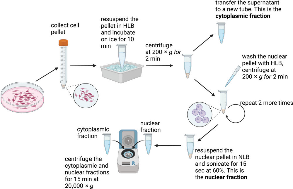
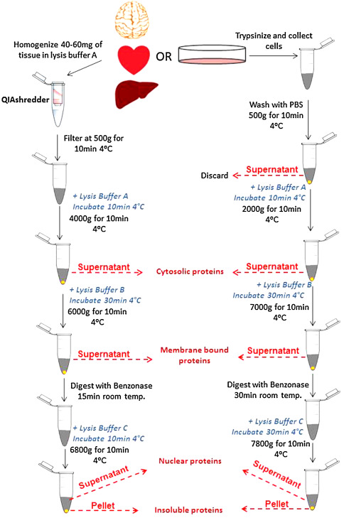
| Sequential or selective lysis | Buffers and detergent strength release compartments in stages. | Separating soluble cytosol, membrane-bound organelles, and nuclear proteins or comparing cytoplasmic and nuclear pools. | Incomplete lysis, leakage, and detergent carryover can bias both recovery and downstream assays. |
| Affinity or immunoisolation | A ligand, antibody, or tag captures a selected structure or complex. | Enriching a defined organelle subpopulation, tagged compartment, or interaction complex. | Capture depends on accessibility and specificity and can perturb composition or lose unlabeled material. |
| Microfluidic, magnetic, or dielectrophoretic separation | Particles are sorted by electrical, magnetic, hydrodynamic, or label-dependent properties. | Small inputs, specialized organelles, or workflows requiring reduced centrifugation. | Platform-specific calibration, throughput, recovery, and biological validation are required. |
These strategies can be combined. A crude differential pellet may be followed by a density gradient, while a nuclear fraction may undergo additional washing or selective lysis. Label a fraction as crude, enriched, or purified according to the marker and contamination evidence actually obtained.
Plan and perform the workflow
The following sequence is a planning framework rather than a universal protocol. Centrifugal force, time, buffer composition, temperature, and homogenization settings must be optimized for the sample, rotor, tube, and target compartment. Keep a complete fraction map so every pellet, wash, and supernatant can be traced.
- Define the sample and endpoint. Record species, tissue or cell type, culture or treatment state, input amount, target compartment, and downstream assay.
- Choose an appropriate buffer. Match osmolarity, pH, salts, detergents, reducing agents, chelators, and inhibitors to the organelle and downstream readout. Avoid components that interfere with activity assays, nucleic-acid extraction, chromatography, or MS.
- Collect and cool the sample. Minimize delay, keep the sample cold when compatible with the experiment, and record the handling interval. Process treated and control samples in parallel where possible.
- Disrupt the cells gently enough to preserve the target. Use a calibrated homogenization method and monitor the extent of lysis by microscopy, marker release, or another sample-specific check.
- Remove large debris and unbroken cells. Retain the pellet or supernatant according to the experimental question and record each transfer. Do not discard a fraction before deciding whether it contains unbroken cells, nuclei, or aggregates relevant to recovery.
- Separate the target fraction. Use differential spins, a density gradient, sequential lysis, or capture as planned. Record rotor, tube, relative centrifugal force, duration, temperature, brake settings, gradient composition, and fraction volumes.
- Wash or refine the fraction when needed. Resuspend gently, avoid leaving contaminating supernatant around a pellet, and collect washes when they can reveal loss or leakage.
- Measure and normalize the material. Determine protein or nucleic-acid amount with a standard curve compatible with the buffer. Keep an aliquot for marker validation before using the entire fraction in a functional assay.
- Validate the fraction. Probe positive markers for the intended compartment and negative markers for likely contaminants. Compare the fractions and input, not only the target band in one tube.
- Store and analyze consistently. Aliquot when appropriate, record freeze-thaw history, and use matched processing for biological replicates. Confirm that storage and buffer do not compromise the downstream assay.
Organelle and compartment marker panels
Use more than one marker when the conclusion depends on compartment identity. A positive marker should be enriched in the intended fraction, while markers from likely contaminants should be relatively depleted. Marker choice is species-, tissue-, treatment-, and fractionation-dependent; the examples below are starting points, not universal invariants.
| Fraction or compartment | Common marker examples | Contamination to check | Interpretation note |
|---|---|---|---|
| Cytosol | GAPDH, calreticulin (CALR) in suitable workflows | Nuclear Lamin B1, mitochondrial VDAC1/COX IV, membrane or ER markers | Soluble markers can leak after disruption; verify that the cytosolic fraction is not simply the post-lysis supernatant. |
| Nucleus | Lamin B1, Lamin A/C, Histone H3 | Cytosolic GAPDH/CALR and mitochondrial VDAC1 | Check nuclear integrity and wash efficiency; histones can be released by harsh lysis. |
| Mitochondria | VDAC1/porin, COX IV, TOM20 | ER calnexin, Golgi GM130, lysosomal LAMP1, nuclear Lamin B1 | Mitochondrial enrichment does not prove intact respiration or exclude associated membranes. |
| Endoplasmic reticulum | Calnexin, calreticulin, SERCA2 | Mitochondrial and Golgi markers | Microsomal fractions can contain several membrane systems and should be labeled accordingly. |
| Plasma membrane | Na+/K+-ATPase, caveolin, cadherins | Cytosolic and ER markers | Membrane disruption and vesicle resealing can redistribute membrane proteins. |
| Golgi apparatus | GM130, Golgin97 | ER calnexin and plasma-membrane markers | Use a panel suited to the tissue and whether the fraction is crude or gradient-purified. |
| Lysosome | LAMP1, cathepsin B or D | Mitochondrial, ER, and peroxisomal markers | Enzyme activity can be altered by lysis and pH; marker enrichment does not establish intact lysosomes. |
| Peroxisome | ABCD3/PMP70, catalase | Mitochondrial and ER markers | Confirm that the marker is appropriate for the species and tissue being studied. |
Assess enrichment, purity, and recovery
Use three complementary concepts:
- Enrichment: the intended marker is more abundant relative to the starting homogenate or other fractions.
- Purity: markers from other compartments are absent or substantially reduced under a defined assay sensitivity.
- Recovery: the fraction retains a measurable portion of the starting material or activity. High purity can occur with poor recovery, and high recovery can occur with substantial contamination.
Minimum validation set
- Run the starting homogenate, every target fraction, and relevant washes or pellets.
- Measure at least one positive marker for the intended compartment and one negative marker for each likely contaminant.
- Use matched protein loading for western blot comparisons and retain the raw membrane image and exposure information.
- Quantify marker signals with a defined background and linear-range rule; do not infer purity from visual band brightness alone.
- Where localization is central, confirm with microscopy, proteomics, enzyme activity, or another orthogonal method.
Fractionation can change the abundance of soluble proteins, expose protease-sensitive sites, or separate a protein from its binding partners. A protein detected in two fractions may reflect genuine distribution, organelle contact, leakage, or contamination. Interpret the marker pattern together with the fractionation design and a matched biological control.
Prepare fractions for downstream analysis
| Downstream assay | Preparation priorities | Control or compatibility check |
|---|---|---|
| Western blot | Equalize protein input, keep fraction identities and marker panel, and avoid detergent or salt conditions that disrupt electrophoresis or antibody binding. | Positive/negative compartment markers, total-protein or loading reference, same exposure range, and full fraction map. |
| Enzyme or functional assay | Preserve cofactors, pH, membrane integrity, and temperature requirements; avoid inhibitors that block the assay. | Activity control, blank, recovery measurement, and a lysis or detergent control. |
| Proteomics or mass spectrometry | Minimize detergents, keratins, polymers, and salts that interfere with digestion, chromatography, ionization, or database analysis. | Process blank, digest control, marker-based purity assessment, and a documented fraction-to-file map. |
| RNA or microRNA analysis | Prevent RNase exposure, collect fractions consistently, and separate RNA extraction chemistry from protein assays. | RNA integrity, compartment markers, input normalization, and contamination controls. |
| Microscopy or imaging | Use intact or appropriately fixed material and preserve morphology when structure is the endpoint. | Compare with intact-cell localization and document any fixation or permeabilization effect. |
Research applications and interpretation
Fractionation is useful for testing whether a protein or modification is enriched in a compartment, following stimulus-dependent translocation, measuring organelle-associated activity, preparing organelle proteomes, and separating biochemical pools for interaction studies. It can also support reconstitution experiments when the fraction retains the needed components and activity.
For a translocation claim, compare untreated and treated samples with the same fractionation and marker checks. A rise in nuclear signal is more persuasive when cytosolic contamination is low, the nuclear marker is retained, total protein recovery is comparable, and an orthogonal assay supports the movement. For a proteomics claim, report the fractionation scheme and marker-based purity because a partial organelle preparation can produce a mixed proteome.
Clinical or disease-related interpretations require additional evidence. A fraction enriched for a candidate biomarker is not by itself a diagnostic test, and an organelle-associated protein is not automatically a therapeutic target.
Troubleshooting by observed pattern
| Observed pattern | Possible explanations | Next check |
|---|---|---|
| Target appears in every fraction | Broad biological distribution, leakage, cross-contamination, or overloading | Check positive and negative markers, input amount, homogenization severity, and fraction recovery before assigning localization. |
| Nuclear fraction contains abundant cytosolic marker | Incomplete washing, cytosol trapped in the pellet, nuclear rupture, or overloading | Collect washes, inspect nuclei, reduce mechanical stress, and compare marker ratios across fractions. |
| Cytosolic fraction contains nuclear marker | Nuclear breakage, incomplete pelleting, or carryover near the pellet | Optimize disruption and low-speed separation, remove the supernatant near the pellet carefully, and test nuclear integrity. |
| Mitochondrial fraction contains ER or lysosomal markers | Co-sedimentation, membrane contact sites, gradient overlap, or insufficient refinement | Add a negative-marker panel and consider a density-gradient or alternative enrichment strategy. |
| Low target recovery | Loss during transfers, adsorption, degradation, insolubility, or target release into an uncollected fraction | Track pellets, supernatants, and washes; add recovery controls and review buffer compatibility. |
| Fraction yield varies between replicates | Unequal input, tissue disruption, cell state, rotor handling, or fraction-volume differences | Standardize input and timing, record the rotor and settings, and normalize with a pre-defined recovery metric. |
| Marker pattern changes after treatment | True organelle remodeling, marker regulation, altered lysis, or changed contamination | Validate marker stability, use more than one marker, and interpret target distribution with total recovery and orthogonal data. |
Common questions
Does a pellet represent one organelle?
Usually not. Differential centrifugation produces enriched mixtures unless additional purification and marker-based validation show otherwise.
Can western blot prove protein localization?
It can support enrichment in a fraction when positive and negative markers are appropriate. Microscopy or another orthogonal method is useful when the localization claim is central.
Why keep the washes and discarded supernatants?
They reveal material lost during washing, leakage from the target compartment, and carryover around a pellet. They are valuable troubleshooting controls even when they are not part of the final assay.
Should I use the same marker for every cell type?
No. Marker expression and stability depend on species, tissue, treatment, and fractionation method. Use a panel and verify each marker in the samples being studied.
Is higher enrichment always better?
No. A highly enriched fraction can have poor recovery or lose fragile structures. Choose the balance of purity, recovery, integrity, and function required by the downstream experiment.
- Baghirova et al., sequential fractionation of tissue and cultured cells — marker-supported cytosolic, membrane, and nuclear fractions.
- Graham, fractionation of subcellular organelles — differential and density-gradient principles.
- Huynh et al., cytoplasmic and nuclear separation — marker checks and fraction-quality assessment.
References
- Graham JM. Fractionation of subcellular organelles. Curr Protoc Cell Biol. 2015;69:3.1.1–3.1.22. doi:10.1002/0471143030.cb0301s69. PMID: 26621372.
- Baghirova S, Hughes BG, Hendzel MJ, Schulz R. Sequential fractionation and isolation of subcellular proteins from tissue or cultured cells. MethodsX. 2015;2:440–445. doi:10.1016/j.mex.2015.11.001. PMID: 26740924; PMCID: PMC4678309.
- Mueller SJ, Hoernstein SNW, Reski R. Approaches to characterize organelle, compartment, or structure purity. Methods Mol Biol. 2017;1511:13–28. doi:10.1007/978-1-4939-6533-5_2. PMID: 27730599.
- Prajapati P, Wang WX, Nelson PT, Springer JE. Methodology for subcellular fractionation and microRNA examination of mitochondria, mitochondria associated ER membrane, ER, and cytosol from human brain. Methods Mol Biol. 2020;2063:139–154. doi:10.1007/978-1-0716-0138-9_11. PMID: 31667768; PMCID: PMC8919243.
- Huynh HT, Shcherbinina E, Huang HC, et al. Biochemical separation of cytoplasmic and nuclear fraction for downstream molecular analysis. Curr Protoc. 2024;4:e1042. doi:10.1002/cpz1.1042. PMID: 38767195.