
In the fields of biomedical detection and cellular molecular experimental research, fluorescence labeling technology is the core technical means for realizing visual observation, qualitative and quantitative analysis of biological samples. As a classic green fluorescent labeling dye, fluorescein isothiocyanate (FITC), with its mature labeling technology, good versatility, and high cost-effectiveness, is widely applied in various experimental scenarios including immunofluorescence staining, flow cytometric detection, biomolecular tracing, and clinical pathological diagnosis, and is currently one of the most frequently used fluorescent probes in the biological research field.
FITC stands for Fluorescein Isothiocyanate, belonging to the fluorescein derivative family. The pure product is a yellow or orange-yellow crystalline powder, soluble in acetone and DMSO, slightly soluble in water, with stable basic physicochemical properties. This substance was first reported in 1942. Its molecular structure involves replacing one hydrogen atom on the benzene ring at the base of the fluorescein core, introducing the isothiocyanate active group (‑N=C=S). Commercially available FITC is generally a mixture of two isomers: 5-isothiocyanate fluorescein (5-FITC) and 6-isothiocyanate fluorescein, synthesized by Robert Seiwald and Joseph Burckhalter in 1958.
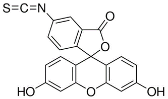
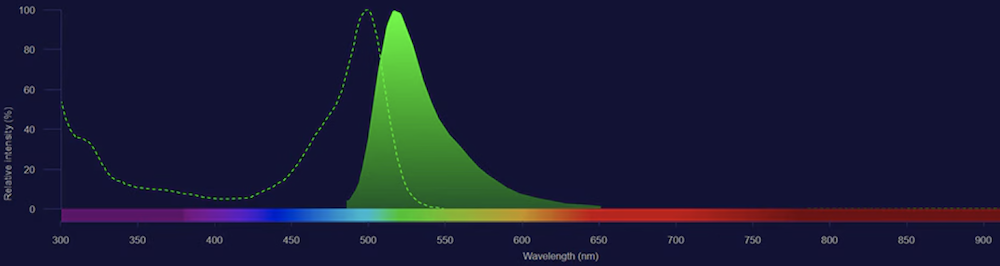
In alkaline environments (pH 8.5 and above), the isothiocyanate group of FITC can react with lysine residues in immunoglobulin molecules. Specifically, the isothiocyanate group of FITC undergoes a carbamylation reaction with the ε-amino group of lysine in immunoglobulins in aqueous solution, forming a stable thiocarbamyl bond, thereby producing fluorescein-labeled immunoglobulin, i.e., fluorescent antibody. Each immunoglobulin molecule can theoretically bind up to 15 to 20 FITC molecules, which gives the fluorescence-labeled antibody high sensitivity and specificity in detection applications.
Through the above covalent coupling reaction, biological samples such as antibodies, functional proteins, cells, and microorganisms that originally have no fluorescent properties can stably bind FITC fluorescent groups, achieving specific fluorescence labeling. Under excitation by the corresponding light source, the samples can present characteristic green fluorescence, and researchers can achieve localization observation, qualitative analysis, and quantitative detection of target biomolecules through the position, intensity, and quantity of fluorescence signals.
Methods for FITC Labeling Antibodies
In fluorescent antibody labeling technology, commonly used methods include the Marshall (1958) method, the Chadwick method, and the dialysis labeling method by Clark et al. (1963). Among them, the Marshall method is a classical labeling method widely used in the preparation of fluorescent antibodies. Additionally, depending on specific experimental conditions and requirements, the Chadwick method and the dialysis labeling method by Clark et al. can also serve as alternative options for the preparation of FITC-labeled antibodies.
Relying on stable labeling performance and broad equipment compatibility, FITC covers the mainstream fluorescence experimental scenarios in basic biological research and clinical detection, with core application areas divided into four categories.
Immunofluorescence staining is the most classic application scenario for FITC. After labeling FITC on primary or secondary antibody molecules, targeted binding to target antigens in cell and tissue samples can be achieved through antigen-antibody specific binding reactions. With the aid of fluorescence microscopes, the expression position and distribution characteristics of target proteins within cells can be clearly observed, enabling visual analysis of cell structures and protein localization.
In flow cytometry detection, FITC fluorescence signal has good recognition and stable performance, and can be used in combination with fluorescent dyes such as PE and PI that have different emission bands. FITC-labeled antibodies can rapidly and specifically recognize target cell surface antigens to complete labeling, thereby enabling detection and analysis of cell typing, cell cycle, and intracellular signaling pathways.
In ELISA detection, FITC-labeled antibodies can be used in detection modes such as indirect or sandwich methods. By combining FITC-labeled antibodies with enzyme-labeled secondary antibodies, the color change generated by enzymatic reactions is used to quantitatively detect antigens or antibodies in samples. This method has high sensitivity and specificity, and is widely applied in clinical diagnosis, food safety detection, environmental monitoring, and other fields.
In radioimmunoassay, FITC-labeled antibodies can competitively bind with radionuclide-labeled antigens or antibodies, and the target substance in the sample is quantitatively analyzed by detecting the radioactivity intensity. Due to its high sensitivity and specificity, radioimmunoassay has important application value in the detection of biologically active substances such as hormones, drugs, and proteins.
Currently, the green fluorescent dyes commonly used in laboratories mainly include three types: FITC, Alexa Fluor 488, and CF488A. The three have similar fluorescence bands and overlapping functions, but their performance and applicable scenarios differ significantly. The specific characteristics and selection criteria are as follows.
FITC
The core advantages of FITC include a mature technical system, comprehensive equipment universality, and low cost, making it the preferred dye for large-scale basic experiments and routine validation experiments. Its shortcomings are quite prominent: poor photostability, with fluorescence quenching prone to occur under intense light irradiation; sensitivity to environmental pH, with fluorescence intensity significantly attenuating in acidic systems; certain non-specific adsorption, resulting in relatively high sample background fluorescence and limited signal-to-noise ratio. This dye is mainly suitable for routine immunofluorescence staining, conventional flow cytometry detection, and basic research experiments with limited budgets.
Alexa Fluor 488
Alexa Fluor 488 is a high-end improved alternative dye to FITC, with excitation and emission wavelengths essentially coinciding with FITC, allowing direct compatibility with FITC-dedicated detection channels without adjusting instrument parameters. Its core advantages include extremely strong photostability, with anti-fluorescence quenching capability far exceeding traditional FITC; a broad pH tolerance range, maintaining stable fluorescence in pH 4.0–10.0 acidic and alkaline environments; and low non-specific adsorption, clean imaging background, and high signal-to-noise ratio. The main drawback of this dye is its high cost, making it unsuitable for large-scale routine experiments. It is primarily adapted for high-precision experimental scenarios such as confocal microscopy high-resolution imaging, long-term dynamic observation, fine experiments with scarce samples, and high-end molecular mechanism research.
CF488A
CF488A is a novel improved green fluorescent dye that balances performance and cost, and has become a popular choice in the scientific research field in recent years. Compared with FITC, its non-specific adsorption is significantly reduced, sample background fluorescence is minimal, and both photostability and pH tolerance are notably improved. The dye itself has extremely low charge, and after labeling proteins, it is less likely to alter the protein isoelectric point. Its limitations include a relatively short application time, fewer mature experimental protocols than FITC, and mediocre compatibility with some older detection equipment. This dye is suitable for fine staining requiring high signal-to-noise ratios, as well as advanced research experiments that balance experimental quality and cost.
As a classic green fluorescent labeling dye, FITC, with its mature labeling technology, good equipment versatility, and extremely high cost-effectiveness, has established its foundational core position in biological fluorescence experiments. Although it has inherent deficiencies such as poor photostability, pH sensitivity, and relatively high background fluorescence, which prevent it from meeting the demands of high-precision, long-term high-end imaging experiments, it still possesses irreplaceable application value in scenarios such as routine basic research, large-scale sample detection, and preliminary clinical screening. During experiments, it is necessary to leverage the physicochemical characteristics of FITC to maximize strengths and minimize weaknesses, strictly standardize operational procedures, and control experimental conditions. Meanwhile, rationally selecting dyes based on experimental precision, sample quantity, and budget cost can effectively avoid experimental errors and improve the stability, accuracy, and reliability of fluorescence detection and imaging results.