
Abstract
Histone post-translational modifications are central to epigenetic regulation, influencing chromatin organization and the recruitment of regulatory proteins. This article summarizes the principal roles and representative markers of histone acetylation, methylation, phosphorylation, and ubiquitination. It also introduces the coordinated actions of histone writers, erasers, and readers; commonly used approaches including ChIP-seq, Western blotting, and immunofluorescence; and the relevance of dysregulated histone modifications to disease mechanisms and therapeutic research. Because the effects of individual marks depend on their genomic context and interactions with other modifications, they should be interpreted using complementary evidence.
Keywords: histone modifications; epigenetics; chromatin; gene expression; ChIP-seq
Part I. What Are Histone Modifications?
In eukaryotic cells, DNA is wrapped around histone proteins to form nucleosomes, which are further organized into chromatin. [18]
Each nucleosome contains a histone octamer composed of two copies each of H2A, H2B, H3, and H4. Histone H1 helps stabilize linker DNA and supports higher-order chromatin organization. [18]
Histones undergo various post-translational modifications, including acetylation, methylation, phosphorylation, and ubiquitination. [18]
These modifications regulate chromatin structure and gene expression by altering histone–DNA interactions or recruiting regulatory proteins. [18]
Loosely packed euchromatin generally allows greater access to transcription machinery, while condensed heterochromatin is commonly associated with transcriptional repression. [18]
Together, different histone modifications form a complex regulatory system known as the “histone code.” [14][18]
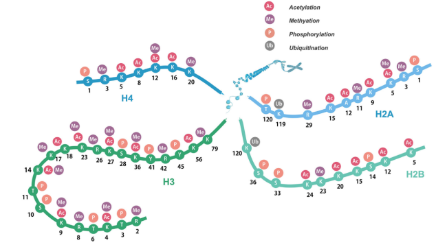
Reference: Post-translational modifications of histones: Mechanisms, biological functions, and therapeutic targets (2023)
Part II. Major Types of Histone Modifications
1. Histone Acetylation
Histone acetylation is one of the most widely studied histone modifications and primarily occurs on lysine residues. [1]
Acetylation neutralizes the positive charge of lysine, weakening electrostatic interactions between histones and negatively charged DNA. This generally promotes chromatin accessibility and gene transcription. [1]
Two major enzyme groups regulate this process: [1]
- Histone acetyltransferases (HATs) add acetyl groups. [1]
- Histone deacetylases (HDACs) remove acetyl groups. [1]
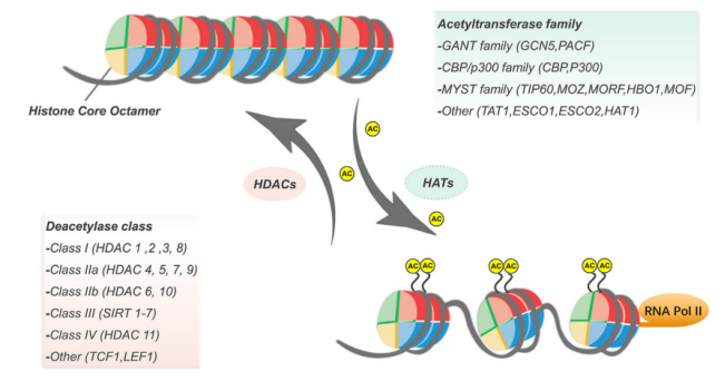
Reference: Post-translational modifications of histones: Mechanisms, biological functions, and therapeutic targets (2023)
H3K9ac and H3K27ac are histone acetylation marks associated with active gene regulatory regions. H3K9ac is preferentially enriched at active promoters and other regulatory elements, while H3K27ac is commonly used to identify active promoters and enhancers. [2][3][22]
Histone acetylation is involved in cell proliferation, differentiation, apoptosis, and DNA repair. Dysregulation of acetylation is also closely associated with cancer development and progression. [1][18]
2. Histone Methylation
Histone methylation primarily occurs on lysine and arginine residues and has diverse regulatory functions. [4]
Unlike acetylation, methylation generally does not change histone residue charge or directly weaken histone–DNA interactions through charge neutralization. Its effects depend on modification site, methylation state, and chromatin context. [4]
Lysine residues can be mono-, di-, or tri-methylated, with different states associated with distinct biological functions. [4]
Representative histone methylation marks associated with transcriptionally active regions include: [4][5][22]
- H3K4me1: Commonly found at enhancers. [5][22]
- H3K4me3: Primarily enriched at gene promoters, particularly those of actively transcribed genes, although its presence does not necessarily indicate high levels of ongoing transcription. [5][6][22]
- H3K36me3: Predominantly enriched across the gene bodies of actively transcribed genes and associated with transcriptional elongation. [5]
Common repressive marks include: [4][5]
- H3K9me3: Associated with heterochromatin formation and repetitive sequence silencing. [5][22]
- H3K27me3: Involved in Polycomb-mediated gene repression and developmental gene regulation. [6][20]
Histone methyltransferases (HMTs) catalyze methylation, while histone demethylases (KDMs) remove certain methylation marks. [4]
Therefore, histone methylation does not simply indicate gene activation or repression. Its function depends on the specific modification and cellular context. [4]
3. Histone Phosphorylation
Histone phosphorylation mainly occurs on serine and threonine residues and participates in cell division, chromatin remodeling, and DNA damage responses. [7]
H3S10ph is a widely used mitotic marker associated with chromosome condensation and is useful for studying mitotic activity during the cell cycle. [7][8]
Another important marker is γH2AX. Following DNA double-strand breaks, serine 139 of the histone variant H2AX can become phosphorylated, generating γH2AX. [9]
This modification helps organize DNA damage signaling and promotes the recruitment of repair-associated proteins. [9][19]
Therefore, γH2AX is widely used to investigate DNA double-strand break-associated damage responses. [9][10]
Histone phosphorylation is regulated by protein kinases and phosphatases. [7]
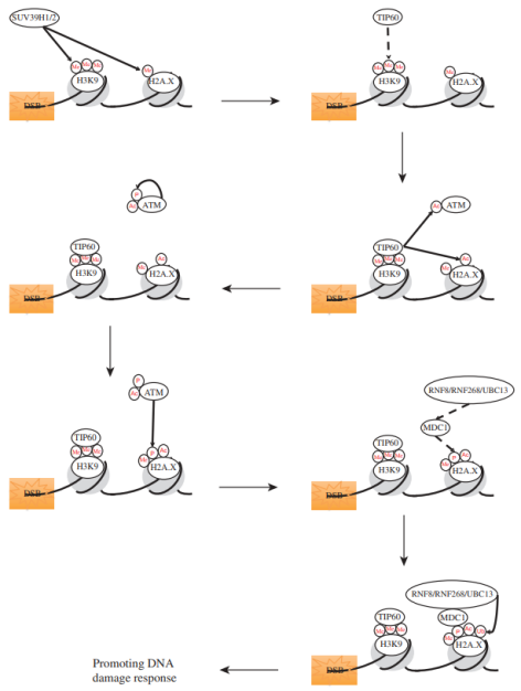
Reference: [19] Van & Santos, 2018
4. Histone Ubiquitination
Histone ubiquitination involves the covalent attachment of ubiquitin to histone proteins, particularly H2A and H2B. [11]
Unlike typical degradation-linked ubiquitination, H2A and H2B monoubiquitination generally does not directly target histones for degradation. Instead, it contributes to transcriptional regulation, chromatin organization, and DNA damage responses; other forms of histone ubiquitination may still be involved in protein degradation. [11][12][13]
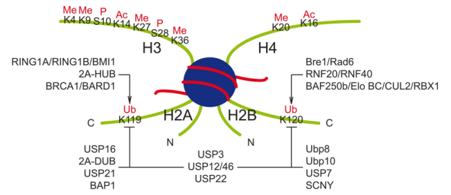
Reference: [11] Cao & Yan, 2012
Common ubiquitination marks include: [11]
- H2AK119ub1: Generally associated with Polycomb-mediated transcriptional repression. [11][20]
- H2BK120ub1: Commonly associated with active transcription and the regulation of other histone modifications. [11][12]
Following DNA damage, ubiquitination signals involving H2A and H2AX can contribute to the recruitment of repair proteins. [11][12][19]
Histone ubiquitination is reversible and is regulated by ubiquitin ligases and deubiquitinating enzymes. [11]
Part III. How Do Histone Modifications Function?
The functions of histone modifications depend not only on the modifications themselves but also on specialized regulatory proteins. [14][15]
These proteins are commonly classified into three groups. [14][15]
Writers add specific modifications to histones, including acetyltransferases and methyltransferases. [14]
Erasers remove these modifications, including deacetylases and demethylases. [14]
Readers recognize histone modifications through specialized binding domains and recruit additional regulatory complexes. [14][15]
For example, bromodomains generally recognize acetylated lysines, chromodomains recognize certain methylated lysines, and some PHD domains recognize specific methylation marks; other PHD domains can recognize unmodified lysines. [14][15]
Together, these proteins form a dynamic epigenetic regulatory network. [14][15]
H3K4me3 and H3K27me3 can coexist in bivalent chromatin domains at certain developmental regulatory genes in embryonic stem cells, which are associated with low expression and potential for subsequent activation. [6]
Therefore, understanding histone function requires examining interactions among multiple modifications rather than interpreting individual marks in isolation. [14]
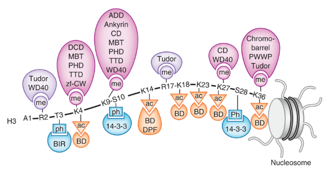
Reference: [15] Musselman et al., 2012
Part IV. How Are Histone Modifications Studied?
Studying histone modifications helps researchers understand gene expression states, chromatin organization, and regulatory mechanisms. [16][17]
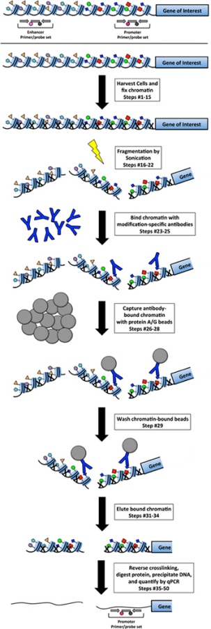
Chromatin immunoprecipitation (ChIP) uses modification-specific antibodies to enrich chromatin complexes containing the target histone modification, followed by isolation and analysis of the associated DNA. [16]
When combined with high-throughput sequencing, ChIP-seq enables genome-wide mapping of histone modifications and identification of regulatory regions. [17]
For example, H3K4me3 is commonly used to study promoters, H3K27ac helps identify active promoters and enhancers, and H3K9me3 is useful for investigating heterochromatin. [2][5][22]
Other methods include Western blotting to examine overall modification levels, immunofluorescence to visualize intracellular distribution, and enzyme activity assays to investigate histone-modifying enzymes. [16][18]
Each technique has distinct advantages and should be selected according to the research objectives, with appropriate experimental controls. [16][17]
Reference: Perceiving the epigenetic landscape through histone readers. (2018)
Part V. Research Significance of Histone Modifications
Histone modifications are essential components of epigenetic regulation and participate in gene expression, cellular differentiation, development, and DNA damage repair. [18][19]
Histone modifications and their regulatory enzymes dynamically contribute to the control of gene expression programs. [18]
Abnormalities in these modifications or their regulatory enzymes can disrupt gene expression programs and are associated with cancer and certain developmental disorders. [20]
Because many histone modifications are reversible, their regulatory enzymes have become important targets in epigenetic drug research. [21]
For example, the histone deacetylase inhibitor vorinostat has been used in the treatment of certain patients with cutaneous T-cell lymphoma. [21]
These findings provide insights into disease mechanisms and support further exploration of biomarkers and targeted therapeutic strategies. [18][20][21]
Quick Reference: Common Histone Modifications
| Histone mark | Main association | Typical distribution or application |
|---|---|---|
| H3K4me1 | Enhancer-associated mark | Enhancers |
| H3K4me3 | Transcription-associated | Promoters |
| H3K9ac | Active transcription-associated | Promoters and enhancers |
| H3K27ac | Active regulatory regions | Active promoters and enhancers |
| H3K36me3 | Transcriptional elongation | Gene bodies |
| H3K9me3 | Transcriptional repression | Heterochromatin and repetitive elements |
| H3K27me3 | Polycomb-associated repression | Developmental regulatory genes, etc. |
| H3S10ph | Mitosis-associated | Condensed mitotic chromosomes |
| γ - H2AX | DNA damage response | Regions associated with DNA double-strand breaks |
| H2AK119ub1 | Transcriptional repression | Polycomb-regulated regions |
| H2BK120ub1 | Transcription and chromatin regulation | Transcription-associated gene regions |
Note: These are common associations rather than absolute functional classifications. The effects of histone marks vary with cell type, genomic context, and other modifications. [5][18]
- γH2AX does not directly quantify the number of DNA double-strand breaks. Its signal also depends on cell state and other stress responses. [10]
- H3K4me1 alone does not establish enhancer activity. Additional evidence, including H3K27ac, is useful. [2][5]
- Histone marks should not be classified simply as activating or repressive; their combinations, genomic context, and reader proteins affect biological outcomes. [14][18]
Part VI. Research Significance of Histone Modifications
EnkiLife offers a range of histone-related antibodies for various experimental applications, including Western blotting (WB), immunohistochemistry (IHC), and immunofluorescence (IF). Our antibody portfolio covers nearly all commonly studied histones, supporting a wide range of histone detection and research needs.
The table below lists some of our histone-related products.
| Products | Cat. No. | Reactivity | Application |
|---|---|---|---|
| Histone H1 Rabbit Polyclonal Antibody | APRab00543 | Human,Mouse | WB,IHC-F,IHC-P,ICC/IF,ELISA |
| Phospho-Histone H1 (Thr17) Rabbit Polyclonal Antibody | APRab04773 | Human,Mouse,Monkey | WB,IHC-P,IF-P,IF-F,ICC/IF,ELISA |
| Acetyl Histone H1 (K25) Rabbit Polyclonal Antibody | APRab04160 | Human,Monkey | WB,IHC-P,IF-P,IF-F,ICC/IF,ELISA |
| Histone 1.0 Rabbit Polyclonal Antibody | APRab12045 | Human,Rat,Mouse | WB,IHC-P,IF-P,IF-F,ICC/IF,ELISA |
| Histone H1.2 Rabbit Monoclonal Antibody | AMRe02816 | Human,Mouse | WB,ICC/IF |
| Histone H1.2 Rabbit Monoclonal Antibody | AMRe85649 | Human,Mouse,Rat | WB,IHC,ICC |
| KD-Validated Histone H1.0 Recombinant Rabbit Monoclonal Antibody | KVAb01812 | Human,Mouse,Rat | WB,ICC |
| KD-Validated H1-3 Recombinant Rabbit Monoclonal Antibody | KVAb00368 | Human | WB,FCM |
| Histone H2A Rabbit Monoclonal Antibody | AMRe21481 | Human,Mouse,Rat | WB,IHC,IF,IP,ELISA,CHIP,Cut&Tag |
| Histone H2A Rabbit Polyclonal Antibody | APRab12055 | Human,Mouse,Rat | WB,IHC-P,IF-P,IF-F,ICC/IF,ELISA |
| Histone H2A(monomethylK118) Rabbit Monoclonal Antibody | AMRe84153 | Human,Mouse,Rat | WB,IHC,ICC/IF,ICC |
| Acetyl-Histone H2A(Lys9) Rabbit Monoclonal Antibody | AMRe83769 | Human,Mouse,Rat | WB,IHC,ICC/IF,ICC |
| Phospho-Histone H2A (Ser129) Rabbit Polyclonal Antibody | APRab00687 | Human,Mouse,Rat | WB |
| Hydroxyl-Histone H2A (Tyr39) Rabbit Monoclonal Antibody | AMRe03278 | Human,Mouse | WB,IHC |
| Acetyl Histone H2A (K5) Rabbit Polyclonal Antibody | APRab04161 | Human,Mouse,Rat | IHC-P,IF-P,IF-F,ICC/IF,WB,ELISA |
| Phospho-Histone H2A (Thr121) Rabbit Polyclonal Antibody | APRab04774 | Human,Mouse,Rat | WB,IHC-P,IF-P,IF-F,ICC/IF,ELISA |
| Histone H2B Mouse Monoclonal Antibody | AMM85023 | Human,Mouse,Rat | WB,IHC |
| Histone H2B Mouse Monoclonal Antibody | AMRe21413 | Human,Mouse,Rat | WB,IHC,IF,IP,ELISA,CHIP,Cut&Tag |
| Histone H2B Rabbit Polyclonal Antibody | APRab00111 | Human,Mouse,Rat | WB,IHC-P,ICC/IF,FC,IP |
| Histone H2B Rabbit Polyclonal Antibody | APRab12063 | Human,Mouse,Monkey | WB,IHC-P,IF-P,IF-F,ICC/IF,ELISA |
| Mouse Histone H2B ELISA Kit | EM21093 | ||
| Histone H2B(crotonylK12) Rabbit Monoclonal Antibody | AMRe84162 | Human,Mouse,Rat | WB,IHC,ICC/IF,ICC |
| Histone H2B(formylK116) Rabbit Monoclonal Antibody | AMRe84155 | Human,Mouse | WB,ICC |
| Histone H2B(formylK108) Rabbit Monoclonal Antibody | AMRe84632 | Human,Mouse,Rat | WB,IHC |
| Histone H2B(monomethylK116) Rabbit Monoclonal Antibody | AMRe84635 | Human,Mouse,Rat | WB,IHC,ICC/IF,ICC |
| Acetyl-Histone H2B (Lys21) Rabbit Polyclonal Antibody | APRab06197 | Human,Rat,Mouse | WB,ELISA |
| Acetyl-Histone H2B(Lys16) Rabbit Monoclonal Antibody | AMRe84169 | Human,Mouse | WB,IHC,ICC/IF,ICC,IP |
| Acetyl-Histone H2B (Lys15) Rabbit Polyclonal Antibody | APRab00853 | Human,Mouse | WB,IHC-F,IHC-P,ICC/IF |
| Acetyl-Histone H2B (Lys5) Rabbit Polyclonal Antibody | APRab00851 | Human,Mouse | WB,IHC-F,IHC-P,ICC/IF |
| Acetyl-Histone H2B (Lys20) Rabbit Polyclonal Antibody | APRab00701 | Human,Mouse,Rat | WB,IHC-P,ICC/IF,FC,IP,ChIP |
| Crotonyl-Histone H2B (Lys12) Rabbit Monoclonal Antibody | AMRe03903 | Human, Mouse, Rat | WB,IHC,ICC/IF |
| MonoMethyl-Histone H2B (Arg79) Rabbit Monoclonal Antibody | AMRe04022 | Human | WB |
| Phospho-HistoneH2B(T129) Rabbit Monoclonal Antibody | AMRe83770 | Yeast | WB |
| Histone H3 Rabbit Monoclonal Antibody | AMRe03904 | Human, Mouse, Rat | WB,ICC/IF,IP |
| Histone H3 Rabbit Monoclonal Antibody | AMRe02091 | Human, Mouse, Rat | WB,IHC |
| Histone H3 Rabbit Monoclonal Antibody | AMRe21336 | Human,Mouse,Rat,Cat | WB,IHC,IF,IP,ELISA,CHIP,Cut&Tag |
| Mouse Histone H3 ELISA Kit | EM21092 | ||
| Histone H3 Recombinant Rabbit Monoclonal Antibody | AMRe86363 | Human, Mouse, Rat | WB,IHC,ICC/IF,FC |
| Histone H3(mutatedK27M) Rabbit Monoclonal Antibody | AMRe84538 | Human | WB,ICC,IP |
| Histone H3(formylK122) Rabbit Monoclonal Antibody | AMRe84637 | Human,Mouse,Rat | WB,IHC |
| Acetyl-Histone H3(Lys18) Rabbit Monoclonal Antibody | AMRe84527 | Human,Mouse,Rat | WB,IHC,ICC/IF,ICC,FC,IP |
| Acetyl-Histone H3(Lys23) Rabbit Monoclonal Antibody | AMRe84536 | Human,Mouse | WB,ICC,IP |
| Acetyl-Histone H3(Lys27) Rabbit Monoclonal Antibody | AMRe84555 | Human,Mouse,Rat | WB,IHC,ICC/IF,ICC,IP |
| Symmetricdimethyl-Histone H3(R17) Rabbit Monoclonal Antibody | AMRe84144 | Human,Mouse | WB,ICC |
| Phospho-Histone H3 (Ser10) Rabbit Polyclonal Antibody | APRab03869 | Human | WB,ICC/IF |
| Phospho-Histone H3 (Ser28) Rabbit Monoclonal Antibody | AMRe02855 | Human,Mouse | WB |
| Phospho-Histone H3 (Thr3) Rabbit Monoclonal Antibody | AMRe03284 | Human,Mouse,Rat | WB,ICC/IF |
| Formyl-Histone H3 (Lys122) Rabbit Monoclonal Antibody | AMRe86229 | Human,Mouse,Rat | WB,IHC |
| Tri-Methyl-Histone H3 (Lys10) Rabbit Polyclonal Antibody | APRab04156 | Human,Rat,Mouse | WB,ELISA |
| Di-Methyl-Histone H3(Lys9) Rabbit Monoclonal Antibody | AMRe83827 | Human,Mouse,Rat | WB,ICC,IP |
| Mono-Methyl-Histone H3 (Lys79) Rabbit Monoclonal Antibody | AMRe84137 | Human,Mouse,Rat | WB,IHC,ICC/IF,ICC,IP |
| Histone H4 Rabbit Monoclonal Antibody | AMRe02093 | Human | WB,IHC,ICC/IF,ChIP |
| Histone H4 Rabbit Monoclonal Antibody | AMRe21614 | Human,Mouse,Rat | WB,IHC,IF,IP,ELISA,CHIP,Cut&Tag |
| Phospho-Histone H4 (Ser1) Rabbit Polyclonal Antibody | APRab00852 | Human,Mouse,Rat | WB,IHC-P |
| Acetyl-Histone H4 (Lys16) Rabbit Polyclonal Antibody | APRab00849 | Human,Mouse,Rat | WB,IHC-F,IHC-P,ICC/IF |
| Acetyl-Histone H4 (Lys5) Rabbit Monoclonal Antibody | AMRe03268 | Human,Mouse | WB,IHC,ICC/IF,IP |
| Acetyl-Histone H4 (Lys16) Rabbit Polyclonal Antibody | APRab06213 | Human,Mouse,Rat | WB,IHC-P,IF-P,IF-F,ICC/IF,ELISA |
| Phospho-Histone H4 (Ser47) Rabbit Polyclonal Antibody | APRab04782 | Human,Mouse,Rat | IF-P,IF-F,ICC/IF,IHC-P,WB,ELISA |
| MonoMethyl-Histone H4 (Lys20) Rabbit Monoclonal Antibody | AMRe03937 | Human, Mouse, Rat | WB,IHC,ICC/IF |
References
[1] Roth SY, Denu JM, Allis CD. Histone acetyltransferases. Annu Rev Biochem. 2001;70:81–120. doi:10.1146/annurev.biochem.70.1.81.
[2] Creyghton MP, et al. Histone H3K27ac separates active from poised enhancers and predicts developmental state. Proc Natl Acad Sci USA. 2010;107(50):21931–21936. doi:10.1073/pnas.1016071107.
[3] Karmodiya K, et al. H3K9 and H3K14 acetylation co-occur at many gene regulatory elements, while H3K14ac marks a subset of inactive inducible promoters in mouse embryonic stem cells. BMC Genomics. 2012;13:424. doi:10.1186/1471-2164-13-424.
[4] Greer EL, Shi Y. Histone methylation: a dynamic mark in health, disease and inheritance. Nat Rev Genet. 2012;13:343–357. doi:10.1038/nrg3173.
[5] Mikkelsen TS, et al. Genome-wide maps of chromatin state in pluripotent and lineage-committed cells. Nature. 2007;448:553–560. doi:10.1038/nature06008.
[6] Bernstein BE, et al. A bivalent chromatin structure marks key developmental genes in embryonic stem cells. Cell. 2006;125(2):315–326. doi:10.1016/j.cell.2006.02.041.
[7] Rossetto D, Avvakumov N, Côté J. Histone phosphorylation: a chromatin modification involved in diverse nuclear events. Epigenetics. 2012;7:1098–1108. doi:10.4161/epi.21975.
[8] Hendzel MJ, et al. Mitosis-specific phosphorylation of histone H3 initiates primarily within pericentromeric heterochromatin during G2 and spreads in an ordered fashion coincident with mitotic chromosome condensation. Chromosoma. 1997;106:348–360. doi:10.1007/s004120050256.
[9] Lowndes NF, Toh GW-L. DNA repair: the importance of phosphorylating histone H2AX. Curr Biol. 2005;15:R99–R102. doi:10.1016/j.cub.2005.01.034.
[10] Turinetto V, Giachino C. Multiple facets of histone variant H2AX: a DNA double-strand-break marker with several biological functions. Nucleic Acids Res. 2015;43:2489–2498. doi:10.1093/nar/gkv061.
[11] Cao J, Yan Q. Histone ubiquitination and deubiquitination in transcription, DNA damage response, and cancer. Front Oncol. 2012;2:26. doi:10.3389/fonc.2012.00026.
[12] Meas R, Mao P. Histone ubiquitylation and its roles in transcription and DNA damage response. DNA Repair (Amst). 2015;36:36–42. doi:10.1016/j.dnarep.2015.09.016.
[13] Shmueli MD, Sheban D, Eisenberg-Lerner A, Merbl Y. Histone degradation by the proteasome regulates chromatin and cellular plasticity. FEBS J. 2022;289:3304–3316. doi:10.1111/febs.15903.
[14] Ruthenburg AJ, Li H, Patel DJ, Allis CD. Multivalent engagement of chromatin modifications by linked binding modules. Nat Rev Mol Cell Biol. 2007;8:983–994. doi:10.1038/nrm2298.
[15] Musselman CA, Lalonde ME, Côté J, Kutateladze TG. Perceiving the epigenetic landscape through histone readers. Nat Struct Mol Biol. 2012;19:1218–1227. doi:10.1038/nsmb.2436.
[16] Wardle FC, Tan H. A ChIP on the shoulder? Chromatin immunoprecipitation and validation strategies for ChIP antibodies. F1000Res. 2015;4:235. doi:10.12688/f1000research.6719.1.
[17] Hino S, Sato T, Nakao M. Chromatin immunoprecipitation sequencing (ChIP-seq) for detecting histone modifications and modifiers. Methods Mol Biol. 2023;2577:55–64. doi:10.1007/978-1-0716-2724-2_4.
[18] Millán-Zambrano G, Burton A, Bannister AJ, Schneider R. Histone post-translational modifications—cause and consequence of genome function. Nat Rev Genet. 2022;23:563–580. doi:10.1038/s41576-022-00468-7.
[19] Van HT, Santos MA. Histone modifications and the DNA double-strand break response. Cell Cycle. 2018;17:2399–2410. doi:10.1080/15384101.2018.1542899.
[20] Janssen SM, Lorincz MC. Interplay between chromatin marks in development and disease. Nat Rev Genet. 2022;23:137–153. doi:10.1038/s41576-021-00416-x.
[21] Karagiannis D, Rampias T. HDAC inhibitors: dissecting mechanisms of action to counter tumor heterogeneity. Cancers (Basel). 2021;13(14):3575. doi:10.3390/cancers13143575.
[22] The ENCODE Project Consortium. An integrated encyclopedia of DNA elements in the human genome. Nature. 2012;489:57–74. doi:10.1038/nature11247.