
Immunoprecipitation (IP) and Co-immunoprecipitation (Co-IP)
01 Principles
Immunoprecipitation (IP) enriches a target protein from cell or tissue lysates through specific antigen–antibody binding. After the antibody binds the target protein, Protein A/G agarose or magnetic beads capture the immune complex. Following washing and elution, the enriched protein can be analyzed by Western blotting (WB) or mass spectrometry.
Co-immunoprecipitation (Co-IP) extends this approach by using an antibody against a “bait” protein to co-enrich potentially associated “prey” proteins, thereby investigating protein–protein interactions. Co-IP indicates an association within the same protein complex; it does not necessarily demonstrate direct binding between two proteins. [1,2]
02 Materials
Cell or tissue samples; a non-denaturing lysis buffer appropriate for the target protein and its subcellular localization; protease inhibitors (plus phosphatase inhibitors when studying phosphorylation); an IP-validated antibody against the target protein; an appropriate isotype-matched control IgG; Protein A/G magnetic or agarose beads; pre-chilled wash buffer; elution buffer; and Western blotting reagents.
Protein A/G captures antibodies primarily through their IgG Fc regions. Binding affinity varies with antibody host species and IgG subclass.
Lysis buffer selection should account for the target protein’s subcellular location and the stability of the protein complex. Nonionic detergents such as NP-40 and Triton X-100 are commonly used for mild lysis. A commonly recommended concentration range for NP-40/Triton X-100 is 0.5%–1%, although the detergent type and concentration should be optimized for the experimental system. [1,2,3]
03 Experimental Procedure
Note: This section provides a concise overview only. Specific experimental conditions should follow the instructions for the selected target protein, antibody, beads and other reagents, with optimization through pilot experiments.
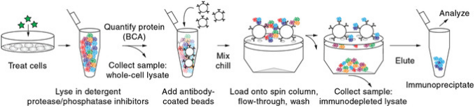
1. Sample Lysis and Protein Quantification
Keep samples cold whenever possible. Add a suitable inhibitor-supplemented lysis buffer, lyse on ice, and centrifuge at 4°C to remove insoluble material. Collect the supernatant and determine protein concentration using a BCA or Bradford assay. Before immunoprecipitation, reserve a small aliquot of total lysate as the Input sample. A common starting amount is 1%–10% of the total lysate, adjusted according to WB detection needs. Input samples intended for WB can be treated separately with SDS sample buffer under denaturing conditions without affecting the remaining lysate used for Co-IP. Mild, non-denaturing lysis conditions are preferred for Co-IP. [1,2]
2. Pre-clearing (Optional)
Pre-incubate the lysate with antibody-free Protein A/G beads to reduce some nonspecific adsorption; this step may be omitted when background is low. Pre-clearing does not replace a separate IgG negative control. Determine whether it is necessary based on sample background and pilot experiments.
3. Antibody Incubation
Two common workflows are used:
Both approaches generally use standard Protein A/G beads and require an appropriate isotype-matched IgG negative control. Optimize incubation time, reagent amounts and order of addition according to product instructions and pilot experiments.
4. Capture and Washing with Protein A/G Beads
Depending on the chosen workflow, add pretreated Protein A/G magnetic or agarose beads, or pre-bind the antibody to the beads before adding the lysate. Confirm the antibody host species, IgG subclass and bead binding capacity beforehand. Separate magnetic beads with a magnetic rack and agarose beads by gentle centrifugation. Avoid aspirating beads when removing the supernatant.
Begin with approximately three washes and, depending on background and interaction stability, adjust to three to five washes. In Co-IP, washing conditions must balance stringency and complex preservation: overly stringent washing may disrupt weak interactions, while insufficient washing increases background. High salt may dissociate some physiological or weak interactions, but increasing NaCl can improve wash stringency. NaCl concentrations of 300–500 mM may be used to reduce background; however, this is a relatively high-salt range and should be considered for gradient optimization only when nonspecific background is substantial. [2,5,6]
5. Elution and Detection
Choose the elution method according to downstream applications. For routine WB, denaturing elution with an SDS-containing sample buffer may be used. This denatures proteins and may inactivate antibodies or affinity ligands immobilized on the beads; it is therefore generally unsuitable for subsequent native-state analyses, and bead reuse should not be assumed.
If native protein complexes must be preserved for further enrichment or functional analysis, use a validated mild elution strategy, such as competitive peptide elution for a compatible epitope-tag system. Low-pH glycine eluates generally require prompt neutralization, although acidic conditions may disrupt some complexes. A near-neutral buffer alone does not necessarily release bound antigen effectively. Mass spectrometry requires a compatible elution and sample-preparation workflow. [1,7]
For Co-IP followed by WB, probe for the candidate interacting protein and confirm successful enrichment of the bait protein.
Additional Note: Epitope-Tagged Co-IP (Optional)
When endogenous bait protein is scarce or no suitable IP antibody is available, a bait protein carrying an HA, Myc or FLAG tag may be expressed and precipitated with the corresponding anti-tag antibody. This approach can help detect candidate protein associations, but exogenous overexpression may introduce nonphysiological interactions. Interpret results with appropriate controls, such as empty-vector or single-expression controls.
For FLAG and other epitope tags, affinity beads bearing immobilized anti-tag antibodies (e.g., anti-FLAG/HA agarose) can also capture tagged proteins directly. These are tag-specific affinity beads rather than general Protein A/G beads. They effectively reduce the risk of free IP antibody being co-eluted with the sample and can inherently avoid heavy- and light-chain contamination. [7]
04 Controls and Key Considerations
Additional Note: Interpreting Co-IP Western Blots
Input confirms that the bait and candidate prey proteins are detectable in the starting sample. IgG or other suitable negative controls help assess nonspecific precipitation. Detection of bait in the IP fraction indicates successful enrichment, whereas detection of candidate prey supports an association under the experimental conditions. When the prey has a molecular weight close to the IP antibody heavy chain (~50 kDa) or light chain (~25 kDa), antibody-chain interference must be considered. Interpret prey bands alongside the IgG negative control and other appropriate controls. Reciprocal Co-IP, endogenous Co-IP, or complementary approaches may provide further validation. A positive Co-IP result alone does not prove direct binding. [2,9]
05 References and Technical Resources
[1] Abcam. Immunoprecipitation & Co-immunoprecipitation Protocol. View source
[2] Thermo Fisher Scientific. Co-Immunoprecipitation (Co-IP). View source
[3] Co-immunoprecipitation and semi-quantitative immunoblotting for the analysis of protein-protein interactions. (2021). View source
[4] Thermo Fisher Scientific. Dynabeads Co-Immunoprecipitation User Guide. View source
[5] Hall RA. Co-immunoprecipitation as a strategy to evaluate receptor–receptor or receptor–protein interactions. In: G Protein-Coupled Receptor–Protein Interactions. Chapter 9, 2005. View source
[6] Coimmunoprecipitation of Proteins from Yeast. (2015). View source
[7] Merck / Sigma-Aldrich. Immunoprecipitation of FLAG Fusion Proteins Using Monoclonal Antibody Affinity Gels. View source
[8] Cattoglio C, et al. Assessing Self-interaction of Mammalian Nuclear Proteins by Co-immunoprecipitation. Bio-protocol. 2020;10(4):e3526. View source
[9] Cell Signaling Technology. Immunoprecipitation Troubleshooting Guide: Target Signal Obscured by Heavy or Light Chain IgG. View source
Disclaimer: This document is a concise experimental guide. The conditions listed are starting references from published protocols or manufacturers’ instructions, not a universal SOP for all target proteins, cell types or antibody systems. Follow the relevant reagent instructions and optimize through pilot experiments.