
This article introduces the fundamental principles, major technical approaches, and applications of multiplex immunohistochemistry (mIHC), with a particular focus on fluorescence-based detection and tyramide signal amplification (TSA).
Multiplex immunohistochemistry (mIHC) is an in situ technique that detects multiple protein biomarkers within a single tissue section. Unlike conventional single-plex IHC, which generally focuses on one antigen per slide, mIHC simultaneously reveals multiple cell populations, functional states, and spatial relationships while preserving tissue morphology. [1,2,3,6]
For example, tumor microenvironment studies may combine tumor markers with CD3, CD8, CD68, PD-1, and PD-L1 to identify immune cell populations and their distribution relative to tumor cells. Beyond detecting more markers, mIHC can help determine whether proteins are co-expressed within the same cell or whether different cell populations are spatially associated. [1,3,4]
mIHC is based on specific antigen–antibody recognition. Detection systems convert these binding events into measurable signals using chromogens, fluorophores, metal isotopes, or DNA barcodes. The signals must be distinguishable so that the distribution of each target can be reconstructed within the tissue. [1,3,6]
The major challenges include antibody cross-reactivity, spectral bleed-through, antigen damage during repeated staining, and channel-to-channel imaging variations. Therefore, mIHC encompasses not only staining but also antibody panel design, assay optimization, imaging, and quantitative analysis. [1,2,8]
mIHC technologies can be classified according to antibody labeling, detection chemistry, staining format, and signal amplification strategy. [1,3,6,7]
| Classification | Main Types | Key Difference |
|---|---|---|
| Antibody labeling | Direct / Indirect | Reporter directly attached to a primary antibody or introduced through secondary detection |
| Detection chemistry | Chromogenic, fluorescent, metal-based, DNA-barcoded | Signals distinguished by color, fluorescence, atomic mass, or nucleic acid sequences |
| Staining format | Simultaneous / Sequential | Multiple markers detected together or through repeated staining cycles |
| Amplification | Secondary antibodies, polymers, TSA | Increasing reporter signal or localized deposition for weak targets |
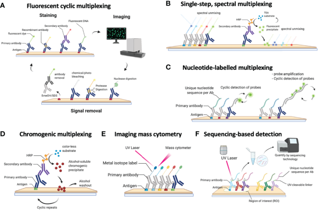
Figure 1. Major mIHC technology categories. Source: Next-Generation Pathology Using Multiplexed Immunohistochemistry: Mapping Tissue Architecture at Single-Cell Level. Front. Oncol. 2022;12:918900.
Direct labeling: Fluorophores, metal tags, or DNA oligonucleotides are directly conjugated to primary antibodies. This reduces secondary-antibody cross-reactivity and is suitable for some high-plex platforms, although each antibody must be individually labeled and validated. Direct fluorescence signals may also be relatively weak. [1,3,6]
Indirect labeling: Unlabeled primary antibodies bind to their antigens, followed by species- or isotype-specific secondary antibodies or enzyme-polymer detection systems. Indirect detection can increase signal intensity, but conventional multicolor indirect IF can suffer from cross-reactivity when several primary antibodies originate from the same species.
In TSA-mIHC, fluorescent deposition is completed before the previous antibodies and HRP components are removed. This allows multiple primary antibodies from the same host species to be used sequentially, improving panel flexibility. [1,6,7,11,12]
Chromogenic mIHC uses horseradish peroxidase (HRP) or alkaline phosphatase (AP) to catalyze the formation of insoluble colored precipitates, such as brown DAB or red Fast Red, at antigen-binding sites. These signals are visible under conventional brightfield microscopy and compatible with established pathology workflows. [1,2,6]
However, chromogenic multiplexing has several limitations. Overlapping absorption spectra and color mixing complicate the interpretation of co-expressed targets, while the number of clearly distinguishable chromogens is limited. This approach is generally more practical for small panels of approximately two to five markers. Cyclic chromogenic staining can extend this capacity, but requires repeated staining, scanning, and image registration. [1,2,6]
Chromogenic methods remain useful for routine pathology and low-plex applications. Nevertheless, fluorescence and other high-dimensional detection strategies are often better suited to complex cellular phenotyping and spatial analysis.
Fluorescence-based mIHC uses fluorophores as reporter molecules. After antibody binding, fluorescent dyes are excited by light of suitable wavelengths and emit light at longer wavelengths. Microscopes use excitation sources, emission filters or spectral detectors, and cameras to collect signals from different markers. Common fluorescent dyes include FITC, Alexa Fluor, and Cy fluorophores. [1,3,6]
Conventional multicolor staining can employ directly labeled primary antibodies or multiple primary antibodies paired with fluorescent secondary antibodies. Different fluorescence channels can be independently recorded and combined to reveal protein co-expression within the same cell and spatial relationships among cell populations. [1,3,6]
Cyclic immunofluorescence (Cyc-IF) extends conventional fluorescence detection through repeated rounds of antibody staining, imaging, fluorescence bleaching or removal, and restaining. Images from successive rounds are registered for integrated analysis. Approaches such as t-CyCIF can detect numerous markers using conventional optical microscopes without relying on TSA-mediated covalent deposition. [16]
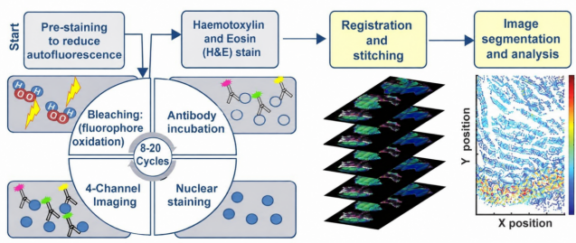
Figure 2. Cyc-IF workflow. Source: Lin JR et al. Highly multiplexed immunofluorescence imaging of human tissues and tumors using t-CyCIF and conventional optical microscopes. eLife. 2018;7:e31657.
Fluorescence-based mIHC is mainly limited by spectral overlap, tissue autofluorescence, and photobleaching. Strong fluorescence from one dye may bleed into another detection channel, while endogenous compounds such as collagen, elastin, and lipofuscin can generate background fluorescence and obscure weak signals. [1,4,6]
Appropriate fluorophore selection, optimized excitation and emission windows, single-stain and unstained controls, sequential scanning, multispectral imaging, and spectral unmixing can reduce these problems. For quantitative comparison, raw channel data should be retained and imaging parameters kept consistent wherever possible. [1,4,6]
The value of fluorescence-based mIHC goes beyond displaying multiple colors: independent detection channels enable the integrated analysis of target expression, cell phenotypes, and spatial organization.
Tyramide signal amplification (TSA), also known as catalyzed reporter deposition (CARD), is an HRP-mediated technique for localized signal amplification. Unlike conventional fluorescent antibody labeling, TSA uses HRP positioned near the antigen to catalyze the deposition of numerous fluorophore-labeled tyramide molecules onto nearby tissue proteins. This makes TSA particularly useful for detecting low-abundance antigens. [1,6,7,9]
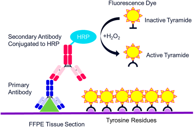
Figure 3. TSA signal amplification mechanism. Source: Multiplex Immunofluorescence and Multispectral Imaging: Forming the Basis of a Clinical Test Platform for Immuno-Oncology. Front. Mol. Biosci. 2021;8:674747.
The TSA reaction consists of five key events: (1) a primary antibody binds the target antigen; (2) an HRP-conjugated secondary antibody or HRP-polymer system localizes enzyme activity; (3) fluorophore-labeled tyramide and hydrogen peroxide are introduced; (4) HRP catalyzes tyramide oxidation, generating highly reactive intermediates; and (5) activated tyramide covalently attaches to nearby electron-rich protein residues, especially tyrosine residues. [6,9,11]
TSA offers two important advantages. First, a single localized HRP site can catalyze the deposition of many fluorescent molecules, enhancing signal intensity and improving the detectability of low-abundance proteins. Second, covalently deposited fluorescence can remain in the tissue after antibody removal. The amplification achieved depends on tissue characteristics, antibody quality, and experimental conditions. [1,6,7,9]
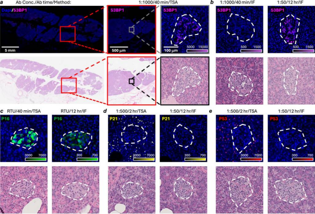
Figure 4. Improved detection of low-abundance antigens using TSA compared with conventional IF. Source: iCLAP: an innovative method for integrable co-detection of low-abundance antigens with high-plex immunostaining. Nat. Commun. 2026;17:3104.
In cyclic TSA-mIHC, markers are labeled sequentially. Each round includes primary antibody incubation, HRP detection, and deposition of one fluorescent tyramide. A validated stripping method then removes the previous antibodies and HRP components while the covalently deposited fluorophore generally remains. Another antibody and fluorescent tyramide are introduced in the following round. Finally, all accumulated fluorescence channels can be imaged together. [1,6,7,11,12]
This strategy allows several primary antibodies from the same host species to be used sequentially, reducing the restrictions associated with conventional multicolor indirect IF. However, incomplete stripping, excessive signal deposition, or repeated tissue processing may compromise staining specificity and antigen preservation. [1,2,6,7]
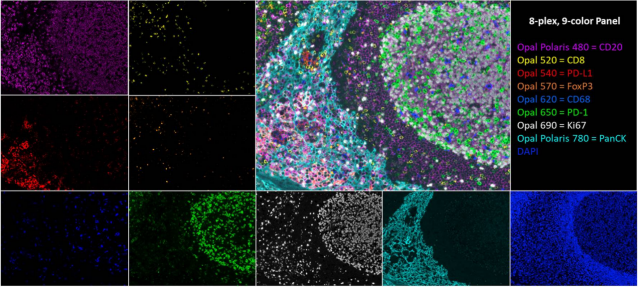
Figure 5. Multiplex immunostaining using TSA. Source: Multiplex Immunofluorescence and Multispectral Imaging: Forming the Basis of a Clinical Test Platform for Immuno-Oncology. Front. Mol. Biosci. 2021;8:674747.
Unlike Cyc-IF, in which fluorescence is generally imaged and then bleached or removed before subsequent rounds, conventional cyclic TSA-mIHC retains fluorescence through covalent deposition while removing antibodies between cycles. These methods therefore differ in their signal retention and image acquisition strategies. [1,7,16]
Polymer-based amplification: Polymer scaffolds carry multiple HRP or AP enzyme molecules, increasing enzymatic activity near antigen-binding sites. These systems can support chromogenic IHC or provide HRP detection for TSA-mIHC. [1,6]
DNA barcoding: Primary antibodies are conjugated to unique DNA oligonucleotides and subsequently detected using complementary fluorescent probes. Repeated hybridization and imaging cycles can substantially increase multiplexing capacity, although they demand specialized reagents and accurate image registration. [3,5,6]
Metal-based detection: Imaging Mass Cytometry (IMC) and Multiplexed Ion Beam Imaging (MIBI) use antibodies labeled with metal isotopes and mass-based imaging methods. These techniques can detect dozens of markers without fluorescence spectral overlap, but require specialized equipment and may consume or destroy tissue during imaging. [1,3,6]
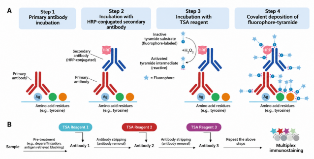
Figure 6. TSA mechanism and sequential staining workflow. Source: EnkiLife.
The following workflow describes a typical cyclic fluorescent TSA-mIHC experiment using formalin-fixed, paraffin-embedded (FFPE) tissue. The principle is to preserve deposited fluorescence while replacing the antibody recognition system between rounds. [1,6,7,11]
Step 1. Tissue preparation: Deparaffinize and rehydrate FFPE sections, ensuring adequate tissue adhesion and integrity. Frozen sections and cellular samples require preparation appropriate to their fixation and permeabilization conditions. [1,6]
Step 2. Antigen retrieval and blocking: Perform antigen retrieval suitable for the target and fixation conditions. Suppress endogenous peroxidase activity to minimize nonspecific TSA deposition and block nonspecific antibody binding sites. [1,2,6]
Step 3. Primary antibody incubation: Apply a primary antibody previously validated under TSA single-stain conditions. Optimize its concentration through titration because amplification may permit greater antibody dilution than conventional IF. [1,2,7]
Step 4. HRP detection and TSA deposition: Introduce a compatible HRP-labeled secondary antibody or polymer detection system, followed by a fluorescent tyramide working solution. HRP catalyzes localized fluorescent deposition. Wash thoroughly to avoid excessive deposition and elevated background. [1,6,9,11]
Step 5. Antibody removal: Use a validated stripping buffer, heat treatment, or other compatible method to remove the previous primary and secondary antibodies and HRP components while retaining deposited fluorescence. Confirm adequate removal before the next round. [1,6,7,11,12]
Step 6. Sequential staining and nuclear counterstaining: Repeat antibody incubation, HRP detection, TSA deposition, and inter-round antibody removal according to the optimized panel design. After all targets are labeled, apply DAPI or another suitable nuclear stain and mount the slide. [1,6,7]
Step 7. Image acquisition: Configure imaging channels according to the excitation and emission spectra of each fluorophore. Avoid detector saturation and excessive bleed-through. Use spectral unmixing when appropriate. [1,4,6]
Step 8. Image analysis: Preserve raw images before tissue annotation, cell segmentation, positivity thresholding, cellular phenotyping, and spatial analysis. Include appropriate negative, single-stain, and stripping controls. [1,4,8]
One major advantage of TSA-mIHC is reduced dependence on the host species of primary antibodies. In conventional multicolor indirect IF, several antibodies raised in the same host species may be cross-recognized by secondary antibodies. In TSA-mIHC, each round deposits fluorescence before antibodies and HRP are removed. Consequently, multiple primary antibodies originating from the same species can be used sequentially, provided that stripping efficiency has been validated. [1,6,7,11,12]
Panel design should begin with the biological question rather than maximizing the number of markers. Antibody selection should consider tissue suitability, specificity, expected cellular localization, and antigen retrieval compatibility.
Each antibody should first be tested individually under TSA conditions, with optimized concentration, retrieval, and fluorescent deposition. The resulting single-stain patterns should be compared with multiplex staining to identify cross-reactivity, signal distortion, or incomplete stripping. [1,2,7,8,11]
Fluorophore selection should account for expression levels, detector sensitivity, spectral separation, and tissue autofluorescence. Low-abundance targets generally benefit from channels with favorable signal-to-background performance, while highly expressed markers should not saturate detection channels. [1,4,6]
Staining order should be optimized according to antigen stability, target abundance, spatial proximity, and tolerance to repeated processing. Recording antibody clones, staining order, fluorophores, imaging settings, and validation results is essential for reproducibility. [1,2,7,8,11]
Fluorophores emit across relatively broad spectral ranges. Consequently, emissions from different dyes may overlap, causing bleed-through between detection channels. Multispectral imaging acquires wavelength-resolved signals and uses reference spectra generated from single-stain controls to separate overlapping fluorescence components. Spectral unmixing can also help distinguish endogenous tissue autofluorescence under suitable conditions. [1,4,6]
Spectral unmixing cannot fully compensate for inappropriate fluorophore combinations, signal saturation, or inaccurate reference spectra. Proper experimental design and imaging remain essential.
Laser scanning confocal microscopy uses point scanning and a pinhole to reject out-of-focus fluorescence, providing improved optical sectioning and contrast. It is particularly useful for subcellular localization, thicker samples, and Z-stack analysis. However, whole-slide fluorescence scanners may be more efficient for large FFPE sections. [4,6]
| Parameter | Practical Considerations |
|---|---|
| Pinhole | Often start near 1 Airy unit. Smaller pinholes improve optical sectioning but reduce collected photons. |
| Laser Power | Use the lowest excitation power providing sufficient signal while minimizing photobleaching. |
| Detector Gain | Adjust detector response without excessive noise amplification or saturation. |
| Sequential Scanning | Acquire channels under separate excitation and detection configurations to reduce some bleed-through. |
| Pixel Size / Dwell Time | Balance sampling density, photon collection, imaging speed, and bleaching. |
| Z-stack | Choose appropriate axial sampling and retain the original image stack for analysis. |
Unlike conventional confocal microscopes with several fixed excitation wavelengths, a tunable white light laser offers flexible excitation wavelength selection within the instrument's supported spectral range. This enables more suitable excitation of multiple fluorophores and facilitates multicolor imaging when combined with adjustable detection windows. [6,13]
WLL improves excitation flexibility but does not directly eliminate fluorescence bleed-through or tissue autofluorescence. Sequential scanning, appropriate emission windows, single-stain controls, and spectral unmixing may still be required.
Fluorescence lifetime imaging microscopy (FLIM) measures the temporal decay of fluorescence following excitation rather than relying only on fluorescence intensity. Fluorophores with overlapping emission spectra may exhibit different lifetimes, allowing certain signals or autofluorescence components to be separated using time-resolved detection and suitable analysis algorithms. [14,15]
FLIM can complement spectral imaging in challenging multicolor experiments but is not a routine requirement for TSA-mIHC. Fluorescence lifetimes depend on fluorophore identity and local environment, and additional acquisition or analysis may increase experimental complexity.
mIHC image analysis typically involves tissue annotation, nuclear and cellular segmentation, marker positivity thresholds, cellular phenotyping, and spatial metrics such as cell density and nearest-neighbor distances. Pixel-level fluorescence overlap, co-expression within a cell, and proximity between different cells are related but distinct concepts. [4,5,8]
Accurate interpretation depends on staining quality, channel separation, segmentation accuracy, and appropriate thresholds. Raw images and unmixed data should be retained, and representative regions should be reviewed to ensure analytical reliability.
mIHC is widely used in tumor microenvironment analysis, immune infiltration studies, tumor heterogeneity research, inflammation and autoimmune disease studies, neuroscience, biomarker discovery, and translational medicine. Its ability to combine cellular identity, protein expression, and spatial organization offers valuable information that is difficult to obtain using individual single-marker stains. [1,3,4,6,7]
| Criterion | Single-plex IHC | Conventional Low-plex IF | TSA-mIHC |
|---|---|---|---|
| Markers per section | Usually one | Usually a few | More markers depending on the panel |
| Co-expression | Often inferred from adjacent sections | Possible within limited panels | Complex cellular phenotypes |
| Spatial analysis | Single-marker distribution | Basic spatial relationships | Integrated phenotyping and spatial analysis |
| Low-abundance targets | Detection-system dependent | May be signal limited | Enhanced by TSA amplification |
| Cost and complexity | Relatively low | Moderate | Higher optimization and analytical demands |
Despite its advantages, TSA-mIHC requires careful panel validation, background control, fluorophore selection, inter-round stripping, and image analysis. Increasing the number of markers does not automatically improve biological reliability. [1,2,4,8]
mIHC integrates multiprotein detection with tissue morphology, cellular phenotypes, and spatial relationships. Fluorescence-based detection allows different targets to be represented in separate channels, while TSA provides localized HRP-mediated signal amplification and supports sequential staining through covalent fluorophore deposition. [1,3,6,7]
Reliable TSA-mIHC requires coordinated optimization of antibodies, staining order, antibody stripping, fluorescence acquisition, and image analysis. Starting with a smaller validated panel is often an effective route toward more complex multiplex assays. [1,4,8,11]
| Catalog No. | Product Name | Representative Image |
|---|---|---|
| RA10008 | 3-Color Multiple Fluorescent Staining Kit (mIHC) | 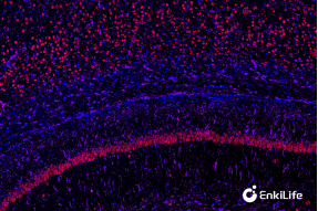 |
| RA10009 | 4-Color Multiple Fluorescent Staining Kit (mIHC) | 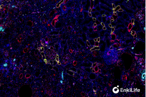 |
| RA10010 | 5-Color Multiple Fluorescent Staining Kit (mIHC) | 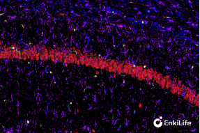 |
| RA10011 | 6-Color Multiple Fluorescent Staining Kit (mIHC) | 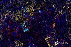 |
| RA10012 | 7-Color Multiple Fluorescent Staining Kit (mIHC) | 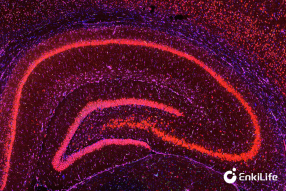 |
[1] Society for Immunotherapy of Cancer (SITC). Best practices for multiplex IHC/IF staining and validation. J Immunother Cancer. 2020;8:e000155. Full text
[2] Exploring Multiplex Immunohistochemistry Techniques and Histopathology Image Analysis. Cancer Medicine. 2025. Full text
[3] Next-Generation Pathology Using Multiplexed Immunohistochemistry: Mapping Tissue Architecture at Single-Cell Level. Front Oncol. 2022. Full text
[4] SITC. Updates and best practices for multiplex IHC/IF image analysis and data sharing. J Immunother Cancer. 2025. Full text
[5] Spatial analysis by current multiplexed imaging technologies for the molecular characterisation of cancer tissues. Br J Cancer. 2024. Article
[6] Abcam. Multiplex Immunohistochemistry (mIHC): A Technical Deep Dive for the Modern Researcher. Original article
[7] Research progress and perspectives on the application of TSA-based multiplex IHC/IF. Front Oncol. 2025. Full text
[8] SITC. Standards for Reporting of Multiplex IHC/IF Assays (STORMI). 2025. Full text
[9] Bobrow MN et al. Catalyzed reporter deposition, a novel method of signal amplification. J Immunol Methods. 1989;125:279–285. DOI
[10] Thermo Fisher Scientific. Multiplexed Tissue Staining with Signal Amplification. Protocol
[11] Buchwalow I et al. Multiple immunolabeling with antibodies from the same host species in combination with TSA. Acta Histochem. 2018;120:405–411. PubMed
[12] Fully automated 5-plex fluorescent immunohistochemistry with TSA and same-species antibodies. 2017. PubMed
[13] Leica Microsystems. The Principles of White Light Laser Confocal Microscopy. Article
[14] Fluorescence Lifetime Imaging Techniques—A Review on Principles, Applications and Clinical Relevance. J Biophotonics. 2025. DOI
[15] Multiplexed imaging in live cells using pulsed interleaved excitation spectral FLIM. 2024. Full text
[16] Lin JR et al. Highly multiplexed immunofluorescence imaging of human tissues and tumors using t-CyCIF and conventional optical microscopes. eLife. 2018;7:e31657. Full text