1. Basic Information of the Article
This is a review article published in 2026. It systematically summarizes the definition, core molecular mechanisms, pathophysiological functions, biomarker applications and therapeutic prospects of a novel form of regulated cell death first named in 2025 — Necrosis by Sodium Overload (NECSO).
2. Research Background and Definition of NECSO
2.1 Research Background
Sodium ion (Na⁺) is the most abundant cation in extracellular fluid. The maintenance of its transmembrane gradient is the basis of cellular function. Under normal conditions, intracellular Na⁺ concentration is tightly regulated by the dynamic balance between active extrusion mediated by Na⁺/K⁺‑ATPase and multiple Na⁺ influx pathways (voltage‑gated Na⁺ channels, Na⁺‑H⁺ exchanger NHE, Na⁺‑Ca²⁺ exchanger NCX, etc.). For a long time, intracellular Ca²⁺ overload has been considered the core driving factor of cell injury and death. However, accumulating evidence indicates that Na⁺ overload can independently trigger irreversible cell injury without Ca²⁺ overload.
2.2 Definition of NECSO
In 2025, Fu et al. first discovered and formally named NECSO through chemical screening — a form of regulated necrosis directly triggered by sustained and excessive Na⁺ influx, mainly mediated by TRPM4 channels. The study found that the small molecule compound Necroside 1 (NC1) can specifically and continuously activate human TRPM4 channels, inducing massive Na⁺ influx and eventual necrotic cell death. This form of death cannot be blocked by apoptosis inhibitors (such as z‑VAD‑fmk), necroptosis inhibitors (necrostatin‑1) or pyroptosis inhibitors (disulfiram), but can be effectively inhibited by Na⁺ channel blockers or Na⁺ chelators, establishing the causal role of Na⁺ overload in this process.
2.3 Morphological Characteristics of NECSO
The morphological features of NECSO are consistent with classic necrosis, including early cell swelling, organelle (especially mitochondrial) edema (usually preceding plasma membrane rupture), late plasma membrane breakdown and release of intracellular contents. Typical apoptotic markers such as chromatin condensation and caspase activation are generally absent. Notably, in some pathological models (e.g., H₂O₂‑induced cardiomyocyte injury), necrotic features of NECSO and apoptotic markers can be observed simultaneously in the same cell, reflecting the complexity of cell death programs.
3. Core Molecular Mechanisms
3.1 TRPM4 Channel: The Trigger Hub of NECSO
TRPM4 (Transient Receptor Potential Melastatin 4) is the molecular switch and core executor of NECSO. It is a Ca²⁺‑activated non‑selective monovalent cation channel, mainly permeable to Na⁺ and K⁺ but impermeable to divalent cations. Channel opening depends on elevated intracellular Ca²⁺ concentration and the auxiliary effect of membrane phosphatidylinositol 4,5‑bisphosphate (PIP₂).
Activation mechanism of NC1: Cryo‑electron microscopy studies have elucidated the detailed structural mechanism by which NC1 activates human TRPM4. As a non‑competitive ligand, NC1 binds to a non‑canonical pocket near the Ca²⁺ site in the voltage‑sensing domain (S1‑S4), induces conformational changes similar to Ca²⁺ binding and promotes channel opening. This activation process also requires PIP₂ to stabilize the open state of the channel.
Molecular basis of species selectivity: NC1 is insensitive to mouse TRPM4, not because the drug fails to bind to the channel, but because the conformational change induced by NC1 destabilizes the selectivity filter of the mouse channel and leads to channel inactivation. However, replacement of key residues can render mouse TRPM4 comparable to human channels in NC1 sensitivity. This species difference is of great significance for preclinical research — results from mouse models may not accurately predict the efficacy of NC1‑based therapeutic strategies in humans.
3.2 Execution Pathway: Mitochondrial Energy Metabolism Collapse
How does Na⁺ overload ultimately lead to cell death? Studies by Qiao et al. have revealed the core execution pathway:
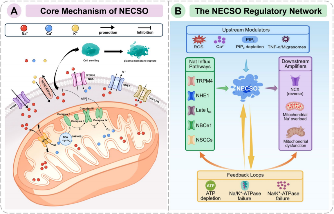
Key steps: Na⁺ influx during NECSO is not merely osmotic imbalance. More importantly, cytoplasmic Na⁺ overload activates the mitochondrial Na⁺‑Ca²⁺ exchanger (NCLX), resulting in elevated Na⁺ and decreased Ca²⁺ concentration in the mitochondrial matrix. Mitochondrial Ca²⁺ is a critical signal for activating key dehydrogenases of the tricarboxylic acid cycle (TCA), and its decline directly suppresses TCA cycle rate and impairs substrate supply for oxidative phosphorylation. Meanwhile, Na⁺ may also directly interfere with the function of mitochondrial electron transport chain complexes. As a result, mitochondrial ATP synthesis is severely impaired and cells fall into an energy crisis.
Lethal vicious cycle: Energy depletion initiates a lethal vicious cycle — plasma membrane Na⁺/K⁺‑ATPase loses function due to ATP deficiency and cannot pump intracellular Na⁺ out, further aggravating Na⁺ overload and cell swelling. At the same time, loss of the transmembrane Na⁺ gradient disrupts all secondary active transport dependent on this gradient, especially the reverse mode operation of NCX, inducing secondary Ca²⁺ overload and amplifying cell injury. Ultimately, complete loss of ion homeostasis and sustained elevation of osmotic pressure lead to extreme swelling of cells and organelles, plasma membrane rupture and necrotic lysis.
Summary of core pathway: Na⁺ influx → elevated mitochondrial Na⁺ and decreased mitochondrial Ca²⁺ → energy metabolism collapse → Na⁺/K⁺‑ATPase failure → complete disruption of ion homeostasis → necrotic cell death.
3.3 Extended Regulatory Network
In addition to TRPM4, other Na⁺ transporters may participate in or exacerbate Na⁺ overload under different pathological contexts:
• NHE1 (Na⁺‑H⁺ exchanger isoform 1): Strongly activated during ischemia and acidosis, importing Na⁺ while extruding H⁺, serving as the major source of Na⁺ overload in early myocardial ischemia.
• Late I_Na: Enhanced under pathological conditions, and its inhibitors such as ranolazine exert cardioprotective effects.
• Reverse‑mode NCX operation: A critical bridge linking Na⁺ overload to lethal Ca²⁺ overload.
• NBCe1 and NSCCs: Also participate in Na⁺ influx under specific conditions.
Upstream signals: Reactive oxygen species (ROS) can directly activate TRPM2 channels or indirectly induce energy dysfunction via mitochondrial damage. In myocardial I/R injury, Tspan4‑positive fibroblasts upregulate CYP11B2, the rate‑limiting enzyme for aldosterone synthesis in cardiomyocytes through TNF‑α autocrine mediated by migrasomes, which may further promote cardiomyocyte NECSO by affecting genes related to Na⁺ reabsorption.

4. Crosstalk with Other Regulated Cell Death Pathways
• Crosstalk with apoptosis: Oxidative stress and energy depletion are common upstream triggers for both. Mild‑to‑moderate Na⁺ overload may trigger apoptosis through mitochondrial outer membrane permeabilization, whereas severe and sustained Na⁺ overload leads to rapid energy collapse and NECSO. ATP depletion in NECSO may inhibit caspase activation and shift the cell death phenotype from apoptosis to necrosis.
• Crosstalk with necroptosis: Both are regulated necrosis, but mediated by TRPM4‑Na⁺ overload and RIPK1/RIPK3‑MLKL signaling respectively. They may converge at common downstream effectors such as mitochondrial dysfunction and ROS production. Combined inhibition of multiple cell death pathways provides better myocardial protection than inhibition of a single pathway.
• Crosstalk with ferroptosis: Na⁺ overload may disrupt cellular iron homeostasis by regulating iron transporters, increasing intracellular iron levels and sensitivity to ferroptosis. Both are classified into the emerging category of “metalloptosis”.
5. Pathophysiological Roles
5.1 Central Roles in Cardiac Diseases
Myocardial ischemia‑reperfusion (I/R) injury is the most well‑studied pathological scenario for NECSO. ATP reduction during ischemia inhibits Na⁺/K⁺‑ATPase, and acidosis activates NHE1, resulting in progressive increase of [Na⁺]i; ROS burst at the early reperfusion stage further activates Na⁺ influx pathways including late sodium current. As a Ca²⁺‑activated channel, TRPM4 may be activated by intracellular Ca²⁺ fluctuations and increased ROS during reperfusion. A large number of intervention strategies — including NHE inhibitors, late sodium current inhibitors, NCX inhibitors, Na⁺ channel blockers and drugs indirectly affecting Na⁺ homeostasis (such as eplerenone) — have effectively reduced myocardial infarct size in preclinical models.
It should be noted that although TRPM4 is the core mediator of NECSO, it is not the only contributor to pathological Na⁺ overload in I/R injury. NHE1 activation and late sodium current may play dominant roles during ischemia and early reperfusion, and combined blockade of NHE1 and Na⁺ channels almost completely eliminates ischemic Na⁺ overload.
Other cardiac diseases: In dilated cardiomyopathy (DCM), bioinformatic analysis and doxorubicin‑induced DCM models have identified core Na⁺ overload‑related death genes (including BACH2, NXPH4, CD1E and LIF). In genetic cardiomyopathy models, NHE1 overexpression drives excessive Na⁺ influx and induces contraction and necrosis via reverse‑mode NCX leading to secondary Ca²⁺ overload. Tissue‑level Na⁺ imbalance is also associated with cardiac remodeling and heart failure progression.
5.2 Dual Roles in Cancer Biology
NECSO and TRPM4 exhibit highly tissue‑specific dual roles in cancer:
Tumor‑promoting function: TRPM4 is overexpressed in multiple cancers such as bladder cancer, cholangiocarcinoma and ovarian cancer, and high expression correlates with poor prognosis in adrenocortical carcinoma, low‑grade glioma, pancreatic cancer, mesothelioma and uveal melanoma. Mechanistically, TRPM4 may promote tumor progression through epigenetic regulation, DNA damage repair and alternative splicing, and shape an immunosuppressive microenvironment.
Tumor‑suppressive function: In kidney clear cell carcinoma (KIRC) and endometrial carcinoma (UCEC), high TRPM4 expression correlates with better prognosis. TRPM4 overexpression inhibits proliferation, migration and colony formation of renal cancer cells and promotes apoptosis. As a multigene risk stratification model, the NECSO score serves as an independent protective prognostic factor in KIRC.
Mechanistic hypothesis: This tissue‑specific dual role may arise from: ① TRPM4 is regulated by tissue‑specific transcription factors and epigenetic modifiers (e.g., VHL pathway suppresses TRPM4 expression, estrogen receptor upregulates TRPM4 transcription); ② downstream signaling networks vary by cell type; ③ modulation of the tumor microenvironment (immunogenic cell death induced by NECSO predominates in “hot” tumors, while TRPM4‑mediated immune escape dominates in “cold” tumors).
5.3 Crosstalk with Tumor Immune Microenvironment
As a lytic form of regulated necrosis, NECSO ultimately disrupts plasma membrane and organelle membrane integrity, which may release mitochondrial DNA (mtDNA) and theoretically promote anti‑tumor immunity. However, tumor cells may also evade immune surveillance or shape an immunosuppressive microenvironment by regulating NECSO‑related genes. NECSO‑related genes (especially TRPM4) are closely associated with immune cell infiltration patterns: high NECSO score in renal carcinoma correlates with increased infiltration of CD8⁺ T cells and Th1 cells, the high‑risk group of HCC shows immune‑activated characteristics, while the high‑risk group of LUAD is associated with an immunosuppressive microenvironment.
6. Biomarker Applications
6.1 Construction of Prognostic Models
Multigene risk scoring models based on NECSO‑related genes (NRGs) have been constructed and validated in multiple cancers, including LUAD, KIRC, HCC and breast cancer. Generally, TRPM4 co‑expression analysis, differential expression analysis and WGCNA are used to screen candidate genes, followed by machine learning algorithms such as Cox regression, LASSO, SVM‑RFE and random forest to build models. The models have good risk stratification capacity but are mainly based on public omics data and require prospective clinical validation.
6.2 Core Biomarkers
• TRPM4: Undoubtedly the core biomarker of NECSO, with highly heterogeneous expression across pan‑cancer, and can serve as a diagnostic and prognostic biomarker for multiple malignancies.
• SLC9A1: Combined with TRPM4, it has significant diagnostic and prognostic value in breast cancer.
• NR1H3: A novel protective biomarker in breast cancer, and high expression predicts favorable overall survival.
• DENND1C: A key tumor suppressor gene and predictive biomarker for immunotherapy response in LUAD.
• ANKRD13B: A gene promoting tumor progression and potential prognostic biomarker in HCC.
6.3 Prediction of Therapeutic Response
NECSO risk models can predict responses to chemotherapy and immunotherapy. LUAD high‑risk patients have different sensitivities to taxanes and EGFR inhibitors; HCC high‑risk patients are more sensitive to 8 chemotherapeutic drugs including cisplatin, doxorubicin, gemcitabine and paclitaxel. In terms of immunotherapy, LUAD low‑risk patients or patients with high DENND1C expression achieve significant survival benefit after immune checkpoint inhibitor treatment.
7. Therapeutic Strategies
7.1 Direct Targeting of TRPM4
• TRPM4 agonists (such as NC1 or its optimized derivatives): Suitable for cancers with low TRPM4 expression or where TRPM4 exerts tumor‑suppressive functions (e.g., renal cell carcinoma), to induce NECSO in tumor cells.
• TRPM4 inhibitors: Suitable for cancers with TRPM4 overexpression and oncogenic functions.
• Cryo‑EM structural analysis provides a basis for rational drug design, facilitating the development of modulators with higher potency, selectivity and targeting different conformational states.
7.2 Indirect Regulatory Strategies
• NHE1 inhibitors (cariporide, amiloride, etc.): Show potent cardioprotective effects in preclinical studies, but clinical trial results are inconsistent.
• Mineralocorticoid receptor antagonists (eplerenone): Improve systemic Na⁺ balance by antagonizing aldosterone.
• NCX inhibitors and NCLX modulation: Target the linkage between Na⁺ overload and downstream injury.
7.3 Natural Products and Drug Repurposing
• Cinobufagin: The active component of traditional Chinese medicine Venenum Bufonis, targets the SEC62/TRPM4 axis, inhibits the ubiquitination and degradation of TRPM4, stabilizes its protein level and overcomes bortezomib resistance.
• Abrine: CTD database analysis shows that it can simultaneously target four core Na⁺ overload‑related hub genes in DCM.
7.4 Combination Therapy
Combination of NECSO modulation with chemotherapy, targeted therapy or immunotherapy holds great potential. NECSO‑induced immunogenic cell death can increase tumor immunogenicity, and combination with immune checkpoint inhibitors is expected to produce synergistic anti‑tumor effects. For cardioprotection, combined blockade of different Na⁺ influx pathways (Na⁺ channels and NHE1) almost completely prevents ischemic Na⁺ overload and produces additive protective effects.
References
- Necrosis by Sodium Overload: Mechanisms, Pathophysiological Roles, and Therapeutic Prospects of a Novel Form of Regulated Cell Death. Qi T, et al. Cell Biochem Biophys. 2026. [PMID: 42709394]