
Research News | Multiplex Immunostaining | Spatial Proteomics
Original Research
Wu F, Zheng S, Chen Y, et al. iCLAP: an innovative method for integrable co-detection of low-abundance antigens with high-plex immunostaining. Nature Communications. 2026;17:3104. Original Article | DOI: 10.1038/s41467-026-69752-y
Advances in spatial proteomics now allow researchers to detect dozens of protein markers in a single tissue section and examine the distribution, functional states, and interactions of different cell populations. However, accurately identifying low-abundance proteins while maintaining high multiplexing capacity remains a major challenge in spatial biology.
Many biologically important proteins, including senescence-associated markers, transcription factors, and certain signaling regulators, are expressed at low levels in tissues. Conventional immunofluorescence and some high-plex imaging methods may not provide sufficient signal, especially in formalin-fixed, paraffin-embedded (FFPE) samples.
Tyramide signal amplification (TSA) offers an effective way to address this challenge.
TSA uses horseradish peroxidase (HRP) to catalyze covalent deposition of fluorescent tyramide near the antigen, thereby enhancing local fluorescence. Compared with conventional immunofluorescence, TSA has important advantages for detecting low-abundance antigens and has been widely adopted in multiplex immunohistochemistry (TSA-mIHC).
As spatial proteomics moves toward increasingly high-dimensional detection, an additional question arises: can TSA be used only for a few low-abundance proteins, with the remaining markers measured by high-plex techniques such as CyCIF, CODEX, or IMC, and can all results be integrated into one spatial map of the same tissue?
On February 24, 2026, Wu and colleagues published "iCLAP: an innovative method for integrable co-detection of low-abundance antigens with high-plex immunostaining" in Nature Communications. The study proposed an integrated protein-detection strategy called iCLAP to address this need.
iCLAP stands for Integrable Co-detection of Low-Abundance Proteins, a method designed to integrate sensitive detection of low-abundance proteins with highly multiplexed immunostaining.
Rather than replacing existing multiplex imaging platforms, the method combines the high sensitivity of TSA signal amplification with other high-plex immunodetection technologies.
Researchers first use TSA to label and image low-abundance proteins with high sensitivity. An optimized fluorescence-inactivation procedure then reduces residual fluorescence in the previously imaged channels, allowing further staining and imaging.
This strategy enables multiple rounds of detection on the same FFPE tissue section and the integration of data generated with CyCIF, CODEX, and IMC.
By combining different imaging platforms, the team demonstrated high-dimensional spatial analysis of more than 40 protein markers.
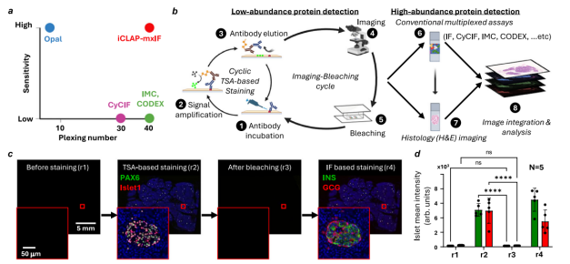
Figure 1. iCLAP workflow and experimental validation of fluorescence bleaching.
Source: Figure 1 of the original article.
Conventional TSA-mIHC generally removes primary antibodies, secondary antibodies, and HRP detection components after each staining round while retaining previously deposited fluorescent signals. This allows multiple primary antibodies, including antibodies raised in the same host species, to be used sequentially on one tissue section.
However, as the number of staining rounds increases, retained fluorescence occupies more detection channels. Spectral overlap between fluorophores also complicates the design of complex antibody panels.
iCLAP takes a different approach: after TSA staining and fluorescence imaging, researchers inactivate the deposited fluorescent labels, reducing residual signals to near-background levels. This frees the detection channels for subsequent staining and enables TSA to participate in longer cyclic imaging workflows.
The aim is not to physically remove every deposited tyramide molecule from the tissue, but to inactivate its fluorescence and minimize interference with later measurements.
Another key capability of iCLAP is integrating images acquired during different staining rounds into a shared spatial coordinate system.
After each imaging round, image registration corrects positional shifts and local tissue deformation, allowing protein-expression data from successive rounds to be combined.
The study used nuclear images for registration, including signals from the DAPI channel or equivalent nuclear-imaging channels. The process involved global rigid registration and grid-based local deformation correction.
This approach enables protein signals captured across different rounds and technologies to be associated with the same cells and tissue regions for downstream single-cell phenotyping and spatial analysis.
TSA fluorophores are covalently deposited near tissue proteins through an enzymatic reaction, so their fluorescent signals cannot be removed simply by conventional antibody stripping.
To address this limitation, researchers developed and optimized a fluorescence-inactivation procedure combining chemical oxidation with illumination.
Under the conditions used in the study, tissue sections were placed in a transparent container with an alkaline bleaching solution containing hydrogen peroxide and EDTA in PBS. Illumination was then used to promote fluorophore inactivation.
The study's fluorescence-inactivation procedure placed tissue sections in a transparent box with bleaching solution containing 2 M H₂O₂ and 3 mM EDTA at pH 12.5, followed by illumination from both sides at 5,000 lux for approximately one hour.
The investigators further tested the treated samples. Their results showed effective reduction of existing TSA fluorescence while preserving the performance needed for subsequent antigen detection and tissue-morphology analysis under the tested conditions.
Thus, TSA need not be limited to a fixed set of fluorescence channels: it may serve as a reusable, high-sensitivity detection module in more complex spatial proteomics workflows.
Before establishing the iCLAP workflow, the team compared TSA with conventional immunofluorescence for low-abundance protein detection.
They selected four important senescence-associated proteins: P16, P21, P53, and 53BP1.
These proteins are involved in cell-cycle regulation, DNA-damage responses, and cellular senescence, but their expression can be low in certain normal tissues, making conventional immunofluorescence challenging.
In human pancreatic FFPE tissue, TSA produced clearer detection of these protein-expression patterns.
Even when conventional IF was enhanced by increasing primary-antibody concentration or extending incubation, its performance for some targets remained inferior to TSA.
For example, quantitative analysis of P16 showed that even with extended antibody incubation, the signal-to-noise ratio in positive tissue regions using conventional IF was approximately one-tenth of that obtained with TSA.
Moreover, for P53 and P21, conventional IF under the tested conditions failed to yield clear positive signals even with longer incubations and higher antibody concentrations, whereas TSA enabled effective detection.
These findings underscore the value of retaining a TSA detection module when investigating low-abundance proteins in high-dimensional spatial analyses.
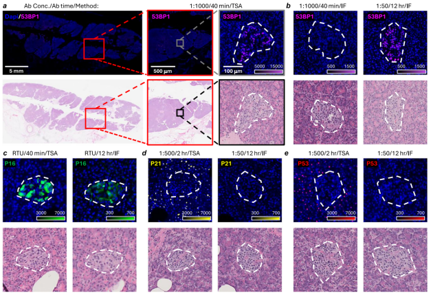
Figure 2. Detection of senescence markers in the iCLAP workflow, demonstrating substantially stronger low-abundance protein signals with TSA than with conventional IF.
Source: Figure 2 of the original article.
To assess the practical utility of iCLAP, the researchers combined it with cyclic immunofluorescence (CyCIF) for multiplex protein detection in human pancreatic tissue.
A total of 12 protein markers were measured in this experiment.
TSA was used to detect four low-abundance senescence-associated proteins: P16, P21, P53, and 53BP1.
The other markers characterized senescence-associated features, pancreatic cell types, and cell function: HMGB1, Lamin B1, Insulin, Glucagon, Somatostatin 28, Lipase, Trypsin, and Carboxypeptidase A.
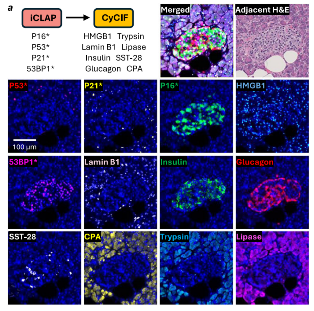
Figure 3. iCLAP can be combined with CyCIF.
Source: Figure 4a of the original article.
By integrating these measurements, the researchers analyzed cell identity, functional markers, and senescence-associated protein expression in the same tissue regions.
The results showed relatively pronounced enrichment of P16 and 53BP1 signals in islet regions, heterogeneity in P16 expression among islet endocrine cell populations, a significant association between higher P16 expression and larger islet area in the analyzed islets, and differing relationships between certain senescence-associated markers and functional markers such as insulin and glucagon.
These observations indicate that senescence-associated proteins are not uniformly distributed across all pancreatic cells, but have complex associations with particular cell populations and their tissue microenvironment.
More importantly, iCLAP allowed difficult-to-detect senescence markers to be analyzed together with cell-identity and functional markers, providing a more comprehensive picture of tissue spatial organization.
In addition to CyCIF, the team assessed the compatibility of iCLAP with other high-dimensional spatial protein-detection platforms.
In the iCLAP-CODEX experiments, researchers combined TSA-detected markers, including P16, P21, and P53, with a high-plex CODEX antibody panel, enabling spatial protein detection of more than 40 markers.
They observed distinct epithelial-marker expression patterns in P21-positive and P53-positive cells within pancreatic acinar regions.
For example, P21-positive cells were more often co-expressed with panCK, whereas P53-positive cells were generally panCK-negative.
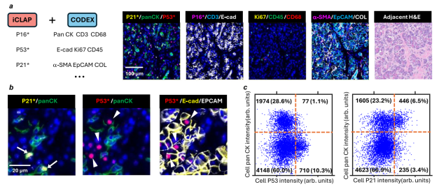
Figure 4. iCLAP can be combined with CODEX.
Source: Figures 5a–c of the original article.
In a separate experiment, researchers combined iCLAP with imaging mass cytometry (IMC), demonstrating the feasibility of integrating TSA detection results with metal-isotope-based imaging data.
Together, these experiments show that iCLAP can extend the cycling capacity of TSA and incorporate it as a high-sensitivity module into different high-plex spatial proteomics platforms.
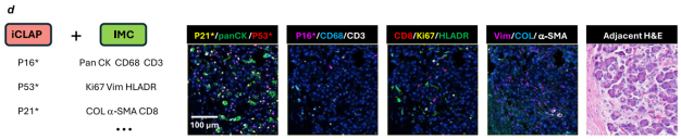
Figure 5. iCLAP can be combined with IMC.
Source: Figure 5d of the original article.
Conventional TSA-mIHC excels at improving detection sensitivity and increasing the number of markers through sequential staining.
CyCIF, CODEX, and IMC, by contrast, emphasize higher-dimensional protein detection and spatial information from tissues.
iCLAP brings these strengths together: TSA provides highly sensitive detection of low-abundance proteins, while fluorescence inactivation, multi-round imaging, and data integration link those proteins to larger panels of cellular phenotype markers.
This provides new possibilities for studying cellular senescence, tumor microenvironments, tissue heterogeneity, and complex disease mechanisms.
Nevertheless, iCLAP requires rigorous methodological validation, including inactivation efficiency, tissue integrity, antigen stability, registration accuracy across imaging rounds, and data consistency between platforms.
At present, this study mainly demonstrates the potential of iCLAP in experimental spatial proteomics workflows. Its applicability to a wider range of tissues and routine research platforms needs further evaluation.
The central innovation of iCLAP is not a new protein-labeling reaction, but a way to make established TSA signal amplification more flexible within high-dimensional spatial proteomics.
By optimizing fluorescence inactivation and combining different high-plex imaging platforms, iCLAP preserves the advantages of TSA for low-abundance proteins while overcoming some limitations imposed by conventional fluorescence-channel capacity.
For researchers using TSA-mIHC to investigate complex tissue microenvironments, this study suggests an important direction: future multiplex immunostaining may benefit not only from adding more markers to an individual staining assay, but also from combining complementary technical modules to better balance detection sensitivity and information depth.