
1. Basic Information of xCT Protein
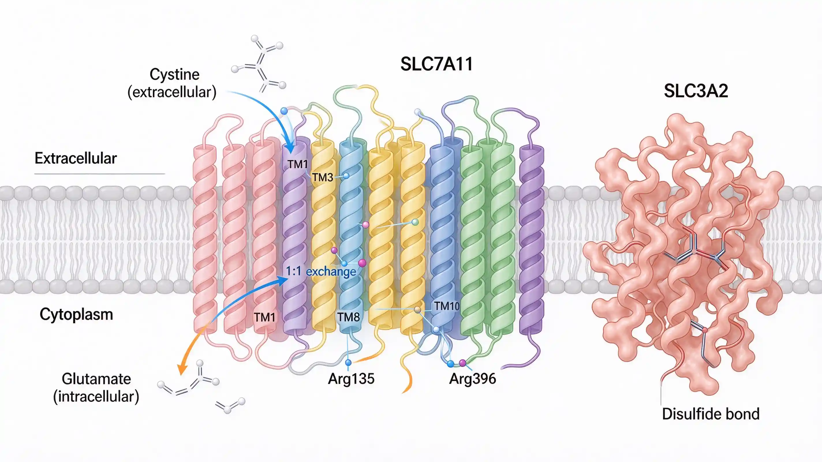
xCT, also known as SLC7A11, is a sodium‑independent cystine‑glutamate antiporter located on the cell membrane. Its predicted molecular weight is about 55 kDa, but observed WB bands usually range from 35‑70 kDa, among which 35 kDa corresponds to the monomer and 70 kDa represents the heterodimer. xCT forms a heterodimer with CD98 (SLC3A2, also named 4F2hc) of the SLC3 family. This xCT/CD98 heterodimer is the well‑known system xc⁻ complex: xCT is a non‑glycosylated light‑chain subunit, while CD98 is a type‑II glycoprotein heavy‑chain. The two subunits interact via disulfide bonds formed by two conserved cysteine residues.
2. Special Notes for Sample Extraction
2.1 Membrane‑protein fraction extraction is critical
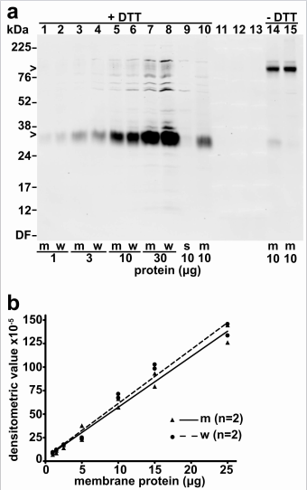
xCT is an integral membrane protein and cannot be detected in soluble protein fractions. In experiments, tissue or cell lysates should be centrifuged to isolate membrane‑bound components. Signal will be lost if only soluble proteins are collected. Published data show that removal of soluble proteins barely affects xCT immunoreactivity, indicating that functional signals originate entirely from the membrane fraction.

2.2 Detergent selection: SDS is preferred over Triton X‑100
Comparative experiments indicate that 1% Triton X‑100 shows poorer solubilization performance for xCT than SDS. For maximal solubilization of membrane proteins, lysis buffer containing SDS is recommended.
2.3 Reduction treatment: a common pitfall
WB detection of xCT is highly sensitive to reducing conditions:
• Room‑temperature (RT) reduction for 15 min shifts xCT to approximately 35 kDa.
• High‑temperature boiling (100 °C, 2 min) gives sub‑optimal reduction and results in higher‑molecular‑weight bands.
• 30 mM DTT is effective for reduction.
⚠️ Unlike standard WB protocols, boiling xCT samples is not advised. Short‑time reduction at room temperature is the preferred approach.
3. Key WB Experimental Tips
• Molecular‑weight interpretation: Bands at 35 kDa or 55 kDa can both represent xCT (monomer with distinct modifications); a 70 kDa band may correspond to the dimer.
• Positive controls: Mouse brain tissue, mouse kidney tissue, HEK‑293, HeLa, HepG2 cells are suitable positive‑control materials.
• Loading controls: Tubulin or GAPDH are commonly used loading controls.
4. Quick Summary Checklist
1. Enrich membrane proteins first. xCT is undetectable in soluble fractions; membrane fractions are mandatory.
2. Use SDS‑containing lysis buffer instead of Triton X‑100.
3. Reduce for 15 min at room temperature with 30 mM DTT. Do NOT boil samples.
4. Expected bands: 35 kDa (fast‑migrating reduced monomer), 45 kDa or 55 kDa (monomer), 70 kDa (dimer).
5. Add protease inhibitors throughout the workflow and perform all operations on ice.
References
- Autophagy is required for proper cysteine homeostasis in pancreatic cancer through regulation of SLC7A11. Mukhopadhyay S, et al. Proc Natl Acad Sci U S A. 2021. [PMID: 33531365]
- The cystine‑glutamate exchanger (xCT, Slc7a11) is expressed in significant concentrations in a subpopulation of astrocytes in the mouse brain. Ottestad‑Hansen S, et al. Glia. 2018. [PMID: 29350434]
- Comparative analysis of antibodies to xCT (Slc7a11): Forewarned is forearmed. Van Liefferinge J, et al. J Comp Neurol. 2016. [PMID: 26494316]