
The core of histopathological diagnosis is to observe cellular morphology, tissue architecture and inter‑cellular relationships under a microscope, so as to distinguish the normal physiological state or pathological changes of tissues. Fresh isolated tissues are rich in water; untreated tissue sections are nearly transparent and lack natural contrast between various cells and stromal structures, making direct microscopic observation impossible. H&E staining uses two dyes with selective staining capacity to differentially label different chemical components in tissues, building a stable, reproducible and globally‑applicable tissue colour development system. For all surgical biopsies and resected specimens, H&E‑stained sections are routinely observed first. Techniques including immunohistochemistry, special staining and molecular pathological tests are all based on morphological evaluation of H&E sections. Therefore, H&E staining is more than a simple colouring procedure; it is a universal morphological language in pathology.
H&E staining is not a single staining step, but a continuous tissue preparation and staining process. From tissue blocks to final microscope‑ready slides, it consists of four major stages: pre‑processing, sectioning, staining and coverslipping.
Isolated tissues are promptly placed in fixative (typically 10 % neutral buffered formalin). The purpose is to rapidly cross‑link intracellular proteins, stop cellular autolysis, preserve the original morphology of cells and stroma, and prevent tissue decay and deformation. Fixation quality directly determines subsequent sectioning and staining outcomes.
Fixed tissue blocks contain large amounts of water, which is immiscible with paraffin. Gradient ethanol is applied sequentially to remove water from tissues. Clearing agents such as xylene then replace ethanol to render tissues translucent. Tissues are subsequently immersed in molten paraffin and cooled to form solid paraffin‑embedded blocks. Paraffin blocks can be stored long‑term for repeated sectioning.
Paraffin blocks are secured on a rotary microtome and cut into ultrathin sections of approximately 3‑5 μm thickness. Sections float on warm‑water surfaces for flattening, then are fished up with clean glass slides and baked in an incubator to firmly adhere tissues onto slides and avoid tissue loss during staining.
Paraffin is insoluble in water and must be removed prior to staining. Slides go through xylene for de‑waxing, followed by descending‑gradient ethanol to displace xylene, and finally into distilled water to return tissues to an aqueous environment for water‑soluble dye staining.
• Hematoxylin staining: Slides are immersed in hematoxylin solution; incubation time depends on hematoxylin type and concentration.
• Differentiation: Hydrochloric‑alcohol solution removes excess hematoxylin from cell nuclei to sharpen nuclear staining.
• Bluing: Weak‑alkaline aqueous solution (ammonia water / lithium carbonate) is used to produce vivid‑blue nuclei.
• Eosin staining: Sections are immersed in eosin solution; incubation time depends on eosin concentration.
• Gradient ethanol dehydration: Remove water from tissue sections.
• Xylene clearing and mounting with neutral balsam: Apply coverslips to isolate air for permanent slide preservation.
H‑E staining is accomplished by two coordinated dyes with distinct targets. Hematoxylin mainly labels cell nuclei; eosin targets cytoplasm, extracellular matrix and other protein‑rich structures. Selective staining arises not from simple physical adsorption but from charge‑driven chemical binding, corresponding to the classic pathological concepts of basophilia and eosinophilia.
Native hematoxylin is nearly colourless and lacks direct staining capacity. During staining, hematoxylin is oxidized to haematein, which combines with aluminium‑salt mordants (typically alum) to form positively‑charged coloured metal complexes. Cell nuclei are rich in deoxyribonucleic acid (DNA), whose phosphate backbone carries abundant negative charges. By electrostatic interaction, positively‑charged haematein‑aluminium complexes tightly bind negatively‑charged nucleic acids, producing stable blue colour.
Chromatin, nucleoli and chromosomes are nucleic‑acid‑rich and show variable staining intensity: condensed chromatin stains darker, whereas euchromatin appears paler. Nucleoli containing abundant RNA also stain blue‑purple. Pathologists evaluate nuclear size, shape, chromatin distribution, nucleolar count and mitotic figures to judge cellular atypia.
Eosin is an acidic dye that dissociates into negatively‑charged dye anions under staining conditions. Cytoplasm, collagen fibres, smooth‑muscle fibres, red blood cells, basement membranes and other structures are mainly composed of proteins. Under H&E staining pH conditions, amino groups on polypeptide chains are protonated and become positively charged. Negatively‑charged eosin anions bind to proteins via electrostatic attraction.
Eosin is a red‑range dye and imparts pink colour upon binding to protein components. Protein type and density produce graded pink hues: collagen fibres show bright pale‑pink; smooth‑muscle fibres stain deep‑pink; haemoglobin within erythrocytes stains bright scarlet. Such colour gradients enable differentiation of diverse extracellular‑matrix types and identification of lesions such as fibrosis, haemorrhage and muscular hyperplasia.
Properly prepared H&E sections display well‑differentiated colour patterns under light microscopy for straightforward identification of tissue components:
Cell nuclei (DNA / RNA), including nuclei of epithelial cells, inflammatory cells and tumour cells; bacteria and some calcified foci may also exhibit basophilic staining.
• Cytoplasm: variable staining intensity across different cell types;
• Extracellular matrix: generally distinct pink to red;
• Red blood cells: characteristic bright‑red;
• Keratin and muscle fibres: variable degrees of red staining.
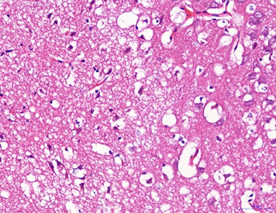
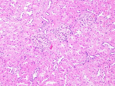
H&E stained sections form a standardized colour‑coding system. Blue‑purple signals mark nucleic‑acid‑rich cell nuclei for cell localization and nuclear‑morphology assessment. Multi‑toned pink signals represent protein‑rich cytoplasm and stroma, outlining cell contours, cellular arrangement and tissue scaffolds.
Normal tissues follow fixed patterns regarding cell count, nuclear size, cytoplasmic proportion and stromal distribution, serving as stable morphological references. In inflammation, hyperplasia, neoplasia or necrosis, this ordered architecture is disturbed: nuclei may enlarge, become hyperchromatic or irregular in shape, with increased mitotic activity; cellular arrangement becomes disorganized; stroma may show oedema, fibrosis and inflammatory‑cell infiltration. Structural abnormalities can be recognised through this blue‑and‑pink microscopic visual map.
This colour‑coding system delivers high reproducibility. Standardised protocols yield consistent staining results across laboratories and operators. Pathologists worldwide share unified H&E interpretation criteria, enabling direct cross‑regional communication of morphological observations. This explains why H&E staining is regarded as the “fundamental language” of pathology.
After more than a century of development, H‑E staining remains the cornerstone of pathological diagnosis. Its major strengths include mature protocols, cost‑effectiveness and excellent reproducibility. It simultaneously visualizes both cell nuclei and extracellular matrix to fully reveal tissue architecture.
Advanced techniques such as immunohistochemistry, in‑situ hybridization and next‑generation sequencing provide protein‑expression and genetic information. Nevertheless, all these assays require prior morphological evaluation of H&E sections. Without H&E‑based morphological context, immunohistochemical and molecular read‑outs lose their tissue‑localization references.
High‑quality ready‑to‑use H&E staining solutions compatible with paraffin sections, frozen sections and cell preparations for human, animal, livestock and aquatic‑sample experiments. Formulations are mercury‑free and methanol‑free for improved laboratory safety, minimising precipitate formation. Reagents are reusable and deliver bright, consistent staining performance.