
Trypan Blue Stain: How to Measure Cell Viability?
In cell culture, cell passaging and cell counting experiments, besides cell concentration, one key parameter needs to be monitored — cell viability.
How many cells in the suspension retain intact plasma membranes? How many have lost membrane integrity? How can we quickly assess cell quality before seeding, passaging or downstream assays? The Trypan Blue Exclusion Assay is a classic, straightforward method for cell viability assessment.
1. What is trypan blue, and how does it distinguish live cells from dead cells?
Trypan Blue is a dye widely used for cell counting and viability testing. Its mechanism relies on differences in plasma membrane integrity to determine dye exclusion. In the standard trypan blue exclusion assay:
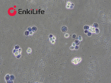
Live cells: Possess intact cell membranes and can exclude trypan blue, appearing unstained under brightfield microscopy.
Dead cells or cells with compromised membrane integrity: Permit dye entry and stain blue.
Cell viability in the suspension can therefore be estimated by microscopic counting of unstained and blue-stained cells separately.
It is important to note that trypan blue only reflects plasma membrane integrity at the time of testing, not the full functional status of cells. Cells with early-stage injury or in special physiological states may remain unstained. Thus, trypan blue results cannot fully replace assays for metabolic activity, apoptosis or other functional readouts.
2. Common applications of trypan blue stain
1. Cell counting and viability evaluation
During routine cell passaging, cryopreservation and thawing, the trypan blue exclusion method estimates total cell count, unstained cell count and viability to support experimental decisions.
2. Pre-seeding quality check
Before proliferation assays, functional studies and other downstream experiments, viability testing verifies whether cell quality meets experimental requirements.
3. Post-thaw cell condition assessment
Cryopreservation and thawing can damage cell membranes. Trypan blue provides a rapid readout of immediate cell status after recovery. However, assays for long-term proliferative capacity, functional recovery or apoptosis are required for deeper characterization.
3. Protocol
Dilute the cell suspension to the appropriate concentration, following standard cell counting guidelines.
Mix cell suspension with fresh trypan blue working solution and incubate at room temperature for 3–5 min.
Load one drop of stained cell mixture onto a slide, apply a coverslip and observe under high magnification.
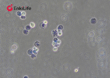
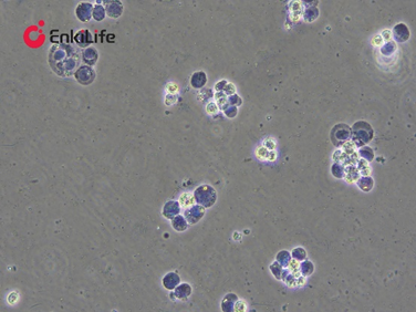
Dead cells appear pale blue, swollen and dull; live cells remain unstained, retain normal morphology and are bright.
Count live and dead cells.
Tally unstained cells and calculate cell viability using the formula.
4. Calculation of trypan blue cell viability
For manual counting with trypan blue exclusion, the standard formula is:
Cell Viability (%) = Number of unstained cells ÷ (Unstained cells + Blue-stained cells) × 100%
Example counting result:
- Unstained cells: 180
- Blue-stained cells: 20
Calculation:
Cell Viability = 180 ÷ (180 + 20) × 100% = 90%
This example is for demonstration only. In practice, count multiple fields or grids and run replicates to improve reliability.
Dilution factors are required when calculating cell concentration, but they do not alter the viability formula above.
5. Critical staining notes
1. Maintain consistent workflow
Keep fixed dye-to-cell ratio, incubation time and counting criteria across all samples.
2. Count promptly after staining
Do not leave stained samples standing for extended periods. Count immediately per product instructions to avoid cell status drift.
3. Mix thoroughly but gently
Homogenize suspensions to prevent cell sedimentation and clumping, while minimizing mechanical damage.
4. Standardize discrimination between stained and unstained events
Cell debris and aggregates interfere with manual or automated counting; apply unified scoring rules.
5. Viability ≠ full cellular function
Trypan blue only reports membrane integrity. An unstained cell is not guaranteed to have normal proliferation, metabolism or specialized functions.
6. Dye quality control
Use commercial stain promptly after opening. In-house prepared solutions must be filter-sterilized to avoid particulate contamination.
Although simple, trypan blue staining is a staple QC tool in cell experiments. From passaging and cryorecovery to pre-assay counting, proper understanding of its principle and limitations supports reliable cell quality evaluation.
| EnkiLife Cat.No. | EnkiLife Product Name |
| RA20141 | Hematoxylin and Eosin (H&E) Staining Kit,Aqueous |
| RA20142 | Hematoxylin and Eosin (H&E) Staining Kit,Alcoholic |
| RA20092 | Eosin Y Staining Kit,Aqueous |
| RA20093 | Eosin Y Staining Kit,Alcoholic |
| RA20094 | Masson Trichrome Staining Kit |
| RA20095 | Modified Safranin O-Fast Green Cartilage Staining Kit |
| RA20096 | Safranin O-Fast Green Plant Tissue Staining Kit |
| RA20097 | Nissl Staining Kit,Cresyl Violet Method |
| RA20098 | Nissl Staining Kit,Toluidine Blue Method |
| RA20099 | Alcian Blue Staining Kit,pH 2.5 |
| RA20100 | Alcian Blue Staining Kit,pH 1.0 |
| RA20101 | Alcian Blue Staining Kit,pH 0.4 |
| RA20102 | Alcian Blue-Periodic Acid-Schiff (AB-PAS) Staining Kit |
| RA20103 | Weigert's Iron Hematoxylin Staining Kit |
| RA20104 | Crystal Violet Staining Kit |
| RA20105 | Trypan Blue Staining Kit, 0.08% |
| RA20106 | Trypan Blue Staining Kit, 0.2% |
| RA20107 | Trypan Blue Staining Kit, 0.4% |
| RA20108 | Ponceau S Staining Kit,1x |
| RA20109 | Ponceau S Staining Kit,10x |
| RA20110 | TTC Staining Kit,0.5% |
| RA20111 | TTC Staining Kit,1% |
| RA20112 | TTC Staining Kit,2% |
| RA20113 | TTC Staining Kit,4% |
| RA20114 | Alizarin Red S Staining Kit,0.1%,pH 4.2 |
| RA20115 | Alizarin Red S Staining Kit,0.2%,pH 4.2 |
| RA20116 | Alizarin Red S Staining Kit,1%,pH 4.2 |
| RA20117 | Alizarin Red S Staining Kit,2%,pH 4.2 |
EnkiLife provides a complete portfolio of well‑validated histochemical staining reagents with stable batch‑to‑batch performance and consistent staining results. Suitable for pathological, animal and plant tissue experiments to meet diverse tissue‑staining research requirements.