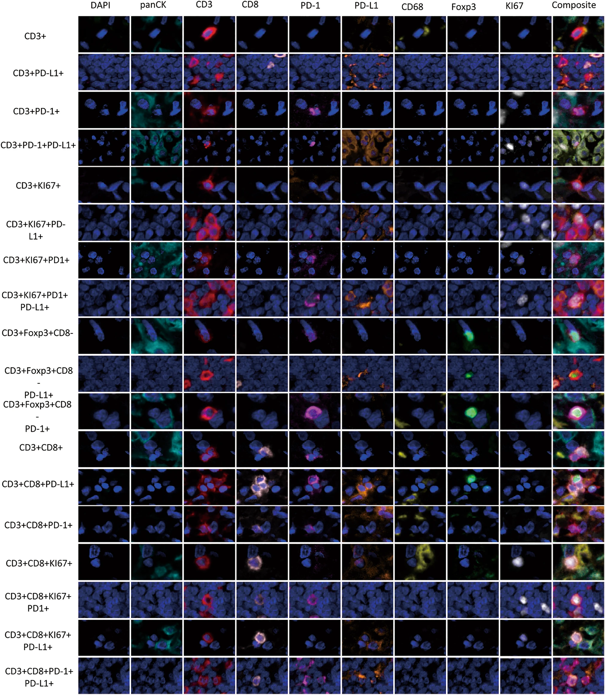
Literature Interpretation: Mapping Tumor Immune Landscapes of Malignant Pleural Mesothelioma Using 9‑Color Multiplex Immunofluorescence
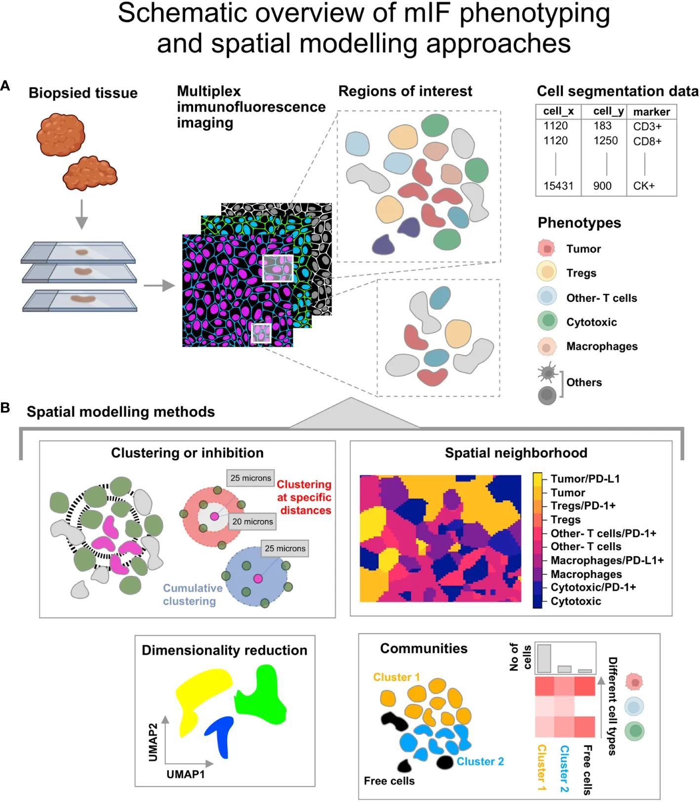
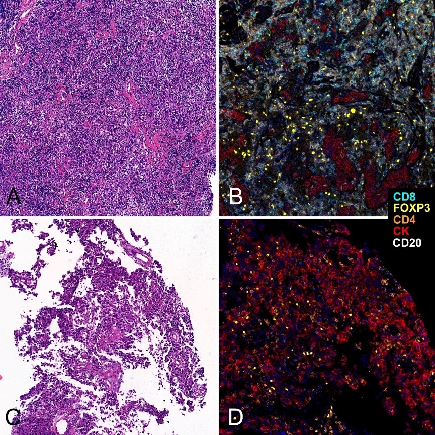
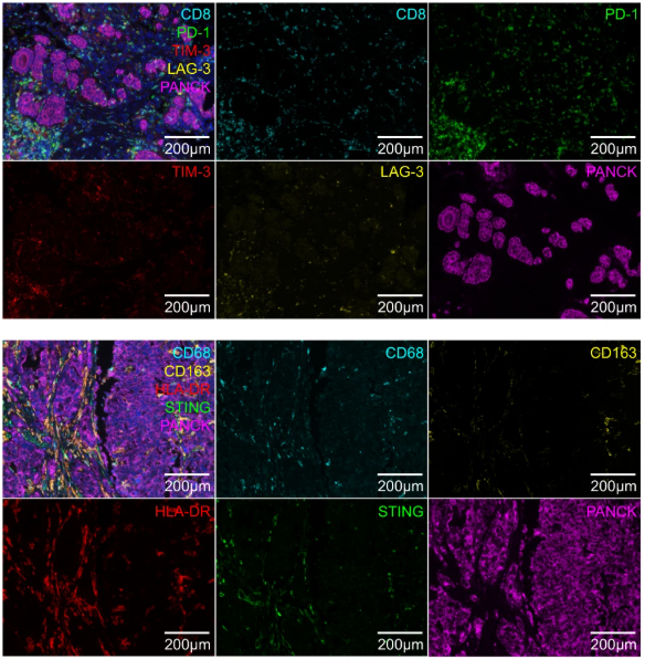
Conventional immunohistochemistry (IHC) only enables detection of 1–2 proteins per tissue section, providing merely cell counts while losing in situ spatial information and protein co‑expression profiles. Standard single‑plex immunofluorescence also fails to resolve complex immune suppressive networks. Multiplex immunofluorescence (mIF) allows simultaneous detection of multiple targets on a single FFPE paraffin section, preserves native tissue architecture, and supports cell phenotyping, cell density quantification and spatial distance analysis, making it a key technique for translational tumor immunology research.