
Article Type: Review Article | Journal: Nature Reviews Molecular Cell Biology | Published: April 09, 2025
DOI: 10.1038/s41580‑025‑00842‑3
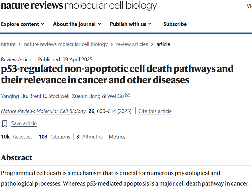
The core insight of this review is to reinterpret the tumour‑suppressive functions of p53. Beyond canonical apoptosis‑driven tumour suppression, p53 can also exert biological effects through multiple non‑apoptotic cell‑death pathways including ferroptosis, necroptosis and pyroptosis, which influence tumorigenesis, tissue injury, neurodegenerative disorders and infections.
This explains an important experimental observation: even when canonical apoptotic pathways are suppressed in certain cells, p53 may still function via alternative cell‑death mechanisms. Conversely, excessive activation of these pathways in normal tissues can aggravate organ damage.
Encoded by the TP53 gene, p53 tumour‑suppressor protein is widely known as “the guardian of the genome”. Upon cellular stresses such as DNA damage, oxidative stress or oncogenic signalling, p53 becomes activated and determines cell fate in a context‑dependent manner.
Traditionally, p53‑mediated tumour suppression has been explained by cell‑cycle arrest, cellular senescence and apoptosis. However, this review points out that these three canonical outputs are not always strictly required for p53‑driven tumour suppression. Non‑apoptotic cell‑death mechanisms provide new perspectives to resolve this puzzle.
Non‑apoptotic cell death (NACD) defines regulated cell‑death modalities distinct from classical apoptosis. Major pathways discussed in this manuscript are listed below:
| Cell‑death type | English term | Core features |
|---|---|---|
| Ferroptosis | Ferroptosis | Iron‑dependent lipid peroxidation |
| Necroptosis | Necroptosis | RIPK3‑MLKL‑dependent regulated necrosis |
| Pyroptosis | Pyroptosis | Gasdermin pore formation, usually pro‑inflammatory |
| Autophagy‑dependent cell death | Autophagy‑dependent cell death | Autophagy machinery is required for cell death execution |
| Entotic cell death | Entotic cell death | Cell‑in‑cell internalization followed by death of internalized cell |
| Parthanatos | Parthanatos | PARP1 hyper‑activation coupled with AIF‑mediated DNA degradation |
| Paraptosis | Paraptosis | Characterized by prominent cytoplasmic vacuolization |
In addition, PANoptosis, cuproptosis and disulfidptosis are briefly discussed as potential p53‑related processes.
Critical conceptual note: NACD does not represent one unified molecular pathway. Ferroptosis relies on lipid peroxidation; necroptosis involves specific kinases and membrane pore assembly; pyroptosis is Gasdermin‑dependent. Cell‑death morphology alone cannot define which mechanism is operational.
Four interrelated research questions are raised:
- (1) What molecular mechanisms enable p53 to regulate distinct forms of non‑apoptotic cell death?
- (2) How can p53 promote cell death in some contexts yet exert cytoprotective effects in others?
- (3) How do these diverse cell‑death pathways influence cancer, organ injury, neurodegeneration and infection?
- (4) Can pharmacological agents selectively modulate p53‑associated cell‑death pathways for therapeutic benefit?
A central theme throughout this article is the strong context‑dependency of p53 biology: activating p53 does not simply equal triggering cell death.
Instead of memorizing individual death pathways in isolation, a unified conceptual framework helps interpret this review. p53 controls cell‑death outputs at four major regulatory layers:
- Layer one: transcriptional regulation
p53 directly activates or represses target genes such as SLC7A11 repression, GLS2 induction and GSDME modulation, rewiring cellular metabolism, redox homeostasis and death‑execution capacity. - Layer two: protein‑protein interaction and subcellular localization
p53 interacts with mitochondrial proteins including CypD and is modulated by factors such as G3BP1 and MYBBP1A. Nuclear versus mitochondrial localization shapes p53 functional outputs. - Layer three: cellular metabolism and stress status
p53 remodels lipid biosynthesis, glutathione (GSH) pools, reactive oxygen species (ROS), iron homeostasis and mitochondrial fitness, thereby setting cellular susceptibility towards distinct death modalities. - Layer four: crosstalk between signalling pathways
Ferroptosis, autophagy, pyroptosis, necroptosis and apoptosis are not fully independent. One pathway can alter activation thresholds of others, producing synergistic or competitive cell‑death networks.
Collectively, p53 functions not merely as an “apoptosis on‑off switch”, but as an integrative signalling hub decoding diverse stress inputs to dictate cellular fate.
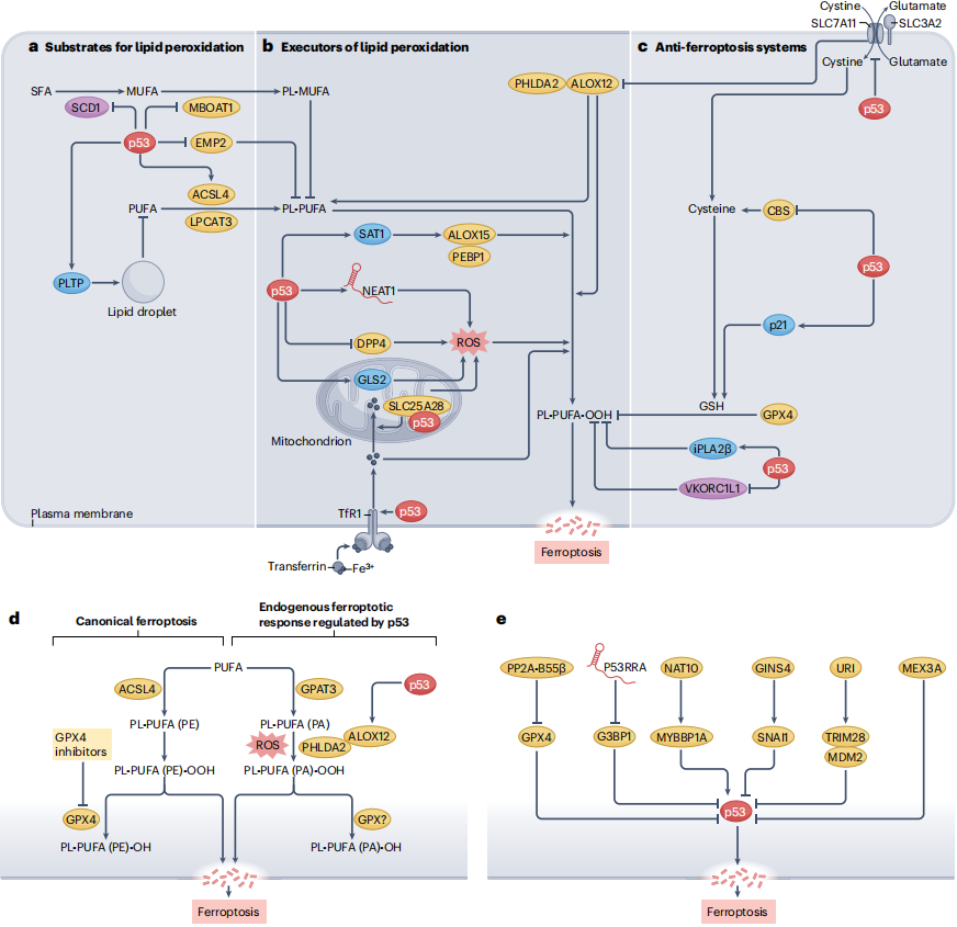
Ferroptosis is an iron‑dependent regulated cell death driven by uncontrolled lipid peroxidation. Polyunsaturated fatty acids (PUFAs) embedded in membrane phospholipids are susceptible to oxidative modification. Cell death ensues when lipid‑peroxide generation outpaces cellular detoxification capacity.
Three conceptual modules define ferroptosis:
- Module 1: Lipid‑peroxidation substrates
PUFA incorporation into membrane phospholipids; enzymes such as ACSL4 and LPCAT3 govern this metabolic step. Abundant substrates generally increase ferroptosis susceptibility under permissive stress. - Module 2: Lipid‑peroxidation execution
Iron + ROS + lipoxygenases drive accumulation of lipid peroxides. Iron catalyses oxidative reactions, ROS provides oxidative burden, and ALOX‑family lipoxygenases contribute to lipid oxidation in certain contexts. - Module 3: Anti‑ferroptosis defence systems
GSH‑GPX4 axis clears toxic lipid peroxides. Ferroptosis is favoured when defence capacity is overwhelmed or suppressed.
These three modules correspond to substrate supply, execution machinery and cytoprotective defence as described in the source review.
Mechanism 1: Repression of SLC7A11 impairs GSH‑GPX4 defensive capacity
SLC7A11 encodes the core subunit of system Xc⁻ cystine‑glutamate antiporter. It imports cystine, which is critical for cysteine and glutathione biosynthesis.
Important distinction: p53 does not directly inhibit GPX4 enzymatic activity. The canonical axis works via transcriptional repression of SLC7A11, lowering cystine uptake, limiting GSH supply and indirectly weakening GPX4‑mediated antioxidant protection.
Mechanism 2: PHLDA2‑ALOX12 axis enhances lipid peroxidation (non‑canonical ferroptosis route)
This non‑canonical pathway is highlighted in the review. Unlike GPX4‑inhibition‑centric ferroptosis models, p53 can drive ferroptosis through crosstalk among SLC7A11, PHLDA2 and ALOX12:
- (1) p53 represses SLC7A11.
- (2) SLC7A11 indirectly restricts substrate accessibility for ALOX12; SLC7A11 removal relieves this constraint.
- (3) PHLDA2 modulates phosphatidic‑acid membrane localization and ALOX12 function.
- (4) GPAT3 generates PUFA‑containing phosphatidic‑acid to supply lipid‑peroxidation substrates.
- (5) Under permissive ROS conditions, amplified lipid peroxidation triggers ferroptosis.
This pathway is pathophysiologically relevant in mouse tumour models and can operate without pharmacological ferroptosis inducers, demonstrating that p53‑driven ferroptosis is not strictly GPX4‑suppression‑dependent.
Mechanism 3: Remodelling lipid composition alters ferroptosis sensitivity
Membrane fatty‑acid composition sets ferroptosis susceptibility:
- ACSL4: Incorporates PUFAs into lipid pools generating peroxidation‑prone membrane phospholipids.
- SCD1: Produces monounsaturated fatty acids (MUFAs). Since MUFAs compete with PUFAs for membrane integration, p53‑mediated SCD1 repression can promote ferroptosis in certain contexts.
- MBOAT1, EMP2: p53‑dependent regulation remodels phospholipid profiles and ferroptosis responsiveness.
- PLTP (counter‑example): p53‑induced PLTP in hepatoma cells promotes lipid‑droplet biogenesis, sequestering PUFAs away from membrane lipid‑peroxidation, thereby suppressing ferroptosis.
Thus p53 modulates both antioxidant defence and the abundance of oxidizable lipid substrates.
Mechanism 4: Modulation of ROS and iron homeostasis via GLS2, TfR1 and other mediators
p53 shapes ferroptosis by tuning oxidative stress and iron metabolism:
| Key molecule | p53‑associated function | Impact on ferroptosis |
|---|---|---|
| GLS2 | Enhances glutamine metabolism and TCA‑cycle activity | Elevates mitochondrial ROS in context‑dependent fashion |
| TfR1 | Promotes cellular iron import | Increases iron availability for oxidative reactions |
| SLC25A28 | Mitochondrial iron importer‑related | Elevated mitochondrial iron fuels ROS generation |
| SAT1 | Regulates polyamine metabolism linked to ALOX15‑driven lipid oxidation | Facilitates ferroptosis under permissive conditions |
| VKORC1L1 | Vitamin‑K reduction and antioxidant function | p53‑dependent repression weakens anti‑ferroptosis defence |
These molecules do not operate simultaneously in every cell type, representing context‑dependent regulatory modules deployed by p53.
Counter‑intuitive protective outputs are well‑documented in the source manuscript:
- p21: p53‑dependent p21 induction can delay cystine‑deprivation‑triggered ferroptosis in selected cellular models.
- iPLA2β (PLA2G6): Under low‑dose doxorubicin or nutlin‑3 treatment, p53 activates PLA2G6. iPLA2β removes oxidized fatty‑acid moieties from membrane phospholipids and halts lipid‑peroxidation chain‑reaction propagation.
- DPP4: In colorectal carcinoma cells, p53 retains DPP4 inside the nucleus, restraining DPP4‑NOX1 complexes and inhibiting specific ferroptosis routes.
- PLTP: Supports lipid‑droplet formation and PUFA sequestration to limit lipid‑peroxidation substrate availability.
Therefore, p53 can toggle between pro‑death and cytoprotective programmes according to stress magnitude, cell type and stimulus duration.
Beneficial outcomes:
- Induction of tumour‑cell death
- Restriction of tumour growth
- Enhanced radiotherapy efficacy in certain settings
- Potential contribution to host defence against particular infections
Detrimental outcomes:
- Exacerbation of ischaemia‑reperfusion‑mediated myocardial injury
- Contribution to acute kidney injury
- Potential neuronal damage
- Normal‑cell injury under certain metabolic or inflammatory milieus
The review also addresses mutant‑p53 effects on ferroptosis. Certain p53 mutants can boost antioxidant capacity or dysregulate SLC7A11 to enable tumour‑cell evasion from ferroptosis. Not all TP53 mutations produce equivalent loss‑of‑function, nor do all mutants modulate ferroptosis identically.
Section take‑home conclusion: p53 can either amplify lipid‑peroxidation stress or reinforce antioxidant/lipid‑sequestration cytoprotective mechanisms. Investigators must evaluate substrates, execution machinery and defensive pathways simultaneously; p53 or GPX4 expression alone cannot prove ferroptosis pathway engagement.
| Category | Target | Catalog# | Product Name | Applications | Reactivity |
|---|---|---|---|---|---|
| Lipid‑peroxidation substrate synthesis | ACSL4 | AMRe21426 | ACSL4 Rabbit Monoclonal Antibody | WB,IHC,IF,IP,ELISA | Human,Mouse,Rat |
| Lipid‑peroxidation substrate synthesis | MBOAT5 | APRab61445 | MBOAT5 Rabbit Polyclonal Antibody | IHC,WB | Human,Mouse,Rat |
| Lipid‑peroxidation substrate synthesis | p53 | AMRe87631 | p53 Rabbit Monoclonal Antibody | WB,IHC,ICC/IF,FC,IP | Human |
| Lipid‑peroxidation execution | ALOX15 | AMRe87733 | ALOX15 Rabbit Monoclonal Antibody | WB,IHC,ICC/IF | Human,Mouse,Rat |
| Lipid‑peroxidation execution | PEBP1/RKIP | AMRe02544 | RKIP Rabbit Monoclonal Antibody | WB,IHC,IP | Human,Mouse,Rat |
| Lipid‑peroxidation execution | GLS2 | AMM86075 | GLS2 Mouse Monoclonal Antibody | WB,IHC | Human, Mouse, Rat |
| Lipid‑peroxidation execution | TfR1 | AMRe86756 | Transferrin Receptor Rabbit Monoclonal Antibody | WB,FC | Human,Mouse |
| Lipid‑peroxidation execution | ALOX12/LOX12 | AMRe84157 | LOX12 Rabbit Monoclonal Antibody | WB | Human,Rat |
| Lipid‑peroxidation execution | PHLDA2/TSSC3 | APRab19374 | TSSC3 Rabbit Polyclonal Antibody | IHC,ICC/IF,ELISA | Human,Rat,Mouse |
| Anti‑ferroptosis defence | xCT/SLC7A11 | AMRe03803 | xCT Rabbit Monoclonal Antibody | WB,IHC,IF,IP,ELISA | Human,Mouse,Rat |
| Anti‑ferroptosis defence | SLC3A2/CD98 | AMRe08492 | CD98 (2G13) Rabbit Monoclonal Antibody | WB,IHC,IP,IF‑P | Human |
| Anti‑ferroptosis defence | CBS | AMRe84468 | CBS Rabbit Monoclonal Antibody | WB,IHC,ICC/IF,ICC | Human |
| Anti‑ferroptosis defence | p21 | AMRe21528 | p21 Rabbit Monoclonal Antibody | WB,IHC,ICC/IF,ELISA,IP | Human,Rat |
| Anti‑ferroptosis defence | GPX4 | AMRe21183 | GPX4 Rabbit Monoclonal Antibody | WB,IHC,IF,ELISA | Human,Mouse,Rat |
| p53‑ferroptosis regulatory network | NAT10 | AMRe84631 | NAT10 Rabbit Monoclonal Antibody | WB,IHC,ICC/IF,ICC,FC,IP | Human,Mouse,Rat |
| p53‑ferroptosis regulatory network | G3BP1 | APRab11212 | G3BP1 Rabbit Polyclonal Antibody | WB,IHC,ICC/IF,ELISA | Human,Mouse,Monkey |
| p53‑ferroptosis regulatory network | MYBBP1A | AMRe14262 | MYBBP1A (14I6) Rabbit Monoclonal Antibody | WB,IHC,ICC/IF,FC | Human |
| p53‑ferroptosis regulatory network | SNAI1 | AMRe86416 | SNAI1 Rabbit Monoclonal Antibody | WB,IP | Human |
| p53‑ferroptosis regulatory network | TRIM28/KAP1 | AMRe02184 | KAP1 Rabbit Monoclonal Antibody | WB,IHC,IF,ELISA | Human,Mouse,Rat |
| p53‑ferroptosis regulatory network | MDM2 | AMRe21162 | MDM2 Rabbit Monoclonal Antibody | WB,IHC,ICC/IF,ELISA,IP | Human,Mouse,Rat |
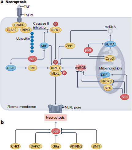
Necroptosis is a form of regulated necrosis associated with inflammation, infection and tissue injury. Its canonical execution axis is RIPK1‑RIPK3‑MLKL.
Canonical necroptosis often manifests under conditions where caspase‑8‑dependent apoptotic signalling is compromised; nevertheless, necroptosis is not absolutely conditional on apoptotic‑pathway suppression in every biological setting.
Mechanism 1: NRF‑miR‑873‑RIPK1/RIPK3 axis
In mouse myocardial ischaemia‑reperfusion injury models, p53 induces long non‑coding RNA NRF.
NRF sequesters miR‑873 and relieves miR‑873‑mediated repression of RIPK1 and RIPK3, thereby enabling necroptosis and exacerbating myocardial damage.
This illustrates that p53 does not necessarily need to directly transactivate RIPK1/RIPK3; indirect regulation via non‑coding RNAs can shape necroptosis‑executor abundance.
Mechanism 2: PUMA‑mitochondrial DNA‑ZBP1‑RIPK3 axis
PUMA is a well‑characterized p53 transcriptional target conventionally linked to mitochondrial apoptosis.
However, in 5‑fluorouracil‑treated colorectal‑cancer cells, the p53‑PUMA axis can trigger mitochondrial‑DNA release into cytosol, engaging ZBP1‑RIPK3 signalling to drive necroptosis.
This mechanism connects classical p53 apoptotic targets to non‑apoptotic cell death and may potentiate anti‑tumour immunity.
Mechanism3: Mitochondrial ROS and p53‑CypD physical interaction
p53 can modulate necroptosis through mitochondrial biology:
- Boost mitochondrial ROS production;
- Alter mitochondrial dynamics via DRP1;
- Translocate to mitochondria and physically interact with cyclophilin D (CypD);
- Promote mitochondrial permeability‑transition pore (mPTP) opening.
These events facilitate cell death, yet execution outputs differ between experimental systems. The review notes that p53‑CypD‑driven mitochondrial death largely manifests as necroptosis.
| Comparison item | Ferroptosis | Necroptosis |
|---|---|---|
| Core event | Uncontrolled iron‑dependent lipid peroxidation | RIPK3‑MLKL‑mediated membrane damage |
| Key effector molecules | SLC7A11, GPX4, ACSL4, ALOX12 | RIPK1, RIPK3, MLKL |
| Major metabolic co‑factors | Iron, PUFA, GSH, ROS | Mitochondrial ROS, death‑receptor signalling |
| p53 regulatory mode | Remodels lipid metabolism, antioxidant defence and iron homeostasis | Regulates non‑coding RNAs, mitochondrial function and death signalling cascades |
| Disease associations | Tumour, organ injury, neurodegeneration | Tumour, inflammation, infection, ischaemic injury |
Necroptosis can exert tumour‑suppressive effects, yet it can also provoke severe damage in non‑malignant tissues.
Examples discussed in source text include CHK1‑p53‑dependent necroptosis in liver‑cancer models, DAPK1‑p53 axis in neuronal ischaemic stress, and BMI1‑mediated suppression of necroptosis in defined neuronal populations. Meanwhile, necroptosis contributes to alcoholic liver disease and certain neurodegenerative pathologies.
Section conclusion: p53‑driven necroptosis cannot be reduced to a simple RIPK1‑RIPK3 linear cascade. Mitochondrial homeostasis, non‑coding RNAs, DNA‑damage‑response circuits and tissue‑specific modulators jointly shape final biological outcomes.
| Target | Catalog# | Product Name | Applications | Reactivity |
|---|---|---|---|---|
| TNF α | AMRe21365 | TNF α Rabbit Monoclonal Antibody | WB,IHC,IF,IP,ELISA | Human,Mouse,Rat |
| TNF‑R1 | APRab19092 | TNF‑R1 Rabbit Polyclonal Antibody | WB,IHC,ICC/IF,ELISA | Human,Mouse,Rat |
| TRADD | AMRe19179 | TRADD (1A3) Rabbit Monoclonal Antibody | WB,IHC,ICC/IF,FC | Human |
| TRAF2 | AMRe86844 | TRAF2 Rabbit Monoclonal Antibody | WB,IHC,ICC/IF,FC,IP | Human,Mouse,Rat |
| RIPK1 | AMM82284 | RIPK1 Mouse Monoclonal Antibody | WB,ELISA | Human |
| RIPK3 | AMM86115 | RIPK3 Mouse Monoclonal Antibody | WB | Human |
| MLKL | AMRe21172 | MLKL Rabbit Monoclonal Antibody | WB,IHC,IF,ELISA | Human,Mouse,Rat,Chicken |
| Caspase‑8 | AMRe07982 | Caspase‑8 (3Q3) Rabbit Monoclonal Antibody | WB,IHC,IF‑P | Human,Mouse,Rat |
| TLR3 | AMM03550 | Toll‑Like Receptor 3 (2F10) Mouse Monoclonal Antibody | WB | Human,Mouse,Rat |
| TRIF | AMRe02715 | TRIF Rabbit Monoclonal Antibody | WB,IP | Human,Mouse,Rat |
| p53 | AMRe02388 | p53 Rabbit Monoclonal antibody | WB,ICC/IF,IP | Mouse |
| PUMA | AMRe83766 | PUMA Rabbit Monoclonal Antibody | WB,IHC,ICC/IF,ICC,FC | Human,Mouse,Rat |
| CypD | APRab09610 | Cyclophilin D Rabbit Polyclonal Antibody | WB,IHC,ICC/IF,ELISA | Human,Mouse,Rat |
| DRP1 | AMRe21581 | DRP1 Rabbit Monoclonal Antibody | WB,IHC,IF,IP,ELISA | Human,Mouse,Rat,Bovine |
| PRDX3 | AMRe03145 | Peroxiredoxin 3 Rabbit Monoclonal Antibody | WB,IHC,ICC/IF,IP | Human |
| Chk1 | AMRe21249 | Chk1 Rabbit Monoclonal Antibody | WB,ICC/IF,ELISA,IP | Human,Mouse,Rat |
| DAPK1 | APRab09786 | DAPK1 Rabbit Polyclonal Antibody | WB,IHC,ICC/IF,ELISA | Human,Mouse |
| EHMT2/G9A | AMRe10354 | EHMT2/G9A (3F1) Rabbit Monoclonal Antibody | WB,IHC,FC,IF‑P | Human |
| BMI1 | AMRe02916 | BMI1 Rabbit Monoclonal Antibody | WB,ICC/IF,IP | Human,Mouse,Rat |
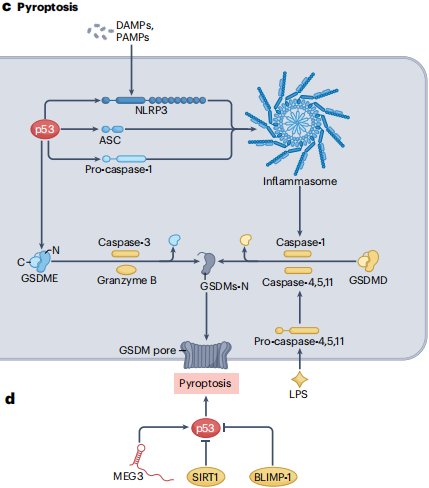
Pyroptosis is an inflammatory regulated cell death driven by Gasdermin‑family pore‑forming proteins.
Beyond canonical caspase‑1‑mediated pyroptosis, non‑canonical pyroptosis is executed by human caspase‑4/5 or mouse caspase‑11 in response to cytosolic lipopolysaccharide, leading to GSDMD cleavage.
The review states that the functional links between p53 and pyroptosis are less well‑characterized compared with ferroptosis and necroptosis, although multiple mechanistic associations have been uncovered.
| p53‑associated molecule | Function and biological relevance |
|---|---|
| NLRP3 | p53 enhances its transcription; NLRP3 can in turn stabilize or activate p53 under defined contexts |
| Caspase‑1 | p53 can up‑regulate its expression to provide pyroptosis execution capacity |
| ASC/PYCARD | p53 can promote ASC expression in non‑small‑cell lung cancer models, influencing inflammasome assembly |
| GSDME | p53 modulates its expression; cleaved GSDME generates membrane pores |
| MEG3 | Stabilizes p53 and thereby facilitates pyroptosis in certain tumour contexts |
| SIRT1 | Represses p53 activity and attenuates pyroptosis in selected experimental models |
Elevated abundance of these molecules does not guarantee pyroptosis execution. Complete proteolytic processing of effector proteins and appropriate upstream stress stimuli are still required.
Yes. In colorectal‑cancer cell studies cited by the manuscript, ginsenoside Rh3 triggers both pyroptosis and ferroptosis via STAT3‑p53‑NRF2 signalling and suppresses tumour‑cell proliferation.
Nevertheless, pyroptosis and ferroptosis represent biochemically distinct effector cascades; shared upstream stress signals can co‑activate both pathways.
Pyroptosis generates pro‑inflammatory signals that can potentiate anti‑tumour immunity in cancer; conversely, aberrant pyroptosis in healthy tissues may amplify inflammatory injury.
Representative scenarios discussed:
- Activation of p53‑dependent pyroptosis to suppress malignant cell growth in tumours;
- Autophagy‑NLRP3‑inflammasome‑driven pyroptosis linked to hyperuricaemic nephropathy;
- Potential p53‑dependent host responses during pathogen infection;
- Protective effects obtained by restraining pathological pyroptosis in selected acute‑organ‑damage models.
Section conclusion: Pyroptosis constitutes one of the critical interfaces connecting p53 with innate immunity, inflammation and tumour immunity. Researchers must discriminate “up‑regulated pyroptosis‑related transcripts/proteins” from bona‑fide pyroptosis execution and validate Gasdermin cleavage and functional pore formation.
| Target | Catalog# | Product Name | Applications | Reactivity |
|---|---|---|---|---|
| NLRP3 | AMRe01571 | NLRP3 Rabbit Monoclonal Antibody | WB,IHC,IF,IP,ELISA | Human,Mouse,Rat |
| ASC | APRab03824 | ASC Rabbit Polyclonal Antibody | WB,IHC,ICC/IF,ELISA | Human,Mouse,Rat |
| Caspase‑1 | AMRe21189 | Caspase‑1 Rabbit Monoclonal Antibody | WB,IHC,ICC/IF,ELISA,IP | Human,Mouse,Rat |
| Cleaved‑Caspase 1 | APRab03387 | Cleaved‑Caspase 1 Rabbit Polyclonal Antibody | WB,IHC,ICC/IF,ELISA | Human,Mouse,Rat |
| Cleaved‑Caspase‑4/5 p20 (D270/D311) | APRab08960 | Cleaved‑Caspase‑4/5 p20 (D270/D311) Rabbit Polyclonal Antibody | WB,ELISA | Human |
| Caspase‑11 | AMRe86937 | Caspase‑11 Rabbit Monoclonal Antibody | WB,IP | Mouse |
| Caspase 3 | AMRe01567 | Caspase 3 Rabbit Monoclonal Antibody | WB,IHC,IP | Human,Mouse,Rat |
| Cleaved‑Caspase 3 p17 | APRab03839 | Cleaved‑Caspase 3 p17 Rabbit Polyclonal Antibody | WB,IHC,ICC/IF,ELISA | Human,Mouse,Rat |
| Cleaved‑Caspase‑3 p12 (D175) | APRab08957 | Cleaved‑Caspase‑3 p12 (D175) Rabbit Polyclonal Antibody | WB,IHC,ICC/IF | Human,Rat,Mouse |
| GSDMD | AMRe87750 | GSDMD Rabbit Monoclonal Antibody | WB,IHC,FC,IP | Mouse,Rat |
| Cleaved N‑terminal GSDMD | AMRe21098 | Cleaved N‑terminal GSDMD Rabbit Monoclonal Antibody | WB,ELISA | Human |
| DFNA5/GSDME | AMRe21257 | DFNA5/GSDME Rabbit Monoclonal Antibody | WB,ICC/IF,ELISA,IP | Human,Mouse,Rat |
| Cleaved N‑terminal GSDME | AMRe87516 | Cleaved N‑terminal GSDME Rabbit Monoclonal Antibody | WB | Mouse |
| Granzyme B | AMRe21246 | Granzyme B Rabbit Monoclonal Antibody | WB,IHC,ICC/IF,ELISA,IP | Human |
| p53 | AMRe02388 | p53 Rabbit Monoclonal antibody | WB,ICC/IF,IP | Mouse |
| SIRT1 | AMRe21606 | SIRT1 Rabbit Monoclonal Antibody | WB,IHC,ICC/IF,ELISA,IP | Human,Rat |
| BLIMP‑1/PRDM1 | AMM81554 | PRDM1 Mouse Monoclonal Antibody | WB,ICC,ELISA,FC | Human,Mouse,Rat,Monkey |
Beyond the three major pathways described above, the manuscript covers autophagy‑dependent cell death, entotic cell death, parthanatos, paraptosis, and briefly discusses PANoptosis, cuproptosis and disulfidptosis.
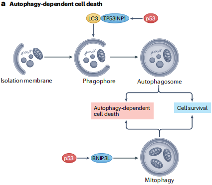
Autophagy is an intracellular degradative and recycling pathway that normally supports cellular adaptation to nutrient deprivation and stress. Under defined contexts, autophagic machinery becomes required for cell‑death execution.
Key mediators mentioned in the text:
- TP53INP1: Transcriptionally activated by p53; physically interacts with LC3 to participate in autophagy‑related processes.
- BNIP3L: Mediates p53‑dependent mitophagy contributing to cell death in certain contexts.
- BECN1 and ATG5: Essential core autophagy components required for p53‑linked autophagy‑dependent cell death in selected experimental models.
Important conceptual pitfall: observation of autophagy occurring alongside cell death does not prove that death is autophagy‑dependent. For instance, autophagy can promote ferroptosis via ferritin degradation. Genetic or pharmacological ablation of autophagy core machinery is necessary to verify causal requirement.
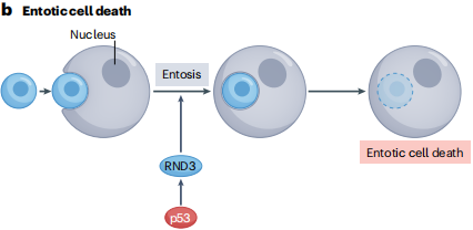
Entosis describes the phenomenon whereby one cell is internalized inside another non‑phagocytic host cell to form cell‑in‑cell structures. Internalized cells may, but do not always, undergo cell death.
According to the review, in DNA‑damaged mitotic epithelial cells, p53 activates RND3 to promote entotic clearance of aneuploid daughter cells as a genome‑maintenance mechanism.
On the other hand, mutant p53 can also foster cell‑in‑cell structures which may promote survival of tumour cells under some circumstances.
Therefore, mere detection of cell‑in‑cell morphology cannot demonstrate entotic cell death, nor automatically indicate tumour‑suppressive function.
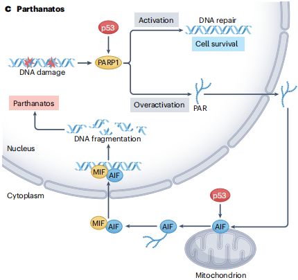
Parthanatos represents a regulated cell‑death modality driven by PARP1 over‑activation coupled with AIF‑mediated nuclear DNA fragmentation.
Core mechanistic sequence:
- DNA damage triggers excessive PARP1 catalytic activity
- Massive accumulation of PAR‑polymer species
- Mitochondrial AIF release
- AIF‑MIF complexes translocate into nucleus
- Large‑scale genomic‑DNA degradation
The review discusses p53’s capacity to enhance PARP1 activity, engage in direct PARP1 protein‑protein interaction and potentially modulate AIF expression to influence parthanatos.
Interestingly, under BRAF V600E‑mutant backgrounds, TP53 loss can sensitize tumour cells to parthanatos‑inducing agents, illustrating strong context‑dependency for p53 regulation of this pathway.
Paraptosis is a caspase‑independent regulated cell‑death form marked by prominent cytoplasmic vacuolization originating from endoplasmic‑reticulum and/or mitochondrial swelling.
The source manuscript cites that ginsenoside Rh2 can trigger both p53‑dependent apoptosis and paraptosis‑like cell death to exert anti‑tumour effects in colorectal‑cancer cells.
Nevertheless, the exact molecular cascades whereby p53 induces paraptotic vacuolization remain incompletely defined.
PANoptosis
PANoptosis denotes an inflammatory regulated cell‑death modality with extensive crosstalk among apoptosis, pyroptosis and necroptosis, assembled via PANoptosome signal complexes. Given p53’s ability to regulate these three death modalities, authors hypothesize p53 may participate in PANoptosis, yet concrete mechanistic proof remains incomplete.
Cuproptosis
Excess intracellular copper triggers aggregation of lipoylated TCA‑cycle proteins and proteotoxic stress. The review notes potential links between p53 and cuproptosis, but direct experimental evidence is still limited.
Disulfidptosis
Disulfide‑stress‑driven cytoskeleton damage occurring under glucose‑deprivation combined with high SLC7A11 expression. Since p53 represses SLC7A11, authors propose p53 could modulate cellular susceptibility towards disulfidptosis; this hypothesis awaits further experimental validation.
These three emerging pathways exemplify how p53, via governing metabolism and cellular redox homeostasis, can impinge on newly discovered cell‑death programmes. However, the review cautions that these should not yet be treated as firmly‑established direct p53‑regulated death cascades.
Collectively, p53‑controlled non‑apoptotic cell‑death programmes are not discrete isolated modules, but interconnected networks sharing common regulatory nodes.
This integrated conceptual diagram is derived by synthesizing multiple sections of the source manuscript rather than representing a single experimentally‑validated unified pathway described in one figure of the original paper.
At least five key variables need to be considered:
- Cell‑type background: Tumour cells, cardiomyocytes, neurons and immune cells have distinct metabolic profiles.
- Type of stress stimulus: DNA‑damage, ROS, TNF signalling and nutrient deprivation engage distinct regulatory modules.
- Stress intensity and duration: Mild stress may trigger adaptive cytoprotective responses; strong or persistent stress can drive cell‑death programmes.
- Molecular status of p53: Protein stability, post‑translational modifications, subcellular localization and mutation types all reshape p53 functions.
- Completeness of downstream execution machinery: Even if p53 is fully activated, cells may not undergo a given type of cell death when essential effector molecules for that pathway are missing.
These five points explain why a drug may exert beneficial effects in tumour cells yet cause adverse effects in normal tissues. They also account for inconsistent experimental observations of p53 function across different research models.
The therapeutic section of the review focuses on ferroptosis, necroptosis and pyroptosis. It emphasizes that therapeutic goals must be determined according to disease context — either enhancing or suppressing specific cell‑death pathways.
Representative pre‑clinical research examples are listed below. These are experimental findings only and should not be interpreted as approved clinical indications or mature therapeutic regimens.
| Representative compounds / agents | Research directions discussed in the review |
|---|---|
| Gambogenic acid | Inhibits melanoma metastasis via p53‑dependent ferroptosis |
| Flubendazole | Promotes p53‑related ferroptosis and suppresses castration‑resistant prostate cancer |
| D13 | Induces p53‑dependent ferroptosis and apoptosis in multi‑drug‑resistant cancer cells |
| Olaparib | Promotes ferroptosis through p53‑related mechanisms in selected BRCA‑proficient ovarian‑cancer models |
| BEBT‑908 | Induces ferroptosis via hyper‑acetylation of p53 |
| Nutlin‑3 | Activates p53‑dependent ferroptosis for studying hepatic‑stage anti‑plasmodium infection under specific experimental settings |
| α‑lipoic acid | Inhibits ferroptosis and alleviates renal injury in folic‑acid‑induced acute‑kidney‑injury models |
| Paeoniflorin | Suppresses neuronal ferroptosis and improves cognitive performance in Alzheimer’s‑disease mouse models |
These examples illustrate that therapeutic aims are not always “to trigger cell death”. For cancer treatment, boosting tumour‑cell ferroptosis may be desirable; for acute kidney injury or neurodegenerative disorders, inhibiting pathological ferroptosis in normal tissues is required.
FTY720: Discussed for its capacity to trigger necroptosis in glioblastoma cells through ROS‑JNK‑p53‑dependent signalling.
Salinomycin: Linked to p53 mitochondrial translocation and CypD‑driven necroptosis in glioma models.
Curcumin: Attenuates hepatocyte necroptosis via NRF2‑p53‑associated mechanisms in alcoholic‑liver‑disease experimental models.
Cyclosporin A: The review addresses its potential research value in interfering with CypD‑p53 protein‑protein interaction.
This set of examples highlights opposing therapeutic goals for the same necroptosis pathway in tumours versus normal organs: induction may be pursued for cancer, while inhibition is needed for tissue‑injury scenarios.
Compounds mentioned in the manuscript:
Ginsenoside Rh3: Simultaneously induces pyroptosis and ferroptosis in colorectal‑cancer cells.
3‑methyladenine: Mitigates pyroptosis‑associated injury by interfering with autophagy‑related processes in hyperuricaemic‑nephropathy models.
CBL0137: Triggers pyroptosis in ovarian‑cancer cells through ROS/BAX‑caspase‑3/GSDME‑dependent signalling cascades.
These studies indicate that pyroptosis‑targeting strategies must balance tumour‑cell elimination against inflammatory tissue damage.
The authors specifically emphasise the tumour‑selectivity problem.
A pharmacological agent that activates p53 and triggers tumour‑cell ferroptosis may be theoretically anti‑tumour. However, if it simultaneously drives deleterious cell death in immune cells or healthy organs, the overall therapeutic benefit can be counteracted.
Accordingly, future therapeutic development should consider:
- Tumour‑specific drug delivery;
- Stratification based on p53 status and completeness of downstream death pathways;
- Accurate identification of actual cell‑death modalities;
- Whether combination therapy improves efficacy or overcomes drug resistance;
- Balancing normal‑tissue toxicity and anti‑tumour immunity.
The review also points out that ferroptosis‑tumour‑immunity relationships are not uniformly favourable: under certain circumstances, ferroptosis in pathologically‑activated neutrophils can dampen anti‑tumour immune responses.
This constitutes a critical concluding section of the review. Rather than simply concluding that p53 regulates many distinct death pathways, the authors stress that systematic understanding of pathway‑selection mechanisms, tissue specificity and pathway crosstalk remains incomplete.
This represents one of the major conceptual questions raised in this paper.
One plausible hypothesis: a physiological function of p53 is to eliminate severely damaged cells, and multiple cell‑death modalities provide alternative routes for such clearance.
For instance:
- When lipid‑peroxidation pressure dominates, ferroptosis may serve as the major execution route;
- When RIPK3‑MLKL signalling becomes activated, necroptosis can occur;
- When inflammasome and Gasdermin execution machineries are permissive, pyroptosis may take place.
Nevertheless, authors note that it remains incompletely understood why particular stresses preferentially engage one death pathway instead of others in given cell contexts.
Past research has largely focused on genes transcriptionally activated by p53. This review highlights that p53‑mediated gene repression is equally biologically important.
Examples include:
- SLC7A11;
- VKORC1L1;
- PHGDH.
Down‑regulation of these genes remodels antioxidant defence, metabolite biosynthesis and ferroptosis susceptibility.
Therefore, future studies should systematically identify both directly activated and directly repressed p53 targets, as well as indirect downstream transcriptional alterations.
Investigators need to identify key molecular determinants governing cell‑death outcome, and clarify contributions of stress signals, cell‑type‑specific and tissue‑specific regulatory factors.
For example, DNA damage and ROS can both influence multiple cell‑death pathways but do not inevitably produce identical cellular fate. TNF‑ and TLR‑related signalling also impinge on diverse death programmes.
In addition, whether p53 post‑translational modifications, binding partners and subcellular localization can specifically bias towards one particular cell‑death mode remains an important open research question.
Distinct cell‑death machineries exert mutual crosstalk.
Consequently, up‑regulation of markers for one death pathway in an experiment does not prove that the whole cell‑death process is driven solely by that pathway. Researchers need to address these questions:
- Is this pathway truly required for observed cell death?
- Are multiple death pathways activated simultaneously?
- Is the observed pathway the primary execution driver or merely a secondary accompanying event?
- Does genetic/pharmacological perturbation of key molecules switch cells into an alternative death modality?
Addressing these questions is especially critical for developing precision therapeutic strategies targeting p53.
p53 is a master tumour‑suppressor factor that orchestrates multiple cell‑death programmes according to cellular stress, metabolic milieu and tissue context. Beyond canonical apoptosis for eliminating damaged cells, p53 can deploy ferroptosis, necroptosis, pyroptosis and other non‑apoptotic cell‑death pathways. However, these outputs are not universally beneficial: they may suppress tumour progression in cancer, yet amplify inflammation, organ injury and neurodegeneration in normal tissues. Understanding how p53 selects specific cell‑death routes and achieving disease‑ and tissue‑specific modulation represents a key direction for future basic research and therapeutic development.
References
- Yanqing Liu, Brent R. Stockwell, Xuejun Jiang & Wei Gu. p53‑regulated non‑apoptotic cell death pathways and their relevance in cancer and other diseases. Nat Rev Mol Cell Biol. 2025;26:600‑614. DOI:10.1038/s41580‑025‑00842‑3
Related Products