Literature Sharing: Manganese Exposure Regulates Apoptosis and Ferroptosis through cGAS-STING/ROS Pathway
1. Research Background
Manganese (Mn) is an essential trace element for humans and mammals, widely involved in various physiological processes in the body, including bone formation, energy metabolism, neurotransmitter synthesis, and immune function regulation, playing an irreplaceable role in maintaining normal physiological functions of the body. However, with the rapid advancement of industrialization, the large-scale development of industries such as manganese mining, metallurgical smelting, and battery production has led to an annual increase in environmental manganese exposure levels. Manganese poisoning incidents caused by occupational and environmental exposure occur frequently, and its environmental toxicity risk has become a global public health hotspot. Among them, the neurotoxicity caused by manganese exposure is particularly prominent. Long-term excessive exposure can lead to structural and functional damage to the nervous system, causing a series of neurodegenerative diseases such as Parkinsonism-like symptoms, cognitive impairment, and emotional abnormalities, which seriously threaten human health. Currently, effective targets and methods for the prevention, control, and treatment of manganese neurotoxicity are still lacking, and its underlying molecular mechanisms urgently need in-depth analysis.
Existing studies have shown that manganese has rich immunomodulatory functions and can regulate innate and adaptive immune responses. The cGAS-STING pathway, as a core pathway in the innate immune system, plays a key role in recognizing cytoplasmic DNA, initiating antiviral immune responses, regulating inflammatory factor release, and determining cell fate. In recent years, an increasing number of studies have found that abnormal activation of the cGAS-STING pathway is closely related to various neurodegenerative diseases, oxidative stress damage, and cell apoptosis. However, to date, the specific role and regulatory patterns of the cGAS-STING pathway in manganese exposure-induced neurotoxicity, as well as its association with damage mechanisms such as oxidative stress, cell apoptosis, and ferroptosis, have not been clearly elucidated, which has become an important gap in the current field of manganese neurotoxicity research.
To address this research gap, Zhang et al. published a research paper titled "Mn exposure mediates ROS generation and further induces apoptosis and ferroptosis through activation of the cGAS-STING pathway" in Redox Biology. Using a combination of in vitro and in vivo experiments, with BV2 microglial cells and mice as research models, they systematically investigated the regulatory effect of manganese exposure on the cGAS-STING pathway and the intrinsic association between this pathway and ROS, cell apoptosis, and ferroptosis. They deeply revealed the potential molecular mechanisms of manganese exposure-induced neurotoxicity, providing new research perspectives and solid theoretical basis for the prevention, control, target screening, and clinical intervention of manganese neurotoxicity-related diseases.
2. Research Methods
This study adopted an integrated model of BV2 microglial cells and mouse brain tissue for cross-validation. First, different concentrations of MnCl₂·4H₂O were used to treat BV2 microglial cells to construct a cytotoxicity model, while mice were treated with manganese exposure to construct an in vivo model. Then, Western blot was used to detect the expression levels of cGAS, STING and other related proteins; DCFH-DA fluorescent probe combined with flow cytometry and fluorescence microplate reader was used to detect intracellular ROS levels; biochemical kits were used to detect GSH-Px, SOD activities and MDA, Fe²⁺ contents; JC-1 staining combined with flow cytometry was used to detect mitochondrial membrane potential; Annexin V-FITC/PI double staining was used to detect cell apoptosis rate; transmission electron microscopy was used to observe the ultrastructure of mitochondria in mouse hippocampus; immunofluorescence was used to detect the expression of ACSL4 in brain tissue; RNA-seq was used to screen differentially expressed genes. Meanwhile, cGAS⁻/⁻ and STING⁻/⁻ BV2 cell lines were constructed, and ROS inhibitor co-treatment was used to explore the regulatory relationship between cGAS-STING pathway, ROS and cell apoptosis, ferroptosis. All experiments were repeated
3. Key Results Analysis
In vitro Core Basis: Manganese Exposure Activates cGAS-STING Pathway and Triggers Cellular Oxidative Stress
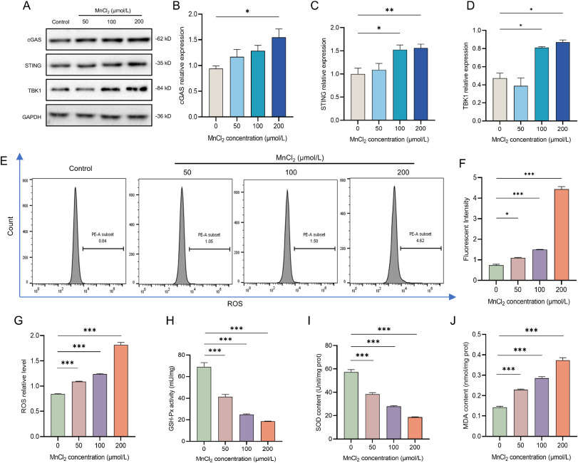
As the starting point of the entire in vitro experiment, the researchers first took BV2 microglial cells as the research object to explore the effects of manganese exposure on key intracellular pathways and oxidative status, laying a solid foundation for subsequent mechanism analysis. After treating cells with different concentrations of manganese chloride, the researchers found through Western blot detection that with the gradient increase of treatment concentration, the expression levels of three key proteins (cGAS, STING, and TBK1) in cells also showed a concentration-dependent significant increase, and all differences reached statistically significant standards. This result clearly confirmed that manganese exposure can specifically activate the cGAS-STING pathway in microglial cells, and this activation effect is closely related to manganese exposure dose. On this basis, further detection of intracellular reactive oxygen species levels through fluorescent probes combined with flow cytometry, as well as detection of antioxidant enzyme activities and lipid peroxidation marker contents, revealed that after manganese treatment, the fluorescence intensity of intracellular reactive oxygen species was significantly enhanced, the activities of GSH-Px and SOD in the antioxidant system were significantly decreased, while the content of lipid peroxidation product MDA continued to increase. This means that while activating the cGAS-STING pathway, manganese will simultaneously trigger oxidative stress responses in cells, which are interrelated and synergistic, jointly forming the initial link of manganese-induced cell damage.
In vivo Experimental Verification: Reproduction of Manganese Exposure-induced Neurotoxicity and Pathway Activation Effects in Animals
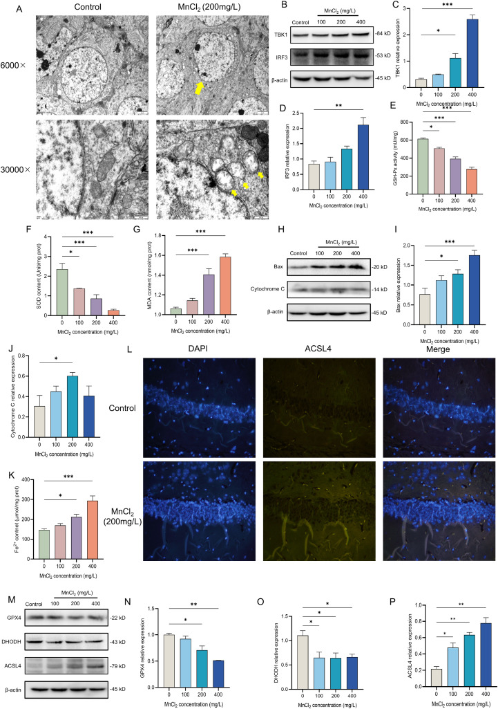
To verify the reliability of the in vitro experimental results and clarify the neurotoxic effects of manganese exposure in vivo, the researchers constructed a manganese-exposed mouse model, taking the mouse hippocampus as the core research site to carry out in vivo verification experiments. Through transmission electron microscopy observation, it was found that the hippocampal cells of mice treated with manganese showed obvious structural damage, including cell membrane shrinkage, and mitochondria, as the core of cellular energy metabolism, showed typical damage characteristics such as matrix swelling, vacuolation, and cristae shortening, which intuitively demonstrated the direct damage effect of manganese exposure on nerve tissue. Further detection of pathway-related proteins in mouse brain tissue through Western blot showed results highly consistent with in vitro experiments: with the increase of manganese exposure concentration, the expression levels of TBK1 and IRF3 in brain tissue significantly increased, confirming that the cGAS-STING pathway is also activated by manganese exposure in vivo. In addition, the researchers also detected indicators related to oxidative stress, apoptosis, and ferroptosis in brain tissue, and found that manganese exposure led to decreased antioxidant enzyme activity, accumulation of lipid peroxidation products, increased expression of pro-apoptotic proteins and ferroptosis-promoting proteins, decreased expression of inhibitory proteins, and a significant increase in iron ion content. This series of results perfectly echoes with in vitro experiments, fully confirming that manganese exposure can induce oxidative stress, cell apoptosis, and ferroptosis through activating the cGAS-STING pathway in vivo, further solidifying the scientific nature and reliability of the research conclusions.
Core Mechanism Analysis: ROS Mediating Role between Pathway and Cell Damage
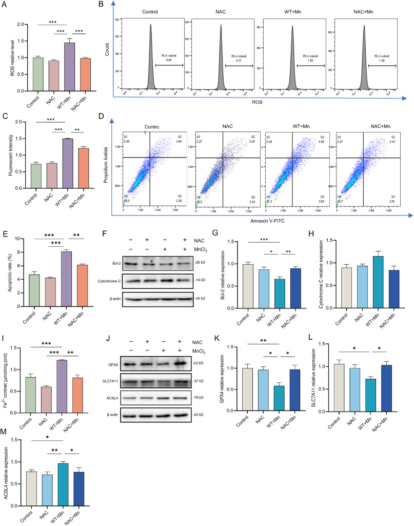
After confirming that manganese exposure can activate the cGAS-STING pathway and induce cell apoptosis and ferroptosis, the researchers further explored the core regulatory relationship among the three, focusing on verifying whether ROS plays a mediating role. The researchers used the ROS inhibitor NAC to co-treat BV2 microglial cells with manganese. First, through detection, it was confirmed that NAC pretreatment could effectively reduce the intracellular ROS levels induced by manganese, proving that NAC can successfully block manganese-mediated ROS generation. On this basis, the cell apoptosis situation was detected, and it was found that after NAC pretreatment, the apoptosis rate of the manganese treatment group significantly decreased, while the expression level of the anti-apoptotic protein Bcl-2 increased, and the expression level of the pro-apoptotic protein cytochrome C decreased. This result indicates that inhibiting ROS generation can effectively alleviate manganese-induced cell apoptosis. Subsequently, the researchers further detected ferroptosis-related indicators and found that NAC pretreatment could also reduce the iron ion content in cells of the manganese treatment group, decrease the expression of the ferroptosis-promoting protein ACSL4, and increase the expression of the ferroptosis-inhibiting proteins GPX4 and SLC7A11, confirming that inhibiting ROS can also alleviate manganese-induced cell ferroptosis. Based on the above experimental results, the researchers clarified the core logic of the entire mechanism: after the cGAS-STING pathway is activated by manganese, it mediates the massive generation of ROS. As a key mediator molecule, ROS further induces cell apoptosis and ferroptosis, becoming the core link connecting the activation of the pathway and cell damage.
Mechanism Summary: Complete Regulatory Network of Manganese Exposure-induced Neurotoxicity
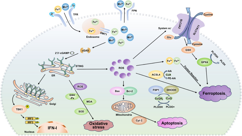
Based on all the above in vitro and in vivo experimental results, the researchers drew a complete mechanism diagram of manganese exposure-induced neurotoxicity, integrating all key regulatory links into a coherent regulatory network, clearly presenting the entire process of manganese-induced neurotoxicity. The entire regulatory process starts with manganese exposure as the initial signal, first stimulating intracellular cGAS to synthesize the second messenger cGAMP, which further binds and activates STING protein. The activated STING will promote the phosphorylation of downstream TBK1 and IRF3, thereby promoting the expression of type I interferons. At the same time, the activated cGAS-STING pathway will trigger obvious oxidative stress responses, specifically manifested as the massive generation of oxidative products such as ROS and MDA, as well as the decrease in the activities of antioxidant enzymes such as GSH-Px and SOD. As a core mediating molecule, ROS will further activate two damage pathways: on the one hand, by regulating the expression of apoptosis-related proteins such as Bax and Bcl-2, it induces mitochondria to release cytochrome C, ultimately triggering cell apoptosis; on the other hand, by disrupting intracellular iron homeostasis, it regulates the expression of ferroptosis key proteins such as GPX4, SLC7A11, and ACSL4, thereby inducing cell ferroptosis. This complete regulatory network organically links the cGAS-STING pathway, ROS, apoptosis and ferroptosis, clearly elucidating the core mechanism of manganese exposure-induced neurotoxicity and condensing the main conclusion of the entire study.
4. Conclusion
This study confirmed through in vitro and in vivo experiments that Mn exposure can activate the cGAS-STING pathway, mediate ROS generation, and further induce apoptosis and ferroptosis in BV2 microglial cells and mouse brain tissue cells, which is the key mechanism of Mn neurotoxicity. The study not only filled the research gap on the role of the cGAS-STING pathway in Mn neurotoxicity but also clarified the mediating role of ROS between the cGAS-STING pathway and apoptosis/ferroptosis, providing new targets and ideas for the prevention and control of Mn neurotoxicity-related diseases. In the future, further in-depth exploration of the specific molecular mechanisms of the interaction between the cGAS-STING pathway and ROS can be conducted, while in vivo verification through inhibitor or gene knockout mouse models, combined with population sample data, to provide more solid theoretical and experimental support for the clinical intervention of Mn neurotoxicity.
References
Zhang Z, Yang J, Zhou Q, Zhong S, Luo J, Chai X, Liu J, Zhang X, Chang X, Wang H. The role and mechanism of the cGAS-STING pathway-mediated ROS in apoptosis and ferroptosis induced by manganese exposure. Redox Biol. 2025 Sep;85:103761. doi: 10.1016/j.redox.2025.103761. Epub 2025 Jul 8. PMID: 40652697; PMCID: PMC12274772.
EnkiLife Western Blot Kits
Product | Catalog Number |
|---|---|
2-Hour Rapid Western Blot Ready-to-Use Complete Workflow Kit | |
4-Hour Fast Western Blot Ready-to-Use Complete Workflow Kit |