mIHC Multiplex Immunohistochemistry TSA Kit
In the fields of biomedical research and clinical diagnosis, accurately capturing the expression levels and spatial distribution of proteins in biological samples is a key prerequisite for understanding disease mechanisms and optimizing treatment plans. Although traditional immunohistochemistry techniques serve as basic detection methods, they often face challenges such as weak signals, insufficient specificity, and limited labeling capacity when detecting low-abundance targets or conducting simultaneous multi-target analysis. TSA (Tyramide Signal Amplification) technology, with its unique enzymatic signal amplification mechanism, breaks through the bottlenecks of traditional techniques. The TSA kit developed based on this technology transforms complex technical principles into standardized, easy-to-operate experimental tools, bringing high-sensitivity, high-specificity multicolor fluorescence detection to ordinary laboratories. From paraffin sections, frozen tissues to cell samples and organoids, the TSA kit plays an irreplaceable role in spatial proteomics research, tumor immune microenvironment analysis, clinical pathological precision diagnosis, and other fields with its wide sample adaptability and stable detection performance.
Our TSA kit has a full range of product components, including TSA-labeled fluorophores, TSA dilution buffer, 3% H₂O₂, blocking solution, primary antibody dilution buffer, antibody elution buffer, HRP-goat anti-rabbit/mouse IgG, DAPI staining solution, and anti-fluorescence quenching mounting medium, providing users with a one-stop experimental solution and greatly enhancing experimental convenience. The kit is equipped with 7-color fluorescent substrates covering the 480–750 nm wavelength range, which can be freely combined as needed to achieve simultaneous acquisition of 7-plex information on a single section. At the same time, the supporting secondary antibodies are compatible with rabbit/mouse multi-species primary antibodies, breaking through traditional species limitations. Combined with specifically developed gentle antibody elution buffer, it can remove antibodies while preserving antigens and fluorescent signals, allowing for 5–7 rounds of cyclic staining while maintaining tissue structure integrity, significantly reducing sample damage compared to microwave heat repair. As a universal kit, it has a wide range of applications and can be adapted to paraffin sections, frozen sections, cell smears, organoids, and other tissue samples, and with professional technical support and quality assurance systems, users can receive timely and effective solutions to various problems encountered during use.
Table 1. Fluorescent Dyes for Multiplex Immunohistochemistry Detection
Fluorescent Dye | Excitation Wavelength λ (nm) | Emission Wavelength λ (nm) | Color | Filter |
|---|---|---|---|---|
TSA-430 | 430 | 480 | Blue | Aqua Channel |
TSA-FITC | 490 | 520 | Green | FITC Channel |
TSA-CY3 | 550 | 570 | Orange-Red | Cy3 Channel |
TSA-594 | 590 | 620 | Red | Red Channel |
TSA-680 | 680 | 700 | Dark Red | Cy5/Cy5.5 Channel |
TSA-CY7 | 750 | 780 | Deeper Red | Cy7 Channel |
Table 2. TSA Complete Kit Components
Component | Specification | Dilution Ratio | ||
|---|---|---|---|---|
20T | 50T | 100T | ||
TSA Dilution Buffer | 12mL | 30mL | 60mL | Ready-to-use |
3%H2O2 | 2mL | 5mL | 10mL | Ready-to-use |
Blocking Solution | 12mL | 30mL | 60mL | Ready-to-use |
Primary Antibody Dilution Buffer | 12mL | 30mL | 60mL | Ready-to-use |
Antibody Elution Buffer | 10mL | 25mL | 50mL | Ready-to-use |
HRP-goat anti-rabbit/mouse IgG | 12mL | 30mL | 60mL | Ready-to-use |
DAPI Staining Solution | 2mL | 5mL | 10mL | Ready-to-use |
Anti-Fluorescence Quenching Mounting Medium | 2mL | 5mL | 10mL | Ready-to-use |
Kit Operating Procedure (Paraffin Sections as Example):
1. Deparaffinization: Immerse the sections sequentially in Xylene 1 (15min), Xylene 2 (15min), Absolute Ethanol 1 (5min), Absolute Ethanol 2 (5min), 95% Ethanol (5min), 85% Ethanol (5min), 75% Ethanol (5min), and finally rinse the slides with water.
2. Antigen Retrieval: Typically use 1× Citrate buffer (pH 6.0) as retrieval solution for high-temperature and high-pressure retrieval. Place sections in a pressure cooker, add appropriate amount of retrieval solution, close the pressure cooker, start timing for 2min after steam rises, cool to room temperature to complete retrieval. [For targets with weak expression, EDTA (pH 9.0) can be used as retrieval solution. For tissues prone to detachment such as bone and brain tissues, microwave retrieval is recommended, with retrieval temperature controlled at around 80°C, and 2× Citrate buffer (pH 6.0) as retrieval solution.]
3. Endogenous Enzyme Blocking: Prepare 3% H2O2 solution with pure water, place sections in the solution, and incubate at room temperature for 20min. For tissues prone to detachment, H2O2 concentration and incubation time can be appropriately reduced.
4. Blocking: Add blocking solution dropwise onto the tissue and incubate at 37°C for 30min.
5. Primary Antibody Incubation: Dilute the primary antibody to appropriate concentration using antibody dilution buffer, add the antibody dropwise onto the tissue, and incubate overnight at 4°C or for 1h at 37°C.
6. Secondary Antibody Incubation: Add HRP-labeled secondary antibody dropwise onto the tissue and incubate at 37°C for 1h.
7. TSA Reagent Incubation: Add TSA-XXX fluorescent dye dropwise onto the tissue, incubate at 37°C for 30min, then wash with PBST 3 times, 5min each time.
8. Antibody Elution: Add antibody elution buffer dropwise onto the tissue, incubate at room temperature for 15min (37°C incubation is better).
9. Repeat steps 4-8 (for incubating other TSA-XXX fluorescent dyes).
10. DAPI Nuclear Staining: Add DAPI working solution dropwise onto the tissue, incubate at room temperature for 5min, then wash with PBS 3 times, 5min each time. Spin dry the liquid, add anti-fluorescence quenching mounting medium dropwise onto the tissue, cover with a coverslip, and observe and photograph under microscope.
Experimental Notes
1. Monoclonal antibodies are preferred as primary antibodies, followed by polyclonal antibodies. For mouse samples, avoid choosing mouse-derived primary antibodies as much as possible. If mouse primary antibodies are chosen, secondary antibodies will bind not only to primary antibodies but also to endogenous IgG in the tissue, resulting in non-specific staining.
2. If the tissue easily detaches, repair can be performed using a 60°C water bath method.
3. Compared to fluorescent secondary antibodies, the TSA kit has higher sensitivity and stronger signals. Therefore, the concentration of primary antibody needs to be reduced,
generally appropriately increasing the dilution ratio based on the dilution ratio recommended in the antibody instructions to reduce background fluorescence caused by non-specific binding. It is recommended to set up a gradient concentration of primary antibody to obtain the best results.
4. If the background fluorescence is strong, it is recommended to add a tissue autofluorescence quenching step.
5. To ensure antibody elution effect and fluorescent multiplex labeling effect, it is recommended to perform individual TSA single-label tests for each antibody before formal multiplex labeling, and determine the experimental conditions such as antigen repair conditions and antibody sequence for multiplex labeling based on the single-label test results after confirming that each antibody can produce ideal positive single-label results.
6. If some antibodies have high titer and strong affinity and are not easily eluted completely, the number of elution steps can be increased.
7. The antibody elution buffer has strong fluidity. If the slides are not placed horizontally, the reagent may flow out of the circle, affecting the elution effect. Pay attention to keeping the slides flat during operation.
Table 3. Related Products
Product | Catalog Number |
|---|---|
TSA Six-Label Seven-Color Multiplex Immunohistochemistry Kit | |
TSA Five-Label Six-Color Multiplex Immunohistochemistry Kit | |
TSA Four-Label Five-Color Multiplex Immunohistochemistry Kit | |
TSA Three-Label Four-Color Multiplex Immunohistochemistry Kit | |
TSA Two-Label Three-Color Multiplex Immunohistochemistry Kit |
In addition, we provide a full range of TSA labeling services. We can select the best detection technology based on target abundance and use the most suitable detection wavelength to match your multicolor labeling experiments.
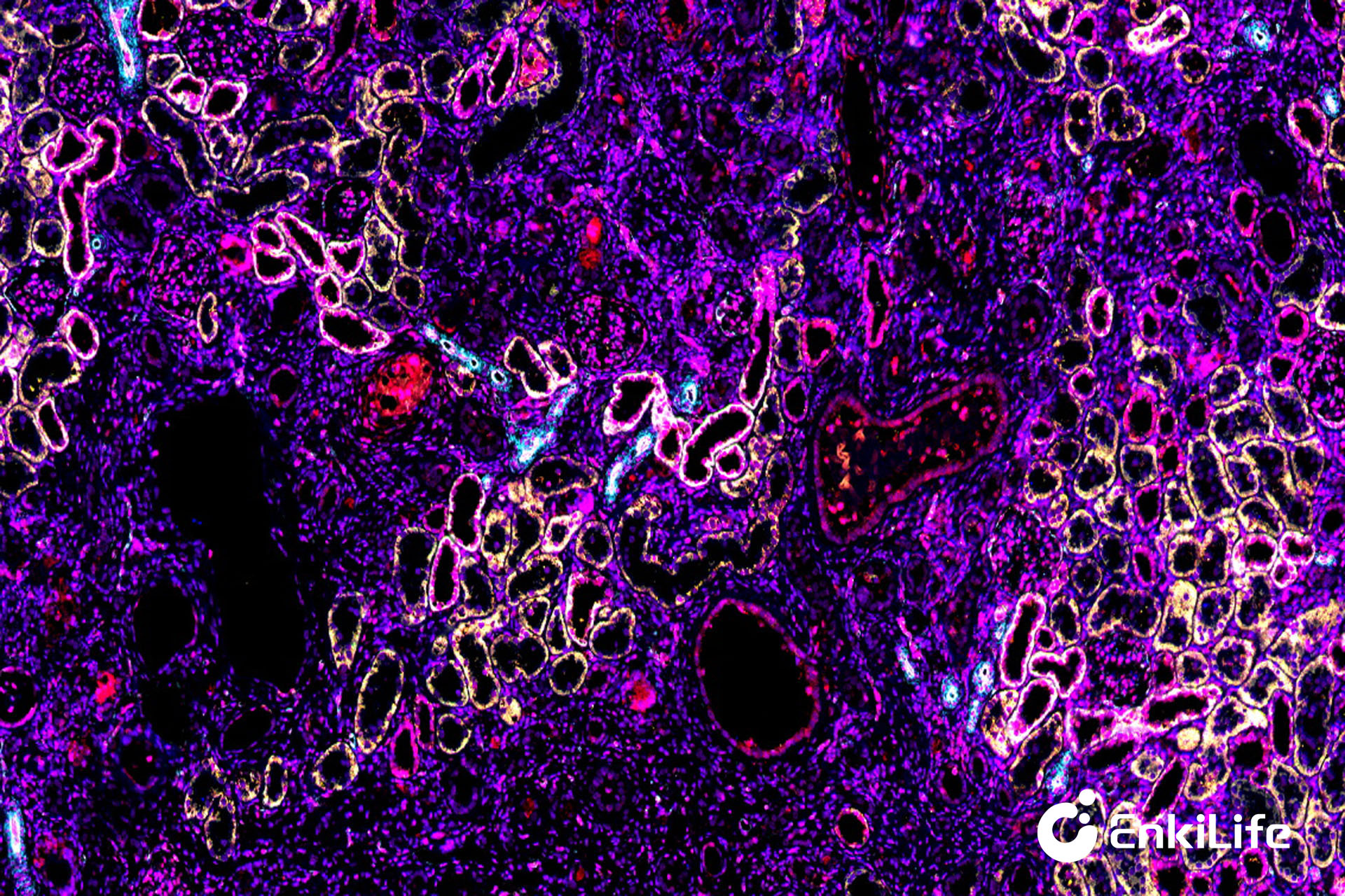
Six-Label Seven-Color Results Display:
![]()
Rat Kidney (Six-Label Seven-Color) — 10x
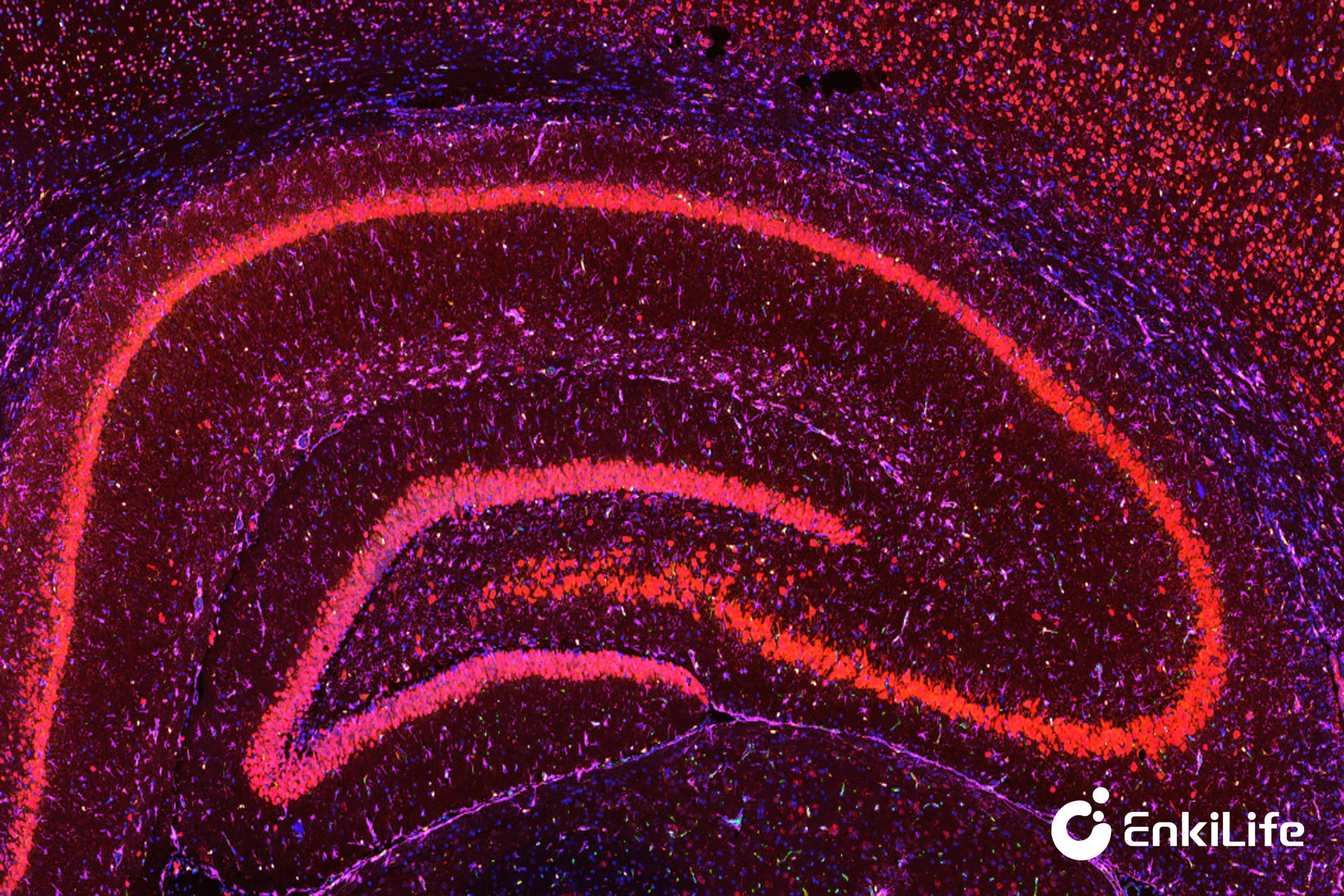
Rat Brain (Six-Label Seven-Color) — 5x