
Contents
- Basic Regulation of the Cell Cycle
- Sources and Types of DNA Damage
- The DNA Damage Response and Cell Cycle Checkpoints
- Major DNA Repair Pathways
- Links Between DNA Repair and the Cell Cycle
- DNA Damage and Cell Fate
- Cell Cycle Dysregulation and Tumorigenesis
- Targeting the DNA Damage Response in Cancer Therapy
- Markers of DNA Damage and Repair
- Conclusions and Research Applications
- References
1. Basic Regulation of the Cell Cycle
The cell cycle is an ordered process through which cells coordinate genome duplication, chromosome segregation, and cell division. It is generally divided into G1, S, G2, and M phases. G1 is a key stage at which cells decide whether to enter another proliferative cycle; S phase is devoted to DNA replication; G2 prepares the cell for mitosis; and M phase comprises chromosome segregation and cytokinesis. Some cells can also leave the proliferative cycle and enter the quiescent G0 state. [1, 2]
Cell cycle progression is coordinated by cyclins, cyclin-dependent kinases (CDKs), the RB–E2F transcriptional network, and protein degradation systems. Different cyclins accumulate at specific stages and form active complexes with their corresponding CDKs, controlling cell cycle events by regulating substrate phosphorylation. [1, 3]
1.1 G1/S Regulation and Cell Cycle Entry
Core references for this section: 1, 3.
During G1, cells integrate growth factors, nutrient availability, and other environmental signals to decide whether to continue proliferating or exit the cell cycle. Acquiring the conditions needed to enter a new cycle, passing the G1 restriction point, and subsequently initiating DNA replication and committing to complete that cycle are distinct regulatory events. These processes are not determined by a single cyclin–CDK complex.
This transition is influenced primarily by regulatory networks involving cyclin D–CDK4/6, RB–E2F, and cyclin E–CDK2.
Retinoblastoma protein (RB) limits the expression of genes required for DNA replication and S phase by inhibiting E2F transcription factors.
In response to appropriate proliferative signals, cyclin D-associated CDK activity regulates RB and other cell cycle proteins. As the cycle progresses, RB-mediated inhibition of E2F weakens and E2F-dependent transcription increases, promoting the expression of cyclin E, cyclin A, and DNA replication proteins.
Cyclin E–CDK2 primarily regulates the G1/S transition, whereas cyclin A–CDK2 contributes to S-phase progression and DNA replication. Replication origin licensing and origin firing are distinct regulatory events and should not be equated with cyclin A accumulation or a change in a single CDK activity.
Late G1 is also regulated by the anaphase-promoting complex/cyclosome (APC/C). APC/C–Cdh1 promotes the degradation of selected cell cycle proteins. A decline in its activity allows cyclin A to accumulate and supports the regulatory transition required for S-phase entry.
Different experimental models have been proposed for the precise relationship between cyclin D–CDK4/6, cyclin E–CDK2, and RB phosphorylation. The G1/S transition should therefore not be viewed as a fixed linear sequence that applies to every cell type.
1.2 The G2/M Transition and Mitotic Entry
Core references for this section: 1.
After DNA replication is completed, cells enter G2 and prepare for mitosis. [4]
Cyclin B–CDK1 is a major regulator of mitotic entry, and its activity is jointly controlled by proteins including WEE1, MYT1, and CDC25. [4]
WEE1 and MYT1 restrain CDK1 activity through inhibitory phosphorylation, whereas CDC25 phosphatases remove these inhibitory modifications to promote CDK1 activation. [4]
When cyclin B–CDK1 activity reaches an appropriate level, cells initiate mitotic events, including chromosome condensation, nuclear envelope breakdown, centrosome separation, and mitotic spindle formation. [5]
During this process, CDK1 works with mitotic regulatory kinases such as PLK1 and Aurora kinases to coordinate chromosome condensation, spindle assembly, and other mitotic events, supporting orderly progression. [4]
1.3 Mitotic Exit and Cell Cycle Resetting
Core references for this section: 1.
Once cells enter mitosis, protein degradation and changes in phosphorylation are required to control chromosome segregation and mitotic exit.
APC/C coordinates mitotic progression and exit by promoting the ubiquitination and degradation of different cell cycle proteins at specific stages. Cyclin A can be degraded early in mitosis, whereas the degradation of securin and cyclin B is closely associated with anaphase onset and mitotic exit.
As cyclin B is degraded, CDK1 activity declines. Protein phosphatases then promote the dephosphorylation of CDK1 substrates, allowing cells to return progressively to interphase.
Meanwhile, APC/C-mediated securin degradation helps activate separase and promotes sister chromatid separation.
Together, these mechanisms enable chromosome segregation, mitotic exit, and the resetting of the cell cycle.
2. Sources and Types of DNA Damage
Core references for this section: 4, 6.
DNA is continuously exposed to endogenous and exogenous challenges. Endogenous sources include spontaneous hydrolytic depurination, base oxidation caused by reactive oxygen species (ROS), replication mismatches, and replication fork abnormalities. Exogenous sources include ultraviolet radiation, ionizing radiation, and certain DNA-reactive chemicals. These factors can cause base modifications, single-strand breaks, double-strand breaks, interstrand crosslinks, and abnormal DNA structures associated with replication. [7]
Different lesions have distinct repair requirements and differ substantially in their effects on genome stability.
2.1 DNA Base Damage
Core references for this section: 4, 6.
DNA base damage primarily involves changes to the chemical structure of bases, including oxidation, deamination, and other modifications.
For example, ROS can oxidize guanine to form 8-oxoguanine (8-oxoG). If this lesion is not repaired promptly, it may mispair with adenine during DNA replication, increasing the risk of base substitution mutations. [8, 9]
Ultraviolet radiation can also induce pyrimidine dimers, while certain chemicals generate bulky DNA adducts. These lesions can alter the DNA double helix and impede DNA or RNA polymerase activity.
Different types of chemical DNA damage are generally processed by different pathways. Base excision repair (BER), for example, handles oxidative base lesions such as 8-oxoG, whereas nucleotide excision repair (NER) primarily removes bulky lesions caused by ultraviolet radiation and other factors, including pyrimidine dimers.
NER comprises global genome NER (GG-NER) and transcription-coupled NER (TC-NER). GG-NER identifies and repairs relevant lesions throughout the genome and is not confined to a particular genomic region or cell cycle phase. TC-NER primarily repairs lesions in actively transcribed genes that obstruct RNA polymerase progression. The two modes differ mainly in lesion recognition but share downstream molecular repair mechanisms.
Cells can also use damage tolerance mechanisms, such as translesion synthesis (TLS), to sustain replication when some template-strand lesions remain. TLS bypasses particular DNA template lesions rather than directly removing them, and it should not be interpreted as a mechanism capable of bypassing every type of DNA break.
2.2 DNA Single-Strand Breaks
Core references for this section: 4, 6.
A DNA single-strand break (SSB) is a discontinuity in the phosphodiester backbone of one strand of the double helix, while the complementary strand usually remains intact.
SSBs can arise from ROS, DNA damage processing, and other endogenous or exogenous factors.
Because the intact complementary strand generally retains the sequence information, most SSBs can be processed by single-strand break repair mechanisms. The exact process depends on the chemical structure of the broken ends and the functional state of repair factors.
However, an SSB is not harmless. If it remains unrepaired before DNA replication, passage of a replication fork through the lesion can cause fork collapse and generate a DNA double-strand break.
Timely SSB repair is therefore important for maintaining stable DNA replication.
2.3 DNA Double-Strand Breaks
Core references for this section: 4.
A DNA double-strand break (DSB) occurs when both strands of the DNA double helix are broken.
DSBs can result from ionizing radiation, certain DNA-damaging drugs, and replication fork collapse.
Compared with SSBs, DSBs pose a greater threat to genome integrity and require more complex repair. Incorrectly repaired DSBs can cause the loss of chromosome fragments, chromosome rearrangements, or translocations.
DSBs can also be classified as two-ended or one-ended breaks according to their structure and origin.
A typical two-ended DSB has two broken ends and can be repaired by non-homologous end joining (NHEJ) or by template-directed homologous recombination (HR).
One-ended DSBs generated by replication fork collapse generally require HR-related processes to restore DNA replication. Inappropriate joining of unrelated broken ends can produce chromosome rearrangements.
Because DSBs can severely disrupt the genome, cells use the DNA damage response (DDR) network to detect damage, activate checkpoints, and coordinate DNA repair with cell cycle progression.
2.4 DNA Interstrand Crosslinks
Core references for this section: 3, 4.
A DNA interstrand crosslink (ICL) is a covalent linkage between the two complementary strands of the DNA double helix.
Because replication and transcription require local DNA unwinding, ICLs can obstruct both DNA and RNA polymerases.
ICLs require several coordinated repair mechanisms. The Fanconi anemia (FA) pathway is particularly important for replication-associated ICL repair.
Certain ICLs can also be processed by other mechanisms, including those involving NEIL3. Different ICLs therefore do not necessarily depend on an identical repair process.
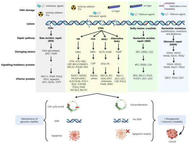
Raimundo L, Calheiros J, Saraiva L. Exploiting DNA Damage Repair in Precision Cancer Therapy: BRCA1 as a Prime Therapeutic Target. Cancers. 2021;13(14):3438. Figure 1. doi:10.3390/cancers13143438.
3. The DNA Damage Response and Cell Cycle Checkpoints
Core references for this section: 4, 6.
The DNA damage response (DDR) is a complex signaling network that helps cells maintain genome stability.
DDR encompasses not only damage recognition and repair but also chromatin regulation, checkpoint activation, and cell fate decisions.
Its basic organization can be summarized as follows:
DNA damage or replication abnormalities → damage recognition and sensing → DDR initiation and signal amplification → DNA repair, cell cycle regulation, and cell fate responses.
DNA repair, cell cycle control, and cell fate decisions are interconnected and do not necessarily occur in a strict sequence.
Cell cycle checkpoints primarily delay or prevent inappropriate progression when DNA damage or other cell cycle abnormalities occur, providing time for damage processing or error correction.
Checkpoint activation generally involves four basic steps:
- Damage recognition: Sensor proteins recognize DNA breaks, single-stranded DNA, or abnormal replication structures.
- Signal initiation: Sensor proteins promote the recruitment and activation of major kinases such as ATM and ATR.
- Signal transmission: Activated kinases phosphorylate CHK1, CHK2, and other downstream proteins to transmit and amplify signals.
- Cell cycle regulation: Checkpoint signaling influences regulators such as CDC25, CDKs, and p53 to delay cell cycle progression.
3.1 Activation of ATM, ATR, and DNA-PK
Core references for this section: 4, 6.
ATM, ATR, and DNA-PK are important DDR protein kinases, but they differ in the DNA structures to which they primarily respond and in their downstream biological functions.
ATM primarily responds to DNA double-strand breaks
Following a DSB, the MRN complex (MRE11–RAD50–NBS1) participates in break recognition and promotes ATM recruitment and activation at the damage site.
Activated ATM can phosphorylate numerous downstream proteins, including CHK2, p53, and histone H2AX.
Phosphorylation of H2AX at Ser139 generates γH2AX, which promotes the recruitment of mediator proteins such as MDC1 and contributes to the organization and amplification of damage-associated chromatin signaling. [10]
ATM also regulates cell cycle checkpoints through factors including CHK2 and p53.
Its downstream functions can therefore be summarized as two interconnected branches:
- ATM–γH2AX–MDC1: Organization of signaling at damage sites and recruitment of repair factors.
- ATM–CHK2/p53: Regulation of cell cycle checkpoints and other cellular responses. ATM can phosphorylate p53 directly and can also regulate it through CHK2 and other factors; these proteins do not form a single obligatory linear sequence.
ATR primarily responds to single-stranded DNA and replication stress
Whereas ATM primarily responds to DSBs, ATR mainly participates in DNA damage and replication stress responses triggered by structures containing single-stranded DNA.
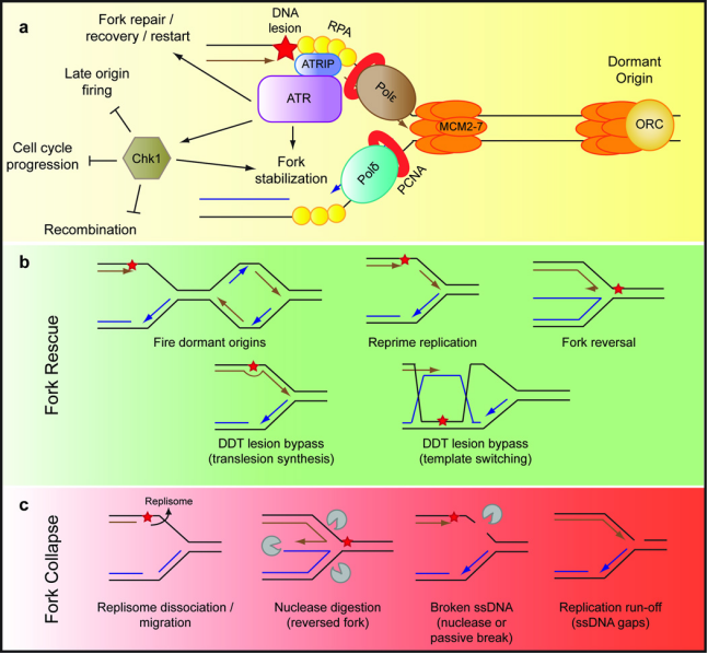
Zeman MK, Cimprich KA. Causes and consequences of replication stress. Nat Cell Biol. 2014;16:2–9. doi:10.1038/ncb2897.
Only Figure 2a is discussed here; the processes shown in Figures 2b and 2c are addressed in later sections.
When replication forks stall, or DSB end resection generates single-stranded DNA, replication protein A (RPA) can bind the exposed DNA.
ATR-interacting protein (ATRIP) promotes ATR recruitment through its interaction with RPA-coated single-stranded DNA.
Factors such as TOPBP1 or ETAA1 then help activate ATR, enabling it to phosphorylate CHK1 and other downstream substrates.
ATR–CHK1 signaling regulates replication activity, restricts inappropriate replication origin firing, and coordinates the replication stress response with cell cycle progression.
ATM and ATR are not completely independent sensing systems. For example, single-stranded DNA generated by DSB end resection can also activate ATR-related signaling.
DNA-PK primarily participates in DSB recognition and NHEJ
DNA-dependent protein kinase (DNA-PK) primarily participates in the recognition and repair of DNA double-strand breaks.
After a DSB, the Ku70/Ku80 heterodimer binds the broken ends and promotes recruitment of the DNA-PK catalytic subunit (DNA-PKcs), forming an active DNA-PK complex.
DNA-PK contributes to end protection, repair complex organization, and non-homologous end joining.
Although DNA-PK is an important DDR kinase, its principal functions should not be equated with classical ATM–CHK2- or ATR–CHK1-mediated cell cycle checkpoint signaling.
3.2 The G1/S DNA Damage Checkpoint
Core references for this section: 4.
The G1/S checkpoint primarily prevents cells carrying DNA damage from entering DNA replication prematurely. [6]
When DSBs occur in G1, the MRN complex contributes to ATM activation, followed by cell cycle regulation through CHK2- and p53-related signaling. [6]
ATM and CHK2 can enhance p53 stability and activity through phosphorylation, thereby inducing transcription of the CDKN1A gene. [6]
CDKN1A encodes the CDK inhibitor p21. [1]
p21 inhibits selected cyclin–CDK complexes, maintaining RB-mediated repression of E2F-dependent transcription and thereby delaying or preventing S-phase entry. [1]
The main purpose of this checkpoint is to prevent genome replication from starting before DNA damage has been appropriately processed. [1]
Its principal signaling events can be summarized as follows: [1]
DSB → MRN → ATM → p53 → p21 → reduced CDK activity → G1/S arrest. ATM can also promote p53 regulation through CHK2 and other factors. This is a representative signaling network rather than a single obligatory linear pathway. [1]
3.3 The S-Phase Replication Stress Checkpoint
Core references for this section: 1, 4.
The S-phase checkpoint primarily monitors abnormalities during DNA replication and helps prevent replication stress from developing into severe DNA damage.
Replication forks may slow or stall because of template lesions, abnormal DNA structures, or other replication barriers.
At stalled forks, DNA unwinding and synthesis can become uncoupled, causing single-stranded DNA to accumulate.
RPA binds this single-stranded DNA, and ATRIP promotes ATR recruitment. TOPBP1, ETAA1, and other factors then support ATR activation.
In addition to influencing CDC25 and CDK activity through downstream factors such as CHK1 and restricting inappropriate origin firing, ATR supports stalled fork stability and recovery through other replication regulators. Fork protection therefore does not depend entirely on CHK1–CDC25–CDK signaling.
The main signaling sequence can be summarized as follows:
Fork stalling → ssDNA accumulation → RPA–ATRIP → ATR → CHK1 → regulation of replication and CDKs.
DNA replication stress must be distinguished from DNA damage.
Replication stress does not necessarily mean that DNA has already broken. A major function of the ATR–CHK1 checkpoint is to preserve replication structures during replication abnormalities and prevent their progression to DNA breakage.
Persistent fork stalling and eventual collapse may generate replication-associated DSBs and activate damage responses involving ATM and other factors.
3.4 The G2/M DNA Damage Checkpoint
Core references for this section: 1, 4.
The G2/M checkpoint primarily prevents premature mitotic entry when DNA damage remains unresolved or DNA replication is in an inappropriate state.
DSBs arising in G2 can primarily trigger ATM-related responses, whereas incomplete replication or replication stress commonly involves ATR–CHK1 signaling. These triggers differ, but their downstream regulation can overlap and influence CDC25 and CDK1 through CHK1, CHK2, and other factors.
Under normal conditions, CDC25 removes inhibitory phosphorylation at CDK1 Thr14/Tyr15, activating CDK1 and allowing the cyclin B–CDK1 complex to initiate mitosis.
Checkpoint activation inhibits CDC25, preserving the inhibitory phosphorylation of CDK1 maintained by kinases such as WEE1.
Consequently, cyclin B–CDK1 fails to become fully active, delaying mitotic entry.
The main mechanism can be summarized as follows:
DSBs or replication abnormalities → ATM/ATR-related responses, depending on the triggering structure → CHK1/CHK2 and related signaling → CDC25 inhibition → restricted CDK1 activity → G2/M arrest.
Because ATM and ATR signaling exhibit crosstalk, the G2/M checkpoint should not be viewed as a process activated exclusively by a single pathway.
3.5 The Mitotic Spindle Assembly Checkpoint
Core references for this section: 1.
In addition to DNA damage and replication stress checkpoints, cells possess a spindle assembly checkpoint (SAC) during mitosis.
SAC primarily monitors the attachment of chromosome kinetochores to spindle microtubules, preventing premature segregation before proper attachment and alignment.
When improperly attached kinetochores remain, factors such as MPS1 transmit checkpoint signals and promote formation of the mitotic checkpoint complex (MCC).
MCC inhibits APC/C–Cdc20, temporarily preventing the onset of anaphase chromosome separation.
Once all kinetochores achieve correct bipolar attachment to spindle microtubules, SAC signaling subsides and MCC disassembles, allowing Cdc20 to activate APC/C. Active APC/C–Cdc20 promotes the ubiquitination and degradation of securin and cyclin B. Securin degradation helps release separase from inhibition and promotes sister chromatid separation, while cyclin B degradation lowers CDK1 activity and drives mitotic exit.
SAC directly monitors chromosome–spindle attachment rather than DNA damage itself. It therefore represents a different type of cell cycle surveillance from ATM/ATR-mediated DNA damage checkpoints.
4. Major DNA Repair Pathways
Core references for this section: 4, 6.
Different types of DNA damage require different repair pathways.
According to their substrates and basic mechanisms, common pathways can be grouped into single-strand break and base damage repair, mismatch repair, double-strand break repair, and repair of complex lesions.
Cells also possess damage tolerance mechanisms, including translesion synthesis and template switching, which allow replication to continue while some lesions remain unresolved.
Together, repair and damage tolerance mechanisms maintain genome stability, although their substrates and molecular processes differ.
4.1 Single-Strand Break Repair
Core references for this section: 4, 6.
Single-strand break repair (SSBR) primarily restores continuity of the phosphodiester backbone of a single DNA strand.
Following an SSB, PARP1 or PARP2 can recognize the break and catalyze poly(ADP-ribosyl)ation (PARylation) of themselves and neighboring target proteins.
PARP1 and its PARylation activity promote recruitment of the scaffold protein XRCC1 to DNA damage sites. [11]
XRCC1 subsequently recruits DNA ligase III (LIG3) and other accessory factors involved in end processing and rejoining.
Poly(ADP-ribose) chains are synthesized and degraded dynamically during this process.
Poly(ADP-ribose) glycohydrolase (PARG) primarily degrades poly(ADP-ribose) chains but cannot directly remove the terminal ADP-ribose moiety that remains attached to a protein. Serine-linked mono-ADP-ribose can be removed by appropriate hydrolases, including ARH3. Together, these processes regulate ADP-ribosylation signaling in the DNA damage response. [12]
SSBR therefore involves break recognition, end processing, and ligation. PARP-mediated PARylation also regulates many SSB responses by promoting repair complex recruitment and organization, but not all SSBR processes are absolutely dependent on PARP.
Key proteins: PARP1, PARP2, XRCC1, LIG3, and PARG.
4.2 Base Excision Repair
Core references for this section: 4.
Base excision repair (BER) primarily processes oxidative, deamination-related, and other small-scale DNA base lesions.
BER usually begins when a lesion-specific DNA glycosylase recognizes and removes a damaged base, generating an abasic (AP) site. [13, 14, 15]
Repair factors such as AP endonucleases then cleave the DNA backbone and process the lesion site. DNA polymerases replace the missing nucleotides, and DNA ligases restore strand continuity.
Oxidative base lesions such as 8-oxoG, for example, can be repaired by specific glycosylases followed by downstream BER steps.
BER can be summarized as follows:
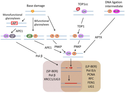
Iyama T, Wilson DM 3rd. DNA repair mechanisms in dividing and non-dividing cells. DNA Repair (Amst). 2013;12(8):620–636. doi:10.1016/j.dnarep.2013.04.015.
Damaged base recognition → base excision → AP site processing → DNA resynthesis → ligation. [6]
BER and SSBR are mechanistically connected and share many pathway components, but they are distinct processes. [11, 14, 15]
BER primarily processes abnormal bases, whereas SSBR primarily repairs breaks in the DNA backbone. BER can generate single-strand break or gap intermediates whose subsequent processing shares factors with SSBR, including XRCC1, POLβ, and DNA ligases. In specific BER branches, SSBR-associated end-processing proteins such as PNKP also participate. [6]
Key proteins: DNA glycosylases, APE1, POLβ, XRCC1, and relevant DNA ligases.
4.3 Nucleotide Excision Repair
Core references for this section: 3, 6.
Nucleotide excision repair (NER) primarily removes lesions that distort the DNA double helix, such as ultraviolet-induced pyrimidine dimers and bulky adducts generated by certain chemicals. [4]
NER comprises two main modes:
Global genome NER (GG-NER)
GG-NER recognizes and repairs relevant lesions in different genomic regions. Its activity is not limited to a particular gene or cell cycle phase.
Protein complexes such as XPC–RAD23–CETN2 participate in initial lesion recognition in GG-NER and promote recruitment of downstream repair proteins.
Transcription-coupled NER (TC-NER)
TC-NER primarily processes DNA lesions that obstruct RNA polymerase progression and is therefore closely linked to damage recognition in actively transcribed genes.
GG-NER and TC-NER use different initial recognition mechanisms but share many proteins during subsequent repair.
Downstream NER involves local DNA unwinding, incisions on both sides of the damaged segment, gap filling, and ligation.
Proteins such as XPA help organize the repair reaction, while endonucleases including ERCC1–XPF and XPG excise the lesion-containing DNA segment.
NER is important in G1, but its activity is not restricted to G1.
Key proteins: Initial GG-NER recognition primarily involves XPC–RAD23–CETN2; TC-NER initiation is associated with stalled RNA polymerase. XPA, ERCC1–XPF, and XPG participate in downstream steps shared by both pathways.
4.4 Mismatch Repair
Core references for this section: 4, 6.
Mismatch repair (MMR) primarily corrects base mismatches that escape DNA polymerase proofreading during replication, as well as small insertion or deletion loops.
In eukaryotic cells, complexes such as MSH2–MSH6 and MSH2–MSH3 recognize different types of mismatch structures.
Repair factors such as MLH1–PMS2 then coordinate processing of the erroneous DNA region, enabling correction through excision, resynthesis, and ligation.
The basic MMR process can be summarized as follows: [16]
Mismatch recognition → processing and excision of the erroneous DNA region → accurate DNA resynthesis → ligation.
Because mismatches commonly arise during replication, MMR is closely associated with S phase. This does not mean that every MMR-related activity occurs exclusively in S phase. [16]
Key proteins: MSH2, MSH6, MSH3, MLH1, and PMS2.
4.5 Homologous Recombination Repair
Core references for this section: 4, 6.
Homologous recombination (HR) repairs DSBs using homologous DNA sequences as templates and functions primarily in S and G2 phases.
Unlike NHEJ, which directly joins broken ends, HR generally uses genetic information supplied by a sister chromatid for template-directed repair.
HR first requires processing of the broken DNA ends.
The MRN complex and CtIP initiate end resection. EXO1- or BLM–DNA2-associated mechanisms then promote long-range resection, producing repair intermediates with 3′ single-stranded DNA tails. Related cell cycle regulation is discussed in Section 5.5.
RPA then binds the exposed single-stranded DNA, helping protect and stabilize these repair intermediates. [17, 18]
BRCA1 subsequently promotes BRCA2 recruitment through interactions with PALB2 and other proteins. BRCA2 facilitates RAD51 loading onto single-stranded DNA, allowing RAD51 to replace RPA and form a nucleoprotein filament.
The RAD51 filament promotes strand invasion into homologous double-stranded DNA and formation of recombination intermediates such as displacement loops (D-loops).
DNA synthesis then uses homologous DNA, typically the sister chromatid, as a template. Depending on intermediate formation and processing, HR can finish through mechanisms such as synthesis-dependent strand annealing (SDSA). Alternatively, double Holliday junctions may form and subsequently undergo dissolution or resolution to complete repair.
The key steps of HR can be summarized as follows:
DSB recognition → end resection → RPA binding → RAD51 loading → homology search and strand invasion → DNA synthesis and recombination intermediate processing.
Because sister chromatids become available after replication, HR is particularly important in S and G2 phases.
End resection is also controlled by CDK activity and repair proteins, giving HR a pronounced dependence on cell cycle stage.
Key proteins: MRN, CtIP, RPA, BRCA1, PALB2, BRCA2, RAD51, EXO1, and other HR-associated proteins.
4.6 Non-Homologous End Joining
Core references for this section: 4, 6.
Non-homologous end joining (NHEJ) is a major pathway for repairing DNA double-strand breaks.
Unlike HR, classical NHEJ does not require an extended homologous DNA template. Instead, it repairs breaks through end recognition, protection, processing, and ligation.
Following a DSB, the Ku70/Ku80 heterodimer initially binds the broken ends. [19]
The Ku complex protects these ends against inappropriate degradation and provides a platform for recruiting other NHEJ proteins.
Ku subsequently recruits DNA-PKcs to form the DNA-PK complex. DNA-PKcs contributes to end synapsis and repair regulation. Its autophosphorylation affects end synapsis and its dissociation from broken ends, thereby regulating subsequent repair steps. [20, 21, 22]
Ends that cannot be ligated directly require appropriate processing. In some repair settings, nucleic acid-processing factors such as Artemis contribute to this step. [23]
Finally, DNA ligase IV (LIG4) cooperates with XRCC4, XLF, and other proteins to join the ends. [17, 18, 24]
The main NHEJ steps can be summarized as follows:
DSB end recognition → Ku70/Ku80 binding → DNA-PKcs recruitment → end processing when necessary → XRCC4–LIG4-associated ligation.
NHEJ can function at several cell cycle stages and is particularly important in G1.
Because NHEJ does not require an extended homologous template, repair events involving end processing may introduce small insertions or deletions. This does not mean that all NHEJ is highly error-prone. Classical NHEJ can also achieve accurate repair when broken ends are suitable for direct ligation.
Incorrect joining of ends from different DNA breaks can generate chromosome translocations or other structural abnormalities.
Key proteins: Ku70, Ku80, DNA-PKcs, Artemis, XRCC4, LIG4, and XLF.
4.7 DNA Interstrand Crosslink Repair
Core references for this section: 3, 4, 176.
Interstrand crosslink (ICL) repair processes lesions in which the two complementary DNA strands are covalently linked.
Because ICLs prevent DNA unwinding, they can stall replication forks and impair transcription. Their repair therefore requires coordination among several pathways.
The Fanconi anemia (FA) pathway is an important mechanism for replication-associated ICL repair.
In the classical FA pathway, the FA core complex promotes monoubiquitination of FANCD2 and FANCI in response to ICL damage.
This is a major regulatory event that helps coordinate repair factors involved in subsequent ICL processing.
During replication-associated ICL repair, scaffold proteins such as SLX4 organize and recruit nucleases, including ERCC1–XPF, to promote DNA incisions near the crosslink and ICL unhooking. This releases the covalent constraint linking the two strands and permits further processing. The residual crosslinked adduct can then be bypassed through mechanisms such as TLS, while associated DSB intermediates can be repaired by HR.
The FA pathway therefore does not execute every ICL repair step independently. It serves as a key regulatory system coordinating multiple repair processes.
Some ICLs can also be processed by alternative mechanisms, including those involving NEIL3. Different ICL types therefore do not necessarily use identical repair pathways. [25]
Key proteins: FANCI, FANCD2, the FA core complex, SLX4, ERCC1–XPF, BRCA1, BRCA2, and associated repair factors.
4.8 Other DNA Repair and Damage Tolerance Mechanisms
Core references for this section: 4, 6.
In addition to the major pathways above, cells use alternative end joining, translesion synthesis, and template switching.
These processes are important in DNA repair or when replication is blocked, although their mechanisms differ from direct lesion excision.
Alternative end joining (alt-EJ)
Alternative end joining is a backup mode of DSB repair. Some alt-EJ mechanisms use short microhomologous sequences near broken ends for alignment and joining. [26]
DNA polymerase theta (POLQ) is an important factor in some alt-EJ pathways, participating in microhomology-mediated joining and potentially providing a major backup repair function in particular HR-deficient contexts. [26, 27, 28]
Compared with classical NHEJ and HR, alt-EJ generally has a stronger mutagenic tendency. Some events introduce deletions, insertions, or other structural changes.
Translesion synthesis (TLS)
TLS is a DNA damage tolerance mechanism.
When replicative DNA polymerases encounter certain unrepaired lesions, specialized polymerases can synthesize across those sites, allowing replication to continue. [29]
TLS does not directly remove the original lesion, and some TLS reactions can increase mutation risk.
Template switching (TS)
Template switching is another replication-associated damage tolerance mechanism. It uses information from newly synthesized sister DNA to bypass lesions in the parental strand and support post-replicative gap filling. [30]
TLS and template switching are closely regulated by post-translational modifications such as PCNA ubiquitination. [31]
Cells therefore require both repair pathways that remove damage and tolerance mechanisms that sustain replication when it encounters obstacles.
Direct DNA repair and DNA damage tolerance should consequently be distinguished.
Supplementary Table A. Proteins involved in DNA repair, damage tolerance, and replication stress responses
Core references for this table: 3, 4, 6.
| Category or mechanism | Key molecules | Functional role |
|---|---|---|
| SSBR | PARP1/2, PAR, XRCC1, LIG3 | SSB recognition, end processing, and ligation |
| BER | DNA glycosylases, APE1, POLβ, XRCC1 | Processing small-scale base lesions |
| NER | XPC, XPA, ERCC1–XPF, XPG | Recognition and excision of bulky DNA lesions |
| MMR | MSH2, MSH6, MLH1, PMS2 | Correction of replication mismatches |
| HR | MRN, CtIP, BRCA1/2, PALB2, RAD51, RPA | End resection and template-directed repair |
| NHEJ | Ku70/80, DNA-PKcs, XRCC4, LIG4, XLF | DNA end recognition and joining |
| FA–ICL | FANCI, FANCD2, BRCA1/2 | Interstrand crosslink responses and repair |
| Replication stress response | ATR, CHK1, RPA, PCNA | Replication fork stress signaling |
5. Links Between DNA Repair and the Cell Cycle
Core references for this section: 6.
DNA repair does not operate independently of the cell cycle. It is jointly regulated by lesion type, cell cycle phase, repair template availability, chromatin structure, and the activity of relevant signaling proteins. [4]
DSB repair, in particular, shows pronounced cell cycle dependence. [32]
In G1, sister chromatids are usually unavailable for HR, limiting template availability. Chromatin structure also affects pathway choice, and NHEJ is generally a major DSB repair pathway at this stage.
As cells enter S phase and replicate DNA, sister chromatids progressively become available. Higher CDK activity in S and G2 favors end resection and subsequent HR, making HR an important repair mechanism in these phases. [33, 34]
Cell cycle specificity does not mean that repair pathways are mutually exclusive. NHEJ can operate at several stages and still repairs two-ended DSBs in S/G2. HR is particularly important for one-ended DSBs caused by fork collapse, whereas inappropriate NHEJ can generate harmful chromosome rearrangements. [4, 35, 36]
5.1 G1 Phase and Damage Repair Before Replication
Core references for this section: 6.
G1 is a preparatory stage before DNA replication. Cells need to process pre-existing lesions before entering S phase wherever possible, preventing them from obstructing replication or being converted into more severe structural abnormalities. [4]
Pre-existing oxidative base lesions, for example, need to be processed during G1 to avoid mispairing or replication blockage in the subsequent S phase. BER and related pathways can handle these lesions. [9]
Ultraviolet-induced pyrimidine dimers can impede DNA polymerase and are primarily processed by NER. NER comprises GG-NER and TC-NER; TC-NER primarily removes lesions that block RNA polymerase II transcription elongation. [7]
If DSBs arise in G1, NHEJ is important because templates for sister chromatid-dependent HR are generally unavailable. [4]
DNA damage can also activate checkpoint signaling involving ATM, CHK2, p53, and p21, inhibiting relevant cyclin–CDK complexes and restricting S-phase entry before lesions have been appropriately processed. [4, 37]
Although BER and NER are important in G1, neither is specific to G1, and both can operate at other cell cycle stages. [4]
5.2 S Phase, Replication Stress, and Replication-Associated Repair
Core references for this section: 6.
S phase is the period of DNA replication and a stage at which repair and replication are particularly closely connected. [1, 4]
Base mismatches and small insertion or deletion loops can arise during replication. These errors are primarily corrected by MMR. [16]
Unrepaired lesions, abnormal DNA structures, and replication–transcription conflicts can interfere with replication and cause forks to slow or stall. [1, 38]
During replication stress, helicase and polymerase activities may become uncoupled, leading to ssDNA accumulation. RPA binds exposed ssDNA, ATRIP contributes to ATR recruitment, and TOPBP1, ETAA1, or other factors promote ATR activation. The mechanism is detailed in Section 3.1. [1, 4, 39, 40, 41]
ATR–CHK1 then regulates DNA replication and cell cycle progression by restricting the firing of some late origins under stress, modulating ongoing replication, and stabilizing stalled forks, thereby supporting resolution of replication barriers and completion of replication. [1, 42, 43, 44, 45, 46, 47, 48, 49]
Replication stress itself is not equivalent to DNA damage. The ATR–CHK1-mediated checkpoint helps prevent replication barriers from progressing to DNA breaks or other more severe lesions. [1]
For some unrepaired lesions that impede replication, cells can use damage tolerance mechanisms such as TLS or template switching to sustain replication.
TLS uses specialized polymerases to bypass selected lesions. Template switching uses information from newly synthesized sister DNA to bypass replication barriers and fill post-replicative gaps. [30]
Both mechanisms help replication continue but should not be equated with direct removal of the original lesion. [1, 4]
Prolonged replication blockage without adequate fork stabilization or recovery can lead to fork collapse and replication-associated DSBs. [1]
Unlike typical two-ended DSBs, collapsed forks can generate one-ended breaks. These lesions generally require HR-related mechanisms to restore replication. [4, 36]
Inappropriate joining of broken ends from different forks can cause chromosome rearrangements and other genomic abnormalities. [4, 36]
S-phase repair must therefore correct replication errors while coordinating replication stress responses, damage tolerance, and replication-associated DSB repair to preserve fork and genome stability. [1, 4]
5.3 G2 Phase, Sister Chromatid-Dependent Repair, and Damage Checkpoints
Core references for this section: 6.
G2 lies between completion of DNA replication and mitotic entry. It is an important stage for processing residual lesions and assessing replication status. [1, 4]
Because replication is complete, sister chromatids are generally available as HR templates. HR therefore has an important role in G2 DSB repair. [1, 4]
However, HR using a sister chromatid also requires the sisters to remain spatially close.
The cohesin complex maintains proximity between sister chromatids after replication, with SMC1 and SMC3 among its major components. Sister chromatid cohesion is normally established during replication, and DNA damage can induce additional cohesion-related processes that support sister chromatid repair in G2. [38, 50, 51, 52]
When DSBs arise in G2, MRN, CtIP, and associated nucleic acid-processing factors can initiate end resection, generating intermediates with 3′ single-stranded DNA tails. [33, 53, 54]
RPA binds the exposed ssDNA and stabilizes these intermediates, while also providing a basis for activation of ATR-related damage checkpoint signaling. [4]
Thus, G2 end resection is closely linked to HR pathway choice and can also influence DNA damage checkpoint signaling. [1, 4]
The G2/M checkpoint involves ATM–CHK2, ATR–CHK1, and related pathways. By regulating CDC25 phosphatases and other cell cycle factors, these pathways restrict CDK1 activation, preserve inhibitory phosphorylation mediated by WEE1 and related kinases, and delay cyclin B–CDK1-driven mitotic entry. [4, 55, 56, 57, 58, 59, 60, 61]
This reduces the risk of premature mitosis while DNA damage remains unresolved or replication status is inappropriate. [1, 4]
HR is not the only G2 DSB repair option. NHEJ can still repair some breaks, with pathway choice depending on break structure, resection extent, template availability, and other regulatory factors. [4, 35]
Persistent or irreparable damage can maintain prolonged checkpoint-mediated arrest and, under particular conditions, lead to senescence or apoptosis. [1]
5.4 M Phase, Chromosome Segregation, and Constraints on DNA Repair
Core references for this section: 4.
M phase is primarily devoted to chromosome segregation and cell division. Compared with interphase, mitotic chromosomes are highly condensed, and DDR and repair are subject to distinct regulation. [6]
The G2/M checkpoint normally delays mitosis when damage or replication abnormalities are present. Nevertheless, cells can enter M phase carrying lesions if damage is not processed in time or checkpoint function is impaired. [55, 62]
Chromosome condensation during mitosis makes homology searching more difficult. Classical DSB repair is also regulated by cell cycle stage: recruitment of some downstream DDR proteins and HR- and NHEJ-related repair processes is suppressed. This regulation helps prevent inappropriate end joining and consequent chromosome structural abnormalities. [6]
Restricted mitotic repair does not mean that cells lose all DDR capacity. Early responses involving ATM, DNA-PK, and H2AX phosphorylation can still occur, and MDC1 can be recruited to damage sites, while selected downstream signaling and repair activities remain constrained. [63, 64]
MDC1 helps organize mitotic DSB responses. TOPBP1 is recruited through interactions with CK2-phosphorylated MDC1, while CIP2A contributes to TOPBP1 localization and associated complex formation at mitotic DSB sites. [64, 65]
Under particular conditions, these protein assemblies help tether broken chromosome fragments and reduce their risk of missegregation. [64, 65]
Because classical DSB repair is restricted in M phase, some unresolved breaks may be deferred for further processing in G1 of the next cell cycle. [6, 33]
The G2/M checkpoint and mitotic DDR regulation therefore have distinct but connected functions. The checkpoint primarily limits entry into M phase before appropriate damage processing, while mitotic DDR helps stabilize damaged chromosomes when classical repair is constrained, reducing genomic abnormalities during segregation. [6, 33, 34]
5.5 DNA End Resection and HR versus NHEJ Pathway Choice
Core references for this section: 4, 6.
DNA end resection is a major regulatory step influencing DSB repair pathway choice. [33]
During typical DSB repair, binding and end protection by Ku70/Ku80 and related factors can support classical NHEJ.
Resection instead creates 3′ single-stranded DNA tails, providing a foundation for subsequent HR. [33]
As described in Section 4.5, HR requires MRN–CtIP- and nuclease-mediated resection to generate 3′ ssDNA. This section focuses on regulation by CDKs and by networks involving BRCA1–BARD1 and 53BP1–RIF1–shieldin. [33, 53, 54]
EXO1- or BLM–DNA2-associated mechanisms then promote long-range resection, producing longer 3′ single-stranded tails. [66, 67]
RPA binds the exposed ssDNA to help protect and stabilize repair intermediates.
Factors including BRCA1, PALB2, and BRCA2 subsequently promote RAD51 loading, enabling filament formation on ssDNA followed by homology searching and strand invasion. [66, 68]
DNA end resection is regulated in a cell cycle-dependent manner. [33]
CDK activity in S and G2 can phosphorylate factors such as CtIP, promoting end processing that favors HR. [6, 33, 51, 53, 54]
BRCA1–BARD1- and 53BP1-associated signaling networks also influence pathway choice.
In general, BRCA1–BARD1-associated mechanisms favor resection and HR, whereas 53BP1 and its partners restrict resection and support NHEJ in particular contexts. [69, 70, 71, 72, 73, 74, 75, 76]
However, HR versus NHEJ choice is not determined by any single protein. It reflects the combined effects of cell cycle phase, lesion structure, end configuration, chromatin environment, and template availability.
Both HR and NHEJ can repair two-ended DSBs. HR is particularly important for one-ended breaks generated by fork collapse, because inappropriate end joining can cause chromosome rearrangements. [4, 36]
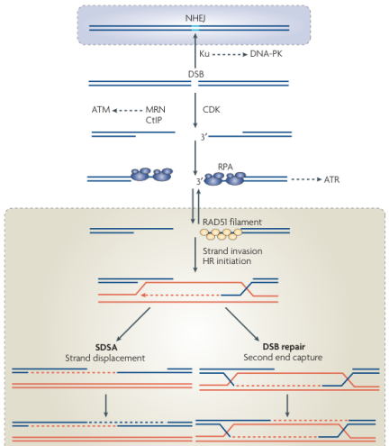
Branzei D, Foiani M. Regulation of DNA repair throughout the cell cycle. Nat Rev Mol Cell Biol. 2008;9:297–308. doi:10.1038/nrm2351.
End resection is therefore both an important early step in HR and a key mechanism linking cell cycle regulation to DSB repair pathway choice. [33]
Section summary: Repair pathway choice is not directly dictated by the cell cycle alone. It depends jointly on cell cycle-associated regulation, lesion structure, template availability, and repair factor activity. The cell cycle dependence of HR and NHEJ illustrates this regulatory network.
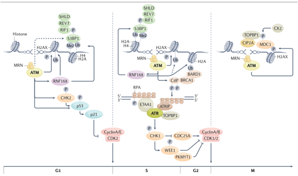
Groelly FJ, Fawkes M, Dagg RA, Blackford AN, Tarsounas M. Targeting DNA damage response pathways in cancer. Nat Rev Cancer. 2023;23:78–94. doi:10.1038/s41568-022-00535-5.
6. DNA Damage and Cell Fate
Core references for this section: 1, 4, 77.
DDR contributes not only to damage recognition and repair but also to the decision to continue proliferating.
Following DNA damage, checkpoint signaling can temporarily delay cell cycle progression while repair factors are recruited to process lesions.
If repair is effective, checkpoint signals generally subside, regulation of CDC25 and related CDKs is restored, and cells can resume progression.
After appropriate damage processing, declining checkpoint signals and active recovery mechanisms allow progression through checkpoint recovery. This differs from checkpoint adaptation, in which progression resumes despite incompletely resolved damage.
If lesions persist or cannot be repaired effectively, checkpoint-mediated arrest may fail to resolve, and cells may progress to sustained arrest, senescence, or programmed cell death. [78]
p53 and its downstream regulators are important in cell fate decisions. p53–p21 signaling primarily supports arrest, while persistent damage can activate additional proapoptotic transcriptional programs that influence entry into programmed cell death. [78]
For example, damage can stabilize p53 through ATM–CHK2 and related signaling, inducing CDKN1A expression. Its protein product, p21, inhibits cyclin–CDK activity and temporarily arrests the cycle. [78]
Cells may resume normal cycling after appropriate damage processing. If damage persists, p53 and other networks can contribute to senescence- or apoptosis-related responses. [78]
Damage responses can also have different outcomes at different cell cycle stages.
In G1, checkpoints can prevent premature replication initiation. In S phase, maintaining fork stability and processing replication-associated lesions are priorities. In G2, the G2/M checkpoint can restrict mitotic entry.
If these abnormalities remain unresolved, cells may undergo sustained arrest and ultimately senescence or apoptosis. [78]
Cell fate is not governed by a fixed rule for each phase. Outcomes depend on lesion type and severity, cell type, and the functional state of regulatory networks. [78]
Distinguishing Quiescence, Senescence, and Apoptosis
Core references for this section: 1.
Quiescence, senescence, and apoptosis can all be associated with cell cycle regulation, but they have different biological meanings. [4, 77]
Quiescence generally involves exit from the proliferative cycle into G0 and is usually reversible. It differs from the more stable arrest of senescence, and neither state is determined solely by DNA damage severity. [3, 77]
Senescence generally involves relatively stable proliferative arrest. Senescent cells usually remain metabolically active and may exhibit changes such as a senescence-associated secretory phenotype (SASP). [3]
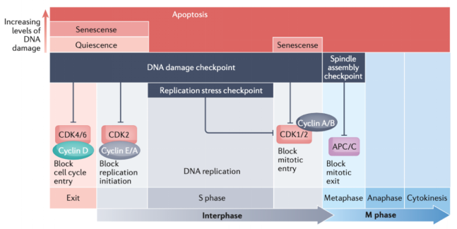
Matthews HK, Bertoli C, de Bruin RAM. Cell cycle control in cancer. Nat Rev Mol Cell Biol. 2022;23:74–88. doi:10.1038/s41580-021-00404-3.
Regulatory networks such as p53–p21 and p16–RB are closely associated with senescence. [3]
Apoptosis is a regulated form of programmed cell death. Severe or persistent DNA damage can activate relevant signaling to eliminate cells unable to maintain normal function. [4, 77]
Together, these cell fate mechanisms limit continued proliferation of abnormal cells and help reduce the accumulation and transmission of genetic errors. [4]
A single molecular marker is generally insufficient to establish senescence or another specific state across all experimental settings. Multiple indicators should therefore be assessed together. [4]
7. Cell Cycle Dysregulation and Tumorigenesis
Core references for this section: 1, 4, 79, 80.
Cell cycle dysregulation is a major feature of tumor initiation and progression.
Normal cells use multiple control mechanisms to enter the proliferative cycle under appropriate conditions and to pause progression or exit proliferation when damage or other abnormalities occur.
Cancer cells have traditionally been described as having uncontrolled cell division.
However, F2 presents a more precise interpretation: sustained cancer cell proliferation does not mean that every cell cycle checkpoint has lost function.
Instead, some cancer-associated genetic alterations mainly disrupt specific controls that restrict cycle entry or promote exit, allowing cells to initiate repeated division cycles.
7.1 Cancer-Associated Cell Cycle Dysregulation
Core references for this section: 1, 4, 81, 82, 83.
In normal cells, G1 cyclin–CDK and RB–E2F networks regulate S-phase entry, while p53-associated DNA damage checkpoints restrict inappropriate progression when damage is present.
Disruption of these mechanisms can allow continued proliferation under inappropriate conditions. [79, 80]
For example, TP53 dysfunction can weaken damage-induced arrest, senescence, or apoptosis, allowing some cells carrying genetic errors to survive and divide.
RB–E2F pathway abnormalities can dysregulate E2F-dependent transcription and promote repeated S-phase entry. Relevant changes include RB1 loss, increased CCND1 or CDK4/6 activity, and CDKN2A (p16) loss. Their types and frequencies vary across cancers and do not imply failure of all cell cycle control.
In addition to RB loss, abnormal cyclin–CDK activity and changes in upstream proliferative signaling can affect RB–E2F regulation.
Disruption of selected key controls can therefore undermine normal proliferative restraint without simultaneous failure of every checkpoint. [79, 80]
7.2 Sustained Proliferation and Dependence on Replication Stress Responses
Core references for this section: 1, 4.
Although some cancer cells lose normal restrictions on proliferation, they still need to complete DNA replication, chromosome segregation, and cell division. [84, 85]
Persistent proliferation and abnormal oncogenic signaling can increase replication stress by altering origin firing, creating replication–transcription conflicts, or reducing fork stability. [84, 85, 86, 87, 88]
These changes increase the risk of DNA damage and genome instability. [84, 85]
To prevent replication stress from becoming intolerable damage, some cancer cells become more dependent on response pathways such as ATR–CHK1. [89]
Similarly, cancer cells may tolerate some chromosome instability, but excessive segregation errors can compromise survival. [90, 91, 92, 93]
Mitotic regulatory mechanisms, including SAC, therefore remain important in some cancer cells. [90]
Cancer cells have consequently not escaped all cell cycle control. Rather, impairment of selected regulators can increase dependence on other protective mechanisms that remain functional. [90]
7.3 Cell Cycle Dysregulation as a Therapeutic Opportunity
Core references for this section: 1, 4.
This selective dysregulation creates opportunities for targeted therapy. [94]
One approach is to restrain proliferation by inhibiting abnormally activated cell cycle networks, for example by using CDK4/6 inhibition to interfere with cell cycle entry in selected cancers. [94]
Another approach exploits dependence on remaining checkpoints, further weakening the ability to cope with replication stress or chromosome segregation. [89, 95, 96]
For example, inhibition strategies targeting ATR, CHK1, or WEE1 can disrupt replication stress tolerance or checkpoint regulation in particular tumor contexts. [89, 95, 96]
Cancer therapy can therefore target both excessive proliferative signaling and the specific dependencies that sustain continued proliferation. [94]
Supplementary Table B. Cell cycle phases and repair priorities
Core references for this table: 1.
| Phase | Primary mechanisms | Interpretation |
|---|---|---|
| G1 | BER / NER / NHEJ | Repair of pre-existing lesions before replication; sister chromatids unavailable. [6] |
| S | MMR / HR / TLS / TS / ATR–CHK1 | Replication error correction and fork protection; one-ended DSBs particularly depend on HR. [4] |
| G2 | HR + NHEJ / G2–M checkpoint | Use of sister chromatids to process residual lesions. [6] |
| M | Selected repair activities restricted / SAC | Chromosome segregation and risks of inappropriate end joining. [4] |
8. Targeting the DNA Damage Response in Cancer Therapy
Core references for this section: 1, 4.
The DNA damage response is not only essential for genome stability but also a major area of anticancer drug research. [97, 98, 99]
Cancer cells frequently exhibit DNA repair defects, dysregulated cell cycle control, and elevated replication stress.
These abnormalities can promote genome instability and tumor progression while increasing dependence on the remaining repair and checkpoint pathways.
Researchers can exploit these dependencies by inhibiting repair, weakening checkpoints, or increasing replication stress to reduce cancer cell survival. [97, 98, 99]
Synthetic lethality is a central rationale for DDR-targeted therapy. [97, 98, 99]
Synthetic lethality describes a relationship in which disruption of either of two functionally connected genes or pathways can be tolerated individually, whereas their combined disruption severely impairs survival or causes cell death. [97, 98, 99]
The sensitivity of BRCA1/2-deficient tumors to PARP inhibitors is a classic example of exploiting repair defects through synthetic lethality. [98, 99]
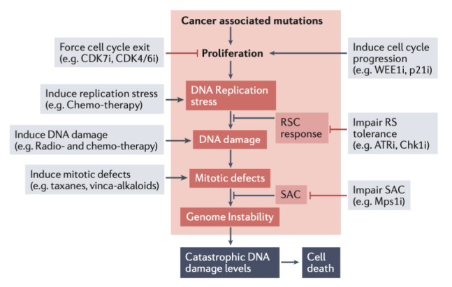
Matthews HK, Bertoli C, de Bruin RAM. Cell cycle control in cancer. Nat Rev Mol Cell Biol. 2022;23:74–88. doi:10.1038/s41580-021-00404-3.
8.1 PARP Inhibitors and Synthetic Lethality in HR-Deficient Tumors
Core references for this section: 1, 4.
PARP1 and PARP2 participate in SSB recognition and related repair processes, regulating recruitment and activity of DDR proteins through PARylation. [98, 99, 100]
PARP inhibitors suppress catalytic activity and, to varying degrees, affect interactions between PARP and DNA. [98, 99, 100]
In the classical model, PARP inhibition impairs processing of some single-strand lesions, which can be converted during replication into more severe replication-associated damage. [98, 99, 100]
In BRCA1- or BRCA2-deficient tumors, reduced HR capacity can compromise repair of these lesions and increase the risk of cell death. [98, 99, 100]
However, this classical model does not fully explain the therapeutic actions of PARP inhibitors. [98, 99, 100]
Some PARP inhibitors also cause PARP trapping, retaining PARP on DNA and generating protein–DNA complexes that obstruct replication or other DNA metabolic processes. [100, 101]
PARP inhibitors differ in their trapping capacity, which contributes to their biological effects. [100, 101]
Experimental studies have also found that PARP inhibition can cause accumulation of post-replicative ssDNA gaps, potentially influencing sensitivity in particular HR-deficient settings. This model should not be treated as a universal lethal mechanism for every drug and tumor. [102, 103, 104]
PARP inhibitor activity therefore involves several interconnected mechanisms, including impaired repair, PARP trapping, and replication-associated DNA abnormalities, rather than SSB accumulation alone. [102, 103, 104]
Representative PARP inhibitors discussed in the literature include olaparib, niraparib, rucaparib, and talazoparib. [98, 99]
Some tumors nevertheless acquire resistance through restoration of HR, altered pathway use, or changes in fork stability. [98, 99]
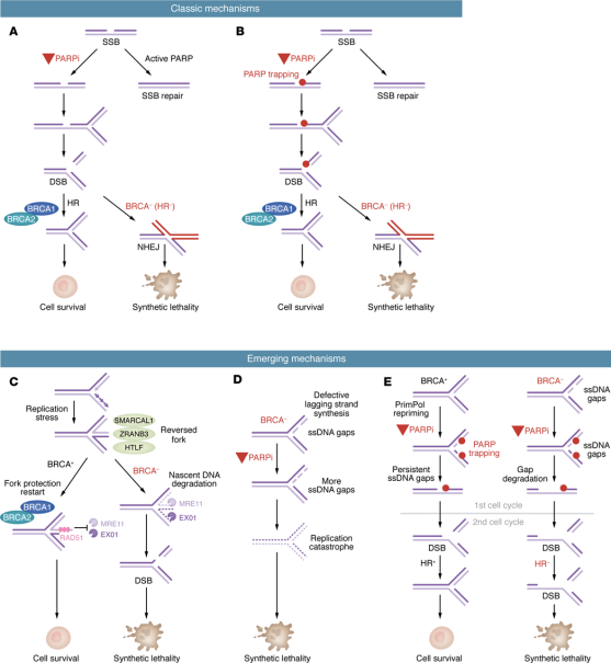
Li X, Zou L. BRCAness, DNA gaps, and gain and loss of PARP inhibitor-induced synthetic lethality. J Clin Invest. 2024;134(14):e181062. doi:10.1172/JCI181062.
8.2 ATM Inhibitors and Double-Strand Break Responses
Core references for this section: 1, 4.
ATM is a major DSB response kinase that regulates damage signaling, checkpoint activation, and repair. [105, 106, 107]
Following DSB formation, MRN promotes ATM recruitment and activation, after which ATM participates in responses involving CHK2, p53, and other downstream factors. [105, 106, 107]
ATM inhibitors aim to weaken tumor responses to DSBs, reducing tolerance of genetic damage caused by radiotherapy or certain DNA-damaging drugs. [105, 106, 107]
They are therefore commonly studied in combination with radiotherapy, chemotherapy, or other targeted treatments. [107, 108, 109, 110, 111]
Representative ATM inhibitors discussed in the literature include AZD0156 and AZD1390. [107, 108, 109, 110, 111]
Because ATM also protects normal cells from DNA damage, treatment selectivity and normal tissue toxicity are important considerations. [112, 113, 114, 115]
8.3 DNA-PK Inhibitors and Non-Homologous End Joining
Core references for this section: 1, 4.
DNA-PK is an important component of classical NHEJ. [116-120]
After a DSB, Ku70/Ku80 binds the broken ends and recruits DNA-PKcs to form DNA-PK, which contributes to end protection, processing, and ligation. [116-120]
By interfering with classical NHEJ, DNA-PKcs inhibitors may reduce the ability of cancer cells to process DSBs. [116-120]
They are consequently being investigated to enhance the antitumor effects of radiotherapy or other DNA-damaging treatments. [116-120]
Representative DNA-PKcs inhibitors discussed in the literature include AZD7648 and M3814. [119-123]
Because NHEJ is also important in normal cells, toxicity and therapeutic windows must be evaluated for each drug and treatment combination. [119-123]
8.4 ATR and CHK1 Inhibitors and Replication Stress Dependence
Core references for this section: 1, 4.
ATR–CHK1 is a major replication stress response network that regulates fork stability, origin firing, and cell cycle checkpoints. [124-128]
Many tumors experience elevated replication stress because of persistent proliferation and oncogenic abnormalities. [124-128]
To prevent this stress from progressing to severe damage, these cells may rely more heavily on ATR–CHK1 for survival. [124-128]
ATR inhibitors weaken the replication stress response, impairing stabilization and processing of abnormal replication structures and promoting accumulation of replication-associated damage. [124-128]
The literature discusses ATR inhibitors such as ceralasertib and berzosertib and their potential combinations with PARP inhibitors, DNA-damaging drugs, and other treatments. [126-128]
CHK1 is an important downstream checkpoint kinase of ATR. [126-128]
CHK1 inhibitors disrupt cell cycle regulation during replication stress and may prevent some cancer cells from effectively processing abnormal replication structures. [129-133]
Representative CHK1 inhibitors discussed in the literature include prexasertib and SRA737. [129-133]
The central rationale for targeting ATR or CHK1 is therefore to exploit reliance on replication stress protection, increasing DNA damage burden and reducing cancer cell survival. [129-133]
8.5 WEE1 and PKMYT1 Inhibitors and Checkpoint Regulation
Core references for this section: 1, 4.
WEE1 regulates replication and mitotic entry through inhibitory phosphorylation of CDK1, CDK2, and related proteins. [58, 59, 134, 135]
WEE1 inhibition can cause excessive CDK activity, increasing replication stress and DNA damage or allowing inappropriate mitotic entry. [58, 59, 134, 135]
WEE1 inhibitors can therefore exploit cancer cell dependence on checkpoints and replication stress regulation. [58, 59, 134, 135]
Adavosertib is the representative WEE1 inhibitor discussed in the literature. [58, 59, 134, 135]
PKMYT1 is a kinase functionally related to WEE1 that also contributes to inhibitory regulation of CDK1. [136]
Studies discussed in F3 found that some cyclin E-overexpressing tumors may develop a particular dependence on PKMYT1. [136]
The PKMYT1 inhibitor RP-6306 promotes abnormal CDK1 activation and showed antitumor activity in relevant experimental models. [136]
PKMYT1 inhibition is therefore an emerging research strategy that exploits dependencies created by cell cycle abnormalities. [136]
8.6 POLQ Inhibitors and Alternative DSB Repair
Core references for this section: 1, 4.
DNA polymerase theta (POLQ) participates in alternative DSB end joining, particularly processes that use short homologous sequences to align and join ends. [26-28, 137-142]
Tumors with BRCA1/2 defects and reduced HR capacity may become more dependent on selected alternative repair mechanisms. [27, 141, 142]
In this setting, POLQ inhibition can further impair remaining DSB repair capacity and produce synthetic lethality. [27, 141, 142]
Within the research context available at its publication in 2023, F3 discussed investigational POLQ-related compounds such as novobiocin, ART558, and ART812, including their potential in selected HR-deficient or PARP inhibitor-resistant models. These compounds differ in target selectivity and clinical development maturity. [143, 144]
The main potential of POLQ inhibition is therefore to target the dependence of HR-deficient tumors on backup DSB repair. [27, 141, 142]
8.7 USP1 Inhibitors, DNA Damage Tolerance, and Replication Stability
Core references for this section: 1, 4, 145-150.
USP1 is a deubiquitinating enzyme that regulates deubiquitination of PCNA and other proteins, influencing damage tolerance and replication-associated repair. [145-150]
PCNA ubiquitination is closely associated with lesion bypass and repair regulation during replication.
Studies described in F3 found that USP1 inhibition can cause persistent accumulation of monoubiquitinated PCNA, affect fork stability, and increase dependence on BRCA1-associated protection in some cancer cells.
USP1 inhibition may therefore be synthetically lethal with BRCA1 deficiency.
The investigational USP1 inhibitor KSQ-4279 described in F3 showed antitumor activity in selected experimental models available at that time and potential for combination studies with PARP inhibitors. These findings should not be extrapolated directly to established clinical efficacy. [151]
USP1 should not be classified simply as a specific DNA repair enzyme. Its therapeutic relevance primarily derives from regulation of repair-associated ubiquitination and replication stability.
8.8 RAD51 Inhibition Strategies and Homologous Recombination
Core references for this section: 1, 4.
RAD51 is a central HR recombinase that forms nucleoprotein filaments on ssDNA and promotes homology searching and strand invasion. [152-155]
Disrupting RAD51 function can therefore weaken HR and reduce the ability of cancer cells to cope with DSBs and replication-associated damage. [152-155]
Research strategies include interfering with filament formation, DNA binding, and other recombination activities. [152-155]
F3 discussed candidate compounds such as CYT-0851 in the context of RAD51-related therapeutic strategies. [155-157]
However, the direct molecular target of CYT-0851 cannot be established solely from its historical description in that review. It should not be presented as a confirmed selective direct RAD51 inhibitor. [155-157]
Because RAD51 is also essential for HR in normal cells, improving therapeutic selectivity remains a major challenge. [152-155]
8.9 WRN Inhibition and Dependencies in MSI Tumors
Core references for this section: 1, 4.
WRN is a DNA helicase involved in processing particular DNA structures and maintaining genome stability. [158-160]
Microsatellite instability (MSI) is commonly associated with defective MMR. [158-160]
Accumulation of repetitive sequences and abnormal DNA structures can make some MSI cancer cells particularly dependent on WRN helicase activity. [161-163]
WRN inhibition may prevent appropriate processing of these structures, leading to DNA breakage, chromosome fragmentation, and cell death. [161-163]
WRN is therefore considered a potential synthetic lethal target in selected MSI tumors. [161-163]
Unlike the relationship between BRCA1/2 deficiency and PARP inhibition, WRN-directed strategies primarily exploit vulnerabilities associated with MMR deficiency and MSI. [161-163]
8.10 Other Cell Cycle-Related Therapeutic Targets
Core references for this section: 1, 4.
In addition to proteins directly involved in DDR and repair, selected cell cycle regulators are important anticancer targets.
- CDK4/6: These kinases regulate G1 progression. Their inhibitors can restrain abnormal proliferation by restricting S-phase entry in selected tumors.
- CDK7: This kinase regulates cell cycle CDKs and transcription. Its inhibition can therefore affect both cycle progression and selected transcriptional dependencies.
- MPS1/TTK: MPS1 is a key SAC regulator. Inhibition may weaken surveillance of chromosome segregation errors and increase chromosome instability beyond tolerable levels in some cancer cells.
These targets primarily concern cell cycle and mitotic regulation and should not be grouped indiscriminately with enzymes that directly repair DNA damage.
8.11 Challenges and Future Directions in DDR-Targeted Therapy
Core references for this section: 1, 4.
Despite substantial progress, DDR-targeted therapies face challenges including resistance, normal tissue toxicity, and patient selection. [101, 164, 165]
First, tumors can acquire resistance by restoring repair capacity, changing pathway use, or adjusting replication stress responses. [101, 164, 165]
For example, restoration of HR in some BRCA1/2-deficient tumors can reduce PARP inhibitor sensitivity. [165, 166]
Second, repair and cell cycle control are also essential in normal cells. Inhibiting DDR proteins can therefore affect normal tissues and limit some treatment strategies. [167-169]
Furthermore, tumors differ in repair defects, replication stress levels, and genetic background, so a particular DDR inhibitor strategy will not be suitable for every patient. [167, 170-173]
Identifying predictive biomarkers and developing combinations with a clear molecular rationale are therefore important research priorities. [167, 170-173]
Future research also includes discovering new synthetic lethal relationships and rationally combining DDR inhibitors with radiotherapy, chemotherapy, other targeted agents, or immunotherapy. [167-169]
Supplementary Table C. Established and emerging DDR targets as of 2023
Core references for this table: 1, 4.
| Target | Candidate agents or strategies | Rationale |
|---|---|---|
| PARP1/2 | Olaparib; Niraparib; Rucaparib; Talazoparib | Clinical applications established; exploits HR deficiency-associated vulnerability |
| ATM | AZD0156; AZD1390 | DSB response signaling |
| DNA-PKcs | AZD7648; M3814 | Classical NHEJ |
| ATR | Ceralasertib; Berzosertib | Replication stress |
| CHK1 | Prexasertib; SRA737 | Replication checkpoint |
| WEE1 | Adavosertib | CDK regulation |
| PKMYT1 | RP-6306 | Cyclin E-associated dependence |
| POLQ | Novobiocin; ART558; ART812 | Investigational strategy; POLQ-associated alternative end joining. Compound selectivity requires verification. |
| USP1 | KSQ-4279 | Investigational strategy; PCNA deubiquitination and replication stress dependence |
| RAD51 | Experimental RAD51-related strategies; CYT-0851 was discussed historically and is not a confirmed direct inhibitor | Investigational HR intervention; direct target assignment remains unconfirmed |
| WRN | Target-based research direction | Investigational strategy; MSI-associated dependence |
9. Markers of DNA Damage and Repair
EnkiLife offers a broad range of high-quality antibodies to support the detection of DNA damage and repair. Selected related products are listed below.
9.1 Markers of DNA Damage and the DNA Damage Response
| Product name | Catalog number | Reactivity | Applications |
|---|---|---|---|
| Damage recognition, replication stress, and checkpoints — 26 products | |||
| 53BP1 Rabbit Monoclonal Antibody | AMRe86743 | Human,Mouse,Rat | WB,IHC,ICC/IF,FC |
| 53BP1 Rabbit Monoclonal Antibody | AMRe87074 | Human | WB,IHC-P,ICC/IF |
| 53BP1 Rabbit Polyclonal Antibody | APRab06336 | Human,Mouse,Rat | WB,IHC-P,IF-P,IF-F,ICC/IF,ELISA |
| ATM Rabbit Monoclonal Antibody | AMRe01480 | Human | WB,ICC/IF |
| ATR Rabbit Polyclonal Antibody | APRab07352 | Human,Rat,Mouse | WB,IHC-P,IF-P,IF-F,ICC/IF,ELISA |
| Chk1 Rabbit Monoclonal Antibody | AMRe86205 | Human | WB,IHC,ICC/IF,FC |
| Chk1 Rabbit Polyclonal Antibody | APRab08763 | Human,Mouse,Rat | WB,IHC-P,IF-P,IF-F,ICC/IF,ELISA |
| Chk2 Rabbit Monoclonal Antibody | AMRe01822 | Human | WB,ICC/IF,IP |
| Chk2 Rabbit Polyclonal Antibody | APRab08765 | Human,Mouse,Rat | WB,IHC-P,IF-P,IF-F,ICC/IF,ELISA |
| Histone H2A.X Rabbit Monoclonal Antibody | AMRe85652 | Human,Mouse,Rat | WB,IHC,ICC,IP |
| Phospho-53BP1 (phospho-Ser1778) Rabbit Polyclonal Antibody | APRab04190 | Human,Rat,Mouse | WB |
| Phospho-53BP1 (Ser6) Rabbit Polyclonal Antibody | APRab04188 | Human,Mouse,Rat,Monkey | WB,IHC,ICC/IF,ELISA |
| Phospho-ATM (S1981) (3F17) Rabbit Monoclonal Antibody | AMRe05856 | Human | WB,IHC-P,FC,IP,IF-P |
| Phospho-Atm (Ser1981) Rabbit Polyclonal Antibody | APRab04283 | Human,Mouse,Rat | WB,IHC-P,IF-P,IF-F,ICC/IF,ELISA |
| Phospho-ATR (Ser428) Rabbit Polyclonal Antibody | APRab04285 | Human,Mouse,Rat | IHC,ICC/IF,ELISA |
| Phospho-Chk1 (Ser280) Rabbit Monoclonal Antibody | AMRe87433 | Human | WB |
| Phospho-Chk1 (Ser286) Rabbit Polyclonal Antibody | APRab04452 | Human,Mouse,Rat | WB,IHC,ICC/IF,ELISA |
| Phospho-Chk1 (Ser301) Rabbit Polyclonal Antibody | APRab04454 | Human,Mouse,Rat | WB,ICC/IF,ELISA |
| Phospho-Chk1 (Ser345) Rabbit Polyclonal Antibody | APRab04456 | Human,Mouse,Rat | IHC-P,IF-P,IF-F,ICC/IF,ELISA |
| Phospho-Chk2 (Thr68) Rabbit Monoclonal Antibody | AMRe21000 | Human,Mouse,Rat | WB,ICC/IF,ELISA,IP |
| Phospho-Histone H2A.X (Ser139) Rabbit Monoclonal Antibody | AMRe03773 | Human,Mouse,Rat | WB,IP |
| Phospho-Histone H2A.X (Ser139) Rabbit Monoclonal Antibody | AMRe21478 | Human,Mouse,Rat,Hamster | WB,IHC,IF,IP,ELISA |
| Phospho-Histone H2A.X (Ser139) (2A9) Mouse Monoclonal Antibody | AMM03679 | Human,Mouse | WB,ICC/IF |
| Phospho-p95/NBS1 (S343) (8Q6) Rabbit Monoclonal Antibody | AMRe05962 | Human | WB,ICC/IF |
| RPA32 Rabbit Monoclonal antibody | AMRe02549 | Human,Mouse,Rat,Hamster | WB,IHC,IP |
| RPA32/RPA2 Rabbit Monoclonal Antibody | AMRe21449 | Human,Mouse,Rat | WB,IHC,IF,IP,ELISA |
Important: γ-H2AX is not an absolutely specific marker of DSBs; 53BP1 foci do not directly demonstrate completed NHEJ; and p-CHK1 alone does not establish that replication forks have recovered.
9.2 Markers Associated with DNA Repair Pathways
| Product name | Catalog number | Reactivity | Applications |
|---|---|---|---|
| SSBR / BER — 8 products | |||
| APE1 Rabbit Monoclonal Antibody | AMRe01660 | Human,Mouse,Rat | WB,IHC,ICC/IF |
| APE1 Rabbit Monoclonal Antibody | AMRe85294 | Human,Mouse,Rat | WB,IHC,ICC |
| KD-Validated APE1 Recombinant Rabbit Monoclonal Antibody | KVAb01320 | Human,Mouse,Rat | WB,FCM,ICC |
| Ogg1 Rabbit Monoclonal Antibody | AMRe86232 | Human | WB |
| PARP Mouse Monoclonal Antibody | AMM80824 | Human | WB,FC,ELISA |
| PARP1 Rabbit Monoclonal Antibody | AMRe02401 | Human,Mouse,Rat | WB,IHC,ICC/IF |
| PARP2 Rabbit Monoclonal Antibody | AMRe84198 | Human,Mouse,Rat | WB,ICC,FC |
| XRCC1 Rabbit Polyclonal Antibody | APRab19971 | Human,Mouse,Rat | WB,IHC-P,IF-P,IF-F,ICC/IF,ELISA |
| NER — 2 products | |||
| ERCC1 Mouse Monoclonal Antibody | AMM82574 | Human,Mouse | WB,IHC,ICC,FC,ELISA |
| XPA Rabbit Polyclonal Antibody | APRab19958 | Human,Mouse | WB,IHC-P,IF-P,IF-F,ICC/IF,ELISA |
| MMR — 1 product | |||
| MSH2 Mouse Monoclonal Antibody | AMM22189 | Human | WB,IHC-P |
| HR — 6 products | |||
| BRCA1 Rabbit Polyclonal Antibody | APRab07642 | Human,Rat | WB,IHC-P,IF-P,IF-F,ICC/IF,ELISA |
| BRCA2 Rabbit Polyclonal Antibody | APRab07643 | Human,Rat | IHC-P,IF-P,IF-F,ICC/IF,ELISA |
| CtIP Rabbit Polyclonal Antibody | APRab09504 | Human,Mouse | IHC-P,IF-P,IF-F,ICC/IF,WB,ELISA |
| Rad51 Rabbit Monoclonal Antibody | AMRe03177 | Human | WB,IHC-F,IHC-P,ICC/IF |
| Rad51 Rabbit Monoclonal Antibody | AMRe03792 | Human,Mouse,Rat | WB,IF,IP,ELISA |
| Rad54 Rabbit Polyclonal Antibody | APRab16849 | Human,Mouse | WB,IHC-P,IF-P,IF-F,ICC/IF,ELISA |
| NHEJ — 8 products | |||
| Artemis Rabbit Polyclonal Antibody | APRab07177 | Human,Mouse,Rat | IHC-P,IF-P,IF-F,ICC/IF,ELISA |
| DNA Ligase IV Rabbit Polyclonal Antibody | APRab10051 | Human,Mouse,Rat | WB,IHC-P,IF-P,IF-F,ICC/IF,ELISA |
| DNA PKcs Rabbit Monoclonal Antibody | AMRe86303 | Human,Mouse,Rat | WB,IHC-P,ICC/IF |
| DNA PKcs Mouse Monoclonal Antibody | AMM85052 | Human | WB,IP,IHC,ICC |
| Ku70 Rabbit Monoclonal Antibody | AMRe03061 | Human | WB,IHC-F,IHC-P,ICC/IF |
| Ku80 Rabbit Monoclonal Antibody | AMRe87254 | Human | WB,IHC-P,FC |
| Phospho-DNA-PKCS (Ser2056) Rabbit Polyclonal Antibody | APRab04552 | Human,Mouse | WB,IHC-P,IF-P,IF-F,ICC/IF,ELISA |
| XLF Rabbit Monoclonal Antibody | AMRe84031 | Human,Mouse,Rat | WB,IHC,ICC,FC,IF |
| Replication and damage tolerance — 2 products | |||
| PCNA Rabbit Monoclonal Antibody | AMRe21497 | Human,Mouse,Rat | WB,IHC,IF,IP,ELISA |
| PCNA Rabbit Monoclonal Antibody | AMRe86349 | Human,Mouse,Rat | WB,IHC-P,ICC/IF,FC,IP |
9.3 Antibodies for Cell Cycle Regulation, Proliferation, and Cell Fate
| Product name | Catalog number | Reactivity | Applications |
|---|---|---|---|
| Mitosis and cell cycle regulation — 24 products | |||
| Aurora A Rabbit Monoclonal Antibody | AMRe21613 | Human | WB,IHC,IF,IP,ELISA |
| Aurora B Rabbit Monoclonal Antibody | AMRe21394 | Human | WB,IHC,IF,IP,ELISA |
| Cdc2 Rabbit Monoclonal Antibody | AMRe21356 | Human,Mouse | WB,IHC,IF,IP,ELISA |
| CDK2 Rabbit Monoclonal Antibody | AMRe04136 | Human,Rat,Hamster | WB,IHC-F,IHC-P,ICC/IF,IP |
| CDK4 Rabbit Monoclonal Antibody | AMRe01808 | Human,Mouse,Rat | WB,IHC-F,IHC-P,ICC/IF,IP |
| CDK6 Rabbit Monoclonal Antibody | AMRe03765 | Human | WB,ICC/IF |
| CDKN2A/p14ARF Rabbit Monoclonal Antibody | AMRe21576 | Human | WB,IF,IP,ELISA |
| CDKN2A/p16INK4a Rabbit Monoclonal Antibody | AMRe87632 | Human | WB,IHC-P,FC,IP |
| CDKN2B/p15 Rabbit Polyclonal Antibody | APRab15574 | Human,Mouse,Rat | WB,IHC-P,IF-P,IF-F,ICC/IF,ELISA |
| CDT1 Rabbit Monoclonal Antibody | AMRe86448 | Human | WB,IHC-P,ICC/IF,IP |
| Cyclin A2 Rabbit Monoclonal Antibody | AMRe87689 | Human,Mouse,Rat | WB,IHC-P,ICC/IF |
| Cyclin B1 Rabbit Monoclonal Antibody | AMRe85222 | Human | WB,IP,ICC |
| Cyclin D1 Rabbit Monoclonal Antibody | AMRe21529 | Human,Mouse,Rat | WB,IHC,IF,IP,ELISA |
| Cyclin E1 Rabbit Monoclonal Antibody | AMRe86268 | Human | WB,IHC-P,ICC/IF,FC,IP |
| Cyclin H Rabbit Monoclonal Antibody | AMRe02961 | Human,Mouse | WB,ICC/IF,IP |
| Geminin Rabbit Monoclonal Antibody | AMRe01408 | Human | WB |
| Histone H3 Rabbit Monoclonal Antibody | AMRe21336 | Human,Mouse,Rat,Cat | WB,IHC,IF,IP,ELISA,CHIP,Cut&Tag |
| Phospho-Cdc2 (Thr161) Rabbit Polyclonal Antibody | APRab04412 | Human,Mouse,Rat | WB,IHC,ICC/IF,ELISA |
| Phospho-Cdc2 (Tyr15) Rabbit Polyclonal Antibody | APRab04413 | Human,Mouse,Rat,Monkey | WB,ELISA |
| Phospho-Histone H3 (Thr3) Rabbit Monoclonal Antibody | AMRe21090 | Human,Mouse,Rat | WB,IHC,IF,IP,ELISA,CHIP,Cut&Tag |
| Phospho-Plk1 (Ser137) Rabbit Polyclonal Antibody | APRab05292 | Human,Mouse,Rat | WB,IHC,ICC/IF,ELISA |
| Phospho-Wee1 (Ser642) Rabbit Polyclonal Antibody | APRab05631 | Human,Mouse,Rat | WB,ELISA |
| PLK1 Rabbit Monoclonal Antibody | AMRe21126 | Human,Mouse,Rat | WB,IHC,IF,IP,ELISA |
| Wee 1 Rabbit Polyclonal Antibody | APRab19893 | Human,Mouse,Rat | WB,ELISA |
| Proliferation, tumor suppression, and cell fate — 14 products | |||
| Ki-67 Rabbit Polyclonal Antibody | APRab13003 | Human | IHC-P,IF-P,IF-F,ICC/IF,ELISA |
| Ki67 Rabbit Monoclonal Antibody | AMRe86305 | Human | WB,IHC,ICC/IF,FC |
| KI67 Mouse Monoclonal Antibody | AMM80747 | Human,Mouse,Rat,Rabbit,Monkey | IHC,ICC,ELISA |
| MDM2 Rabbit Monoclonal Antibody | AMRe21162 | Human,Mouse,Rat | WB,IHC,IF,IP,ELISA |
| MDM2 Rabbit Polyclonal Antibody | APRab13759 | Human,Mouse,Monkey | WB,IHC-P,IF-P,IF-F,ICC/IF,ELISA |
| p21 Rabbit Monoclonal Antibody | AMRe86606 | Human | WB,IHC-P,ICC/IF,FC,IP |
| p27 Kip 1 Rabbit Monoclonal Antibody | AMRe02382 | Human,Mouse,Rat | WB,IHC,ICC/IF,FC,IP |
| p53 Rabbit Monoclonal Antibody | AMRe87631 | Human | WB,IHC-P,ICC/IF,FC,IP |
| p63 Rabbit Monoclonal Antibody | AMRe86214 | Human,Mouse,Rat | WB,IHC-P,ICC/IF,FC,IP |
| p63 Rabbit Polyclonal Antibody | APRab15660 | Human,Mouse,Rat,Monkey | WB,IHC-P,IF-P,IF-F,ICC/IF,ELISA |
| p73 Rabbit Monoclonal Antibody | AMRe03974 | Human,Mouse | WB,IHC-F,IHC-P,ICC/IF,FC |
| Phospho-MDM2 (Ser166) Rabbit Monoclonal Antibody | AMRe01553 | Human,Rat | WB,IHC-P |
| Phospho-p53 (Ser15) Rabbit Polyclonal Antibody | APRab00821 | Human,Rat | WB,IHC-F,IHC-P,ICC/IF,IP,ELISA |
| Phospho-p53 (Ser20) Rabbit Polyclonal Antibody | APRab05164 | Human,Mouse,Rat,Monkey | WB,IHC,ICC/IF,ELISA |
10. Conclusions and Research Applications
Core references for this section: 1, 3, 4, 6.
Cell cycle regulation, DDR, and DNA repair together provide essential protection for genome stability.
Cell cycle networks ensure that replication, chromosome segregation, and division occur in the appropriate order. DDR networks coordinate checkpoints, repair, and cell fate decisions when genetic material is damaged or replication becomes abnormal.
Repair pathway choice depends not only on lesion type but also on cell cycle phase, homologous template availability, chromatin state, and repair factor activity. [32, 33]
For example, sister chromatids are usually unavailable in G1, making NHEJ important at this stage. HR can use sister chromatids in S and G2, while one-ended DSBs caused by fork collapse generally depend more strongly on HR-related processing. [32, 33]
ATM, ATR, and other DDR regulators convert damage or replication abnormalities into checkpoint signals, restricting inappropriate progression and providing opportunities for repair.
Cells may resume cycling after effective damage processing. Persistent or irreparable damage can instead lead to prolonged arrest, senescence, or apoptosis.
In cancer, selected defects in cell cycle control or repair can promote genome instability and abnormal sustained proliferation.
Cancer cells do not necessarily lose all cell cycle control. Instead, impairment of particular protective mechanisms may increase reliance on other repair and checkpoint pathways that remain active.
This combination of selective dysregulation and dependence on remaining pathways provides a molecular basis for PARP inhibitors and other DDR-targeted strategies.
Understanding the interplay between the cell cycle, DDR, and repair therefore helps explain genome maintenance and tumorigenesis and provides a theoretical basis for new treatment strategies and experimental research.
Experimental Markers and Their Interpretation
Studies of DNA damage and repair should distinguish three levels of evidence:
First, evidence of DNA damage.
For example, γ-H2AX and 53BP1 foci reveal damage-associated responses, but γ-H2AX is not absolutely specific for DSBs and 53BP1 foci do not establish successful NHEJ. Cell cycle stratification, post-treatment time courses, and functional repair assays should be combined to distinguish damage signaling, lesion resolution, and repair capacity.
Second, activation of DDR signaling.
Markers such as p-ATM, p-CHK1, and p-CHK2 help assess checkpoint signaling, but their presence does not mean repair is complete.
Third, functional completion of DNA repair.
RAD51 foci, for example, indicate recruitment of HR-associated proteins but do not by themselves demonstrate successful HR. [4, 170, 171, 172, 173]
Experiments should therefore be interpreted in light of the research question, damage induction conditions, treatment duration, cell cycle state, and appropriate functional repair assays.
| Marker | Main interpretation | Limitations |
|---|---|---|
| γ-H2AX (H2A.X Ser139) | Damage-associated chromatin response | Indicates damage signaling; not absolutely DSB-specific and cannot directly count breaks |
| 53BP1 foci | Recruitment of proteins to damage sites | Indicates recruitment; does not establish successful NHEJ |
| p-ATM (Ser1981) | ATM-related signaling | Reflects kinase-related signaling; does not directly quantify DNA breaks |
| p-CHK2 (Thr68) | Checkpoint signaling | Reflects checkpoint signaling only; does not establish completed repair |
| p-ATR (Thr1989) | ATR-related responses | Depends on cell state; interpret in the context of replication stress |
| p-CHK1 (Ser345) | ATR–CHK1-related signaling | Does not establish fork recovery; time-course analysis is recommended |
| RPA / p-RPA32 | ssDNA exposure and replication stress | Affected by multiple DNA metabolic processes; consider cell cycle phase |
| RAD51 foci | HR-associated protein recruitment | Indicates recruitment rather than completed functional HR |
| p-DNA-PKcs (Ser2056) | DNA-PK-related responses | Does not measure NHEJ efficiency or accuracy |
| FANCD2 monoubiquitination | FA pathway activation | Can indicate pathway activation; does not establish ICL removal |
References
References are numbered in order of first citation in the text. Supplementary references from the original manuscript that are not directly cited are listed separately below.
Citation convention: Unless otherwise stated, references listed under “Core references for this section” support all text in the corresponding section.
1. Matthews HK, Bertoli C, de Bruin RAM. Cell cycle control in cancer. Nat Rev Mol Cell Biol. 2022;23:74–88. doi:10.1038/s41580-021-00404-3.
2. Bertoli, C., Skotheim, J. M. & De Bruin, R. A. M. Control of cell cycle transcription during G1 and S phases. Nat. Rev. Mol. Cell Biol. 14, (2013).
3. Cell Signaling Technology. Cell Cycle and DNA Damage and Repair. Technical Brochure. 2022. Publication no. 20-CEP-39959-EN-MAR22.
4. Groelly FJ, Fawkes M, Dagg RA, Blackford AN, Tarsounas M. Targeting DNA damage response pathways in cancer. Nat Rev Cancer. 2023;23:78–94. doi:10.1038/s41568-022-00535-5.
5. Crncec, A. & Hochegger, H. Triggering mitosis. FEBS Letters (2019).
6. Branzei D, Foiani M. Regulation of DNA repair throughout the cell cycle. Nat Rev Mol Cell Biol. 2008;9:297–308. doi:10.1038/nrm2351.
7. Sancar A, Lindsey-Boltz LA, Unsal-Kacmaz K, Linn S. Molecular mechanisms of mammalian DNA repair and the DNA damage checkpoints. Annu Rev Biochem. 2004;73:39–85.
8. Shibutani S, Takeshita M, Grollman AP. Insertion of specific bases during DNA synthesis past the oxidation-damaged base 8-oxodG. Nature. 1991;349:431–434.
9. Russo MT, De Luca G, Degan P, et al. Accumulation of the oxidative base lesion 8-hydroxyguanine in DNA of tumor-prone mice defective in both the Myh and Ogg1 DNA glycosylases. Cancer Res. 2004;64:4411–4414.
10. Kinner A, Wu W, Staudt C, Iliakis G. Gamma-H2AX in recognition and signaling of DNA double-strand breaks in the context of chromatin. Nucleic Acids Res. 2008;36(17):5678–5694. doi:10.1093/nar/gkn550.
11. Campalans A, Kortulewski T, Amouroux R, et al. Distinct spatiotemporal patterns and PARP dependence of XRCC1 recruitment to single-strand break and base excision repair. Nucleic Acids Res. 2013;41(5):3115–3129. doi:10.1093/nar/gkt025.
12. Rack JGM, Liu Q, Zorzini V, et al. Mechanistic insights into the three steps of poly(ADP-ribosylation) reversal. Nat Commun. 2021;12:4581. doi:10.1038/s41467-021-24723-3.
13. Kim YJ, Wilson DM III. Overview of base excision repair biochemistry. Curr Mol Pharmacol. 2012;5(1):3–13. doi:10.2174/1874467211205010003.
14. Gohil D, Sarker AH, Roy R. Base excision repair: mechanisms and impact in biology, disease, and medicine. Int J Mol Sci. 2023;24(18):14186. doi:10.3390/ijms241814186.
15. Almeida KH, Sobol RW. A unified view of base excision repair: lesion-dependent protein complexes regulated by post-translational modification. DNA Repair (Amst). 2007;6(6):695–711. doi:10.1016/j.dnarep.2007.01.009.
16. Jiricny J. The multifaceted mismatch-repair system. Nat Rev Mol Cell Biol. 2006;7:335–346.
17. Critchlow, S. E., Bowater, R. P. & Jackson, S. P. Mammalian DNA double-strand break repair protein XRCC4 interacts with DNA ligase IV. Curr. Biol. 7, 588–598 (1997).
18. Grawunder, U. et al. Activity of DNA ligase IV stimulated by complex formation with XRCC4 protein in mammalian cells. Nature 388, 492–495 (1997).
19. Walker JR, Corpina RA, Goldberg J. Structure of the Ku heterodimer bound to DNA and its implications for double-strand break repair. Nature. 2001;412:607–614.
20. Graham, T. G. W., Walter, J. C. & Loparo, J. J. Two-stage synapsis of DNA ends during non-homologous end joining. Mol. Cell 61, 850–858 (2016).
21. Dobbs, T. A., Tainer, J. A. & Lees-Miller, S. P. A structural model for regulation of NHEJ by DNA-PKcs autophosphorylation. DNA Repair 9, 1307–1314 (2010).
22. Hammel, M. et al. Ku and DNA-dependent protein kinase dynamic conformations and assembly regulate DNA binding and the initial non-homologous end joining complex. J. Biol. Chem. 285, 1414–1423 (2010).
23. Zhao B, Rothenberg E, Ramsden DA, Lieber MR. The molecular basis and disease relevance of non-homologous DNA end joining. Nat Rev Mol Cell Biol. 2020;21:765–781.
24. Ahnesorg, P., Smith, P. & Jackson, S. P. XLF interacts with the XRCC4-DNA ligase IV complex to promote DNA nonhomologous end-joining. Cell 124, 301–313 (2006).
25. Semlow DR, Walter JC. Mechanisms of vertebrate DNA interstrand cross-link repair. Annu Rev Biochem. 2021;90:107–135. doi:10.1146/annurev-biochem-080320-112510.
26. Ramsden DA, Carvajal-Garcia J, Gupta GP. Mechanism, cellular functions and cancer roles of polymerase-theta-mediated DNA end joining. Nat Rev Mol Cell Biol. 2022;23:125–140.
27. Ceccaldi R, Liu JC, Amunugama R, et al. Homologous-recombination-deficient tumours are dependent on Poltheta-mediated repair. Nature. 2015;518:258–262.
28. Kent T, Chandramouly G, McDevitt SM, Ozdemir AY, Pomerantz RT. Mechanism of microhomology-mediated end-joining promoted by human DNA polymerase θ. Nat Struct Mol Biol. 2015;22:230–237.
29. Lehmann AR, Niimi A, Ogi T, et al. Translesion synthesis: Y-family polymerases and the polymerase switch. DNA Repair (Amst). 2007;6:891–899.
30. Branzei D, Foiani M. Interplay of replication checkpoints and repair proteins at stalled replication forks. DNA Repair (Amst). 2007;6:994–1003.
31. Hoege C, Pfander B, Moldovan GL, Pyrowolakis G, Jentsch S. RAD6-dependent DNA repair is linked to modification of PCNA by ubiquitin and SUMO. Nature. 2002;419:135–141.
32. Sonoda E, Hochegger H, Saberi A, Taniguchi Y, Takeda S. Differential usage of non-homologous end-joining and homologous recombination in double strand break repair. DNA Repair (Amst). 2006;5:1021–1029.
33. Ira G, Pellicioli A, Balijja A, et al. DNA end resection, homologous recombination and DNA damage checkpoint activation require CDK1. Nature. 2004;431:1011–1017.
34. Jazayeri A, Falck J, Lukas C, et al. ATM- and cell cycle-dependent regulation of ATR in response to DNA double-strand breaks. Nat Cell Biol. 2006;8:37–45.
35. Karanam K, Kafri R, Loewer A, Lahav G. Quantitative live cell imaging reveals a gradual shift between DNA repair mechanisms and a maximal use of HR in mid S phase. Mol Cell. 2012;47:320–329.
36. Balmus G, Pilger D, Coates J, et al. ATM orchestrates the DNA-damage response to counter toxic non-homologous end-joining at broken replication forks. Nat Commun. 2019;10:87.
37. Kastan MB, Bartek J. Cell-cycle checkpoints and cancer. Nature. 2004;432:316–323.
38. Unal E, Heidinger-Pauli JM, Koshland D. DNA double-strand breaks trigger genome-wide sister-chromatid cohesion through Eco1 (Ctf7). Science. 2007;317:245–248.
39. Abraham, R. T. Cell cycle checkpoint signaling through the ATM and ATR kinases. Genes and Development (2001).
40. Jackson, S. P. & Bartek, J. The DNA-damage response in human biology and disease. Nature (2009).
41. McGowan, C. H. & Russell, P. The DNA damage response: Sensing and signaling. Current Opinion in Cell Biology (2004).
42. Lopes, M. et al. The DNA replication checkpoint response stabilizes stalled replication forks. Nature (2001).
43. Feijoo, C. et al. Activation of mammalian Chk1 during DNA replication arrest: A role for Chk1 in the intra-S phase checkpoint monitoring replication origin firing. J. Cell Biol. (2001).
44. Tercero, J. A. & Diffley, J. F. X. Regulation of DNA replication fork progression through damaged DNA by the Mec1/Rad53 checkpoint. Nature 412, 553–557 (2001).
45. Zachos, G., Rainey, M. D. & Gillespie, D. A. F. Chk1-deficient tumour cells are viable but exhibit multiple checkpoint and survival defects. EMBO J. (2003).
46. Petermann, E. et al. Chk1 requirement for high global rates of replication fork progression during normal vertebrate S phase. Mol. Cell. Biol. 26, 3319–3326 (2006).
47. Katsuno, Y. et al. Cyclin A–Cdk1 regulates the origin firing program in mammalian cells. Proc. Natl. Acad. Sci. 106, 3184 LP – 3189 (2009).
48. Petermann, E., Woodcock, M. & Helleday, T. Chk1 promotes replication fork progression by controlling replication initiation. Proc. Natl. Acad. Sci. U. S. A. (2010).
49. Conti, C., Seiler, J. A. & Pommier, Y. The mammalian DNA replication elongation checkpoint: Implication of Chk1 and relationship with origin firing as determined by single DNA molecule and single cell analyses. Cell Cycle (2007).
50. Hirano T. At the heart of the chromosome: SMC proteins in action. Nat Rev Mol Cell Biol. 2006;7:311–322.
51. Uhlmann F, Nasmyth K. Cohesion between sister chromatids must be established during DNA replication. Curr Biol. 1998;8:1095–1101.
52. Ström L, Lindroos HB, Shirahige K, Sjögren C. Postreplicative formation of cohesion is required for repair and induced by a single DNA break. Science. 2007;317:242–245.
53. Limbo O, Chahwan C, Yamada Y, de Bruin RAM, Wittenberg C, Russell P. Ctp1 is a cell-cycle-regulated protein that functions with Mre11 complex to control double-strand break repair by homologous recombination. Mol Cell. 2007;28:134–146.
54. Sartori AA, Lukas C, Coates J, et al. Human CtIP promotes DNA end resection. Nature. 2007;450:509–514.
55. Mailand, N. et al. Rapid destruction of human Cdc25A in response to DNA damage. Science 288, 1425–1429 (2000).
56. Peng, C. Y. et al. Mitotic and G2 checkpoint control: regulation of 14-3-3 protein binding by phosphorylation of Cdc25C on serine-216. Science 277, 1501–1505 (1997).
57. Sanchez, Y. et al. Conservation of the Chk1 checkpoint pathway in mammals: linkage of DNA damage to Cdk regulation through Cdc25. Science 277, 1497–1501 (1997).
58. Heald, R., McLoughlin, M. & McKeon, F. Human wee1 maintains mitotic timing by protecting the nucleus from cytoplasmically activated Cdc2 kinase. Cell 74, 463–474 (1993).
59. Gu, Y., Rosenblatt, J. & Morgan, D. O. Cell cycle regulation of CDK2 activity by phosphorylation of Thr160 and Tyr15. EMBO J. 11, 3995–4005 (1992).
60. Booher, R. N., Holman, P. S. & Fattaey, A. Human Myt1 is a cell cycle-regulated kinase that inhibits Cdc2 but not Cdk2 activity. J. Biol. Chem. 272, 22300–22306 (1997).
61. Liu, F., Stanton, J. J., Wu, Z. & Piwnica-Worms, H. The human Myt1 kinase preferentially phosphorylates Cdc2 on threonine 14 and localizes to the endoplasmic reticulum and Golgi complex. Mol. Cell Biol. 17, 571–583 (1997).
62. Lecona, E. & Fernandez-Capetillo, O. Targeting ATR in cancer. Nat. Rev. Cancer 18, 586–595 (2018).
63. Blackford AN, Stucki M. How cells respond to DNA breaks in mitosis. Trends Biochem Sci. 2020;45:321–331. doi:10.1016/j.tibs.2019.12.010.
64. Leimbacher PA, Jones SE, Shorrocks AK, et al. MDC1 interacts with TOPBP1 to maintain chromosomal stability during mitosis. Mol Cell. 2019;74:571–583.e8.
65. De Marco Zompit M, et al. The CIP2A–TOPBP1 complex safeguards chromosomal stability during mitosis. Nat Commun. 2022;13:4143.
66. Cejka, P. & Symington, L. S. DNA end resection: mechanism and control. Annu. Rev. Genet. 55, 285–307 (2021).
67. Huertas, P. & Jackson, S. P. Human CtIP mediates cell cycle control of DNA end resection and double strand break repair. J. Biol. Chem. 284, 9558–9565 (2009).
68. West, S. C. et al. Resolution of recombination intermediates: mechanisms and regulation. Cold Spring Harb. Symp. Quant. Biol. 80, 103–109 (2015).
69. Pellegrino, S., Michelena, J., Teloni, F., Imhof, R. & Altmeyer, M. Replication-coupled dilution of H4K20me2 guides 53BP1 to pre-replicative chromatin. Cell Rep. 19, 1819–1831 (2017).
70. Simonetta, M. et al. H4K20me2 distinguishes pre-replicative from post-replicative chromatin to appropriately direct DNA repair pathway choice by 53BP1-RIF1-MAD2L2. Cell Cycle 17, 124–136 (2018).
71. Fradet-Turcotte, A. et al. 53BP1 is a reader of the DNA-damage-induced H2A Lys 15 ubiquitin mark. Nature 499, 50–54 (2013).
72. Botuyan, M. V. et al. Structural basis for the methylation state-specific recognition of histone H4-K20 by 53BP1 and Crb2 in DNA repair. Cell 127, 1361–1373 (2006).
73. Mirman, Z., Sasi, N. K., King, A., Chapman, J. R. & de Lange, T. 53BP1-shieldin-dependent DSB processing in BRCA1-deficient cells requires CST-Polα-primase fill-in synthesis. Nat. Cell Biol. 24, 51–61 (2022).
74. Becker, J. R. et al. BARD1 reads H2A lysine 15 ubiquitination to direct homologous recombination. Nature 596, 433–437 (2021).
75. Krais, J. J. et al. RNF168-mediated localization of BARD1 recruits the BRCA1-PALB2 complex to DNA damage. Nat. Commun. 12, 5016 (2021).
76. Nakamura, K. et al. H4K20me0 recognition by BRCA1-BARD1 directs homologous recombination to sister chromatids. Nat. Cell Biol. 21, 311–318 (2019).
77. Janssen, A. & Medema, R. H. Genetic instability: tipping the balance. Oncogene 32, 4459–4470 (2013).
78. Hafner, A., Bulyk, M. L., Jambhekar, A. & Lahav, G. The multiple mechanisms that regulate p53 activity and cell fate. Nature Reviews Molecular Cell Biology (2019). doi:10.1038/s41580-019-0110-x.
79. Sanchez-Vega, F. et al. Oncogenic Signaling Pathways in The Cancer Genome Atlas. Cell 173, 321–337.e10 (2018).
80. Hanahan, D. & Weinberg, R. A. Hallmarks of cancer: the next generation. Cell 144, 646–674 (2011).
81. Zilfou, J. T. & Lowe, S. W. Tumor suppressive functions of p53. Cold Spring Harb. Perspect. Biol. 1, a001883 (2009).
82. Chen, J. The Cell-Cycle Arrest and Apoptotic Functions of p53 in Tumor Initiation and Progression. Cold Spring Harb. Perspect. Med. 6, a026104 (2016).
83. Chen, H. Z., Tsai, S. Y. & Leone, G. Emerging roles of E2Fs in cancer: An exit from cell cycle control. Nature Reviews Cancer (2009). doi:10.1038/nrc2696.
84. Di Micco, R. et al. Oncogene-induced senescence is a DNA damage response triggered by DNA hyper-replication. Nature (2006). doi:10.1038/nature05327.
85. Dominguez-Sola, D. et al. Non-transcriptional control of DNA replication by c-Myc. Nature 448, 445–451 (2007).
86. Bartkova, J. et al. Oncogene-induced senescence is part of the tumorigenesis barrier imposed by DNA damage checkpoints. Nature (2006). doi:10.1038/nature05268.
87. Jones, R. M. et al. Increased replication initiation and conflicts with transcription underlie Cyclin E-induced replication stress. Oncogene 32, 3744–3753 (2013).
88. Bester, A. C. et al. Nucleotide deficiency promotes genomic instability in early stages of cancer development. Cell (2011). doi:10.1016/j.cell.2011.03.044.
89. Lecona, E. & Fernández-Capetillo, O. Replication stress and cancer: it takes two to tango. Exp. Cell Res. 329, 26–34 (2014).
90. Wilhelm, T., Said, M. & Naim, V. DNA Replication Stress and Chromosomal Instability: Dangerous Liaisons. Genes (Basel). 11, (2020).
91. Funk, L. C., Zasadil, L. M. & Weaver, B. A. Living in CIN: Mitotic Infidelity and Its Consequences for Tumor Promotion and Suppression. Dev. Cell 39, 638–652 (2016).
92. Silk, A. D. et al. Chromosome missegregation rate predicts whether aneuploidy will promote or suppress tumors. Proc. Natl. Acad. Sci. 110, E4134–E4141 (2013).
93. Zasadil, L. M. et al. High rates of chromosome missegregation suppress tumor progression but do not inhibit tumor initiation. Mol. Biol. Cell 27, 1981–1989 (2016).
94. Pearl, L. H., Schierz, A. C., Ward, S. E., Al-Lazikani, B. & Pearl, F. M. G. Therapeutic opportunities within the DNA damage response. Nat. Rev. Cancer 15, 166–180 (2015).
95. Murga, M. et al. Exploiting oncogene-induced replicative stress for the selective killing of Myc-driven tumors. Nat. Struct. Mol. Biol. 18, 1331–1335 (2011).
96. León, T. E. et al. EZH2-Deficient T-cell Acute Lymphoblastic Leukemia Is Sensitized to CHK1 Inhibition through Enhanced Replication Stress. Cancer Discov. (2020). doi:10.1158/2159-8290.CD-19-0789.
97. Lord, C. J. & Ashworth, A. PARP inhibitors: synthetic lethality in the clinic. Science 355, 1152–1158 (2017).
98. Bryant, H. E. et al. Specific killing of BRCA2-deficient tumours with inhibitors of poly(ADP-ribose) polymerase. Nature 434, 913–917 (2005).
99. Farmer, H. et al. Targeting the DNA repair defect in BRCA mutant cells as a therapeutic strategy. Nature 434, 917–921 (2005).
100. Murai, J. et al. Trapping of PARP1 and PARP2 by clinical PARP inhibitors. Cancer Res. 72, 5588–5599 (2012).
101. Pettitt, S. J. et al. Genome-wide and high-density CRISPR-Cas9 screens identify point mutations in PARP1 causing PARP inhibitor resistance. Nat. Commun. 9, 1849 (2018).
102. Zimmermann, M. et al. CRISPR screens identify genomic ribonucleotides as a source of PARP-trapping lesions. Nature 559, 285–289 (2018).
103. Hanzlikova, H. et al. The importance of poly(ADP-ribose) polymerase as a sensor of unligated Okazaki fragments during DNA replication. Mol. Cell 71, e313 (2018).
104. Cong, K. et al. Replication gaps are a key determinant of PARP inhibitor synthetic lethality with BRCA deficiency. Mol. Cell 81, 3128–3144 e3127 (2021).
105. Dupre, A., Boyer-Chatenet, L. & Gautier, J. Two-step activation of ATM by DNA and the Mre11-Rad50-Nbs1 complex. Nat. Struct. Mol. Biol. 13, 451–457 (2006).
106. Lee, J. H. & Paull, T. T. ATM activation by DNA double-strand breaks through the Mre11-Rad50-Nbs1 complex. Science 308, 551–554 (2005).
107. Hickson, I. et al. Identification and characterization of a novel and specific inhibitor of the ataxia-telangiectasia mutated kinase ATM. Cancer Res. 64, 9152–9159 (2004).
108. Bryant, H. E. & Helleday, T. Inhibition of poly (ADP-ribose) polymerase activates ATM which is required for subsequent homologous recombination repair. Nucleic Acids Res. 34, 1685–1691 (2006).
109. Weston, V. J. et al. The PARP inhibitor olaparib induces significant killing of ATM-deficient lymphoid tumor cells in vitro and in vivo. Blood 116, 4578–4587 (2010).
110. Bunting, S. F. et al. 53BP1 inhibits homologous recombination in Brca1-deficient cells by blocking resection of DNA breaks. Cell 141, 243–254 (2010).
111. Xu, G. et al. REV7 counteracts DNA double-strand break resection and affects PARP inhibition. Nature 521, 541–544 (2015).
112. Blackford, A. N. et al. The DNA translocase activity of FANCM protects stalled replication forks. Hum. Mol. Genet. 21, 2005–2016 (2012).
113. Kennedy, R. D. et al. Fanconi anemia pathway-deficient tumor cells are hypersensitive to inhibition of ataxia telangiectasia mutated. J. Clin. Invest. 117, 1440–1449 (2007).
114. Cai, M. Y. et al. Cooperation of the ATM and Fanconi anemia/BRCA pathways in double-strand break end resection. Cell Rep. 30, 2402–2415 e2405 (2020).
115. Wang, C. et al. Genetic vulnerabilities upon inhibition of DNA damage response. Nucleic Acids Res. 49, 8214–8231 (2021).
116. Blunt, T. et al. Defective DNA-dependent protein kinase activity is linked to V(D)J recombination and DNA repair defects associated with the murine scid mutation. Cell 80, 813–823 (1995).
117. Kirchgessner, C. U. et al. DNA-dependent kinase (p350) as a candidate gene for the murine SCID defect. Science 267, 1178–1183 (1995).
118. Peterson, S. R. et al. Loss of the catalytic subunit of the DNA-dependent protein kinase in DNA double-strand-break-repair mutant mammalian cells. Proc. Natl Acad. Sci. USA 92, 3171–3174 (1995).
119. Fok, J. H. L. et al. AZD7648 is a potent and selective DNA-PK inhibitor that enhances radiation, chemotherapy and olaparib activity. Nat. Commun. 10, 5065 (2019).
120. Zhao, Y. et al. Preclinical evaluation of a potent novel DNA-dependent protein kinase inhibitor NU7441. Cancer Res. 66, 5354–5362 (2006).
121. Gurley, K. E. & Kemp, C. J. Synthetic lethality between mutation in Atm and DNA-PK(cs) during murine embryogenesis. Curr. Biol. 11, 191–194 (2001).
122. Callen, E. et al. Essential role for DNA-PKcs in DNA double-strand break repair and apoptosis in ATM-deficient lymphocytes. Mol. Cell 34, 285–297 (2009).
123. Riabinska, A. et al. Therapeutic targeting of a robust non-oncogene addiction to PRKDC in ATM-defective tumors. Sci. Transl. Med. 5, 189ra178 (2013).
124. Cliby, W. A. et al. Overexpression of a kinase-inactive ATR protein causes sensitivity to DNA-damaging agents and defects in cell cycle checkpoints. EMBO J. 17, 159–169 (1998).
125. Nghiem, P., Park, P. K., Kim, Y., Vaziri, C. & Schreiber, S. L. ATR inhibition selectively sensitizes G1 checkpoint-deficient cells to lethal premature chromatin condensation. Proc. Natl Acad. Sci. USA 98, 9092–9097 (2001).
126. Toledo, L. I. et al. A cell-based screen identifies ATR inhibitors with synthetic lethal properties for cancer-associated mutations. Nat. Struct. Mol. Biol. 18, 721–727 (2011).
127. Charrier, J. D. et al. Discovery of potent and selective inhibitors of ataxia telangiectasia mutated and Rad3 related (ATR) protein kinase as potential anticancer agents. J. Med. Chem. 54, 2320–2330 (2011).
128. Foote, K. M. et al. Discovery and characterization of AZD6738, a potent inhibitor of ataxia telangiectasia mutated and Rad3 related (ATR) kinase with application as an anticancer agent. J. Med. Chem. 61, 9889–9907 (2018).
129. Gralewska, P. et al. PARP Inhibition increases the reliance on ATR/CHK1 checkpoint signaling leading to synthetic lethality-an alternative treatment strategy for epithelial ovarian cancer cells independent from HR effectiveness. Int. J. Mol. Sci. https://doi.org/
130. Ferrao, P. T., Bukczynska, E. P., Johnstone, R. W. & McArthur, G. A. Efficacy of CHK inhibitors as single agents in MYC-driven lymphoma cells. Oncogene 31, 1661–1672 (2012).
131. Brooks, K. et al. A potent Chk1 inhibitor is selectively cytotoxic in melanomas with high levels of replicative stress. Oncogene 32, 788–796 (2013).
132. Booth, L. et al. PARP and CHK inhibitors interact to cause DNA damage and cell death in mammary carcinoma cells. Cancer Biol. Ther. 14, 458–465 (2013).
133. Sen, T. et al. Targeting DNA damage response promotes antitumor immunity through STING-mediated T-cell activation in small cell lung cancer. Cancer Discov. 9, 646–661 (2019).
134. Duda, H. et al. A mechanism for controlled breakage of under-replicated chromosomes during mitosis. Dev. Cell 39, 740–755 (2016).
135. Pfister, S. X. et al. Inhibiting WEE1 selectively kills histone H3K36me3-deficient cancers by dNTP starvation. Cancer Cell 28, 557–568 (2015).
136. Gallo, D. et al. CCNE1 amplification is synthetic lethal with PKMYT1 kinase inhibition. Nature 604, 749–756 (2022).
137. Roerink, S. F., van Schendel, R. & Tijsterman, M. Polymerase theta-mediated end joining of replication-associated DNA breaks in C. elegans. Genome Res. 24, 954–962 (2014).
138. Mateos-Gomez, P. A. et al. The helicase domain of Poltheta counteracts RPA to promote alt-NHEJ. Nat. Struct. Mol. Biol. 24, 1116–1123 (2017).
139. Lu, G. et al. Ligase I and ligase III mediate the DNA double-strand break ligation in alternative end-joining. Proc. Natl Acad. Sci. USA 113, 1256–1260 (2016).
140. Wyatt, D. W. et al. Essential roles for polymerase theta-mediated end joining in the repair of chromosome breaks. Mol. Cell 63, 662–673 (2016).
141. Mateos-Gomez, P. A. et al. Mammalian polymerase theta promotes alternative NHEJ and suppresses recombination. Nature 518, 254–257 (2015).
142. Shima, N., Munroe, R. J. & Schimenti, J. C. The mouse genomic instability mutation chaos1 is an allele of Polq that exhibits genetic interaction with Atm. Mol. Cell Biol. 24, 10381–10389 (2004).
143. Zhou, J. et al. A first-in-class polymerase theta inhibitor selectively targets homologous-recombination-deficient tumors. Nat. Cancer 2, 598–610 (2021).
144. Zatreanu, D. et al. Poltheta inhibitors elicit BRCA-gene synthetic lethality and target PARP inhibitor resistance. Nat. Commun. 12, 3636 (2021).
145. Nijman, S. M. et al. The deubiquitinating enzyme USP1 regulates the Fanconi anemia pathway. Mol. Cell 17, 331–339 (2005).
146. Huang, T. T. et al. Regulation of monoubiquitinated PCNA by DUB autocleavage. Nat. Cell Biol. 8, 339–347 (2006).
147. Cohn, M. A. et al. A UAF1-containing multisubunit protein complex regulates the Fanconi anemia pathway. Mol. Cell 28, 786–797 (2007).
148. Lim, K. S. et al. USP1 is required for replication fork protection in BRCA1-deficient tumors. Mol. Cell 72, 925–941 e924 (2018).
149. Ulrich, H. D. & Takahashi, T. Readers of PCNA modifications. Chromosoma 122, 259–274 (2013).
150. Mailand, N., Gibbs-Seymour, I. & Bekker-Jensen, S. Regulation of PCNA-protein interactions for genome stability. Nat. Rev. Mol. Cell Biol. 14, 269–282 (2013).
151. Shenker, S. et al. Abstract 1337: Functional genomic characterization of the USP1 inhibitor KSQ-4279 reveals a distinct mechanism of action and resistance profile relative to other DDR targeting drugs. Cancer Res. 81, 1337–1337 (2021).
152. Turner, N. C. et al. A synthetic lethal siRNA screen identifying genes mediating sensitivity to a PARP inhibitor. EMBO J. 27, 1368–1377 (2008).
153. McCabe, N. et al. Deficiency in the repair of DNA damage by homologous recombination and sensitivity to poly(ADP-ribose) polymerase inhibition. Cancer Res. 66, 8109–8115 (2006).
154. Neijenhuis, S., Bajrami, I., Miller, R., Lord, C. J. & Ashworth, A. Identification of miRNA modulators to PARP inhibitor response. DNA Repair 12, 394–402 (2013).
155. Grundy, M. K., Buckanovich, R. J. & Bernstein, K. A. Regulation and pharmacological targeting of RAD51 in cancer. Nar. Cancer 2, zcaa024 (2020).
156. Lamont, K. R. et al. Attenuating homologous recombination stimulates an AID-induced antileukemic effect. J. Exp. Med. 210, 1021–1033 (2013).
157. Maclay, T. et al. CYT01B, a Novel RAD51 inhibitor, act synergistically with both targeted and chemotherapeutic anti-cancer agents. Blood doi:10.1182/blood-2018-99-119373 (2018).
158. Vilar, E. & Gruber, S. B. Microsatellite instability in colorectal cancer-the stable evidence. Nat. Rev. Clin. Oncol. 7, 153–162 (2010).
159. Cortes-Ciriano, I., Lee, S., Park, W. Y., Kim, T. M. & Park, P. J. A molecular portrait of microsatellite instability across multiple cancers. Nat. Commun. 8, 15180 (2017).
160. Ratti, M., Lampis, A., Hahne, J. C., Passalacqua, R. & Valeri, N. Microsatellite instability in gastric cancer: molecular bases, clinical perspectives, and new treatment approaches. Cell Mol. Life Sci. 75, 4151–4162 (2018).
161. Chan, E. M. et al. WRN helicase is a synthetic lethal target in microsatellite unstable cancers. Nature 568, 551–556 (2019).
162. Behan, F. M. et al. Prioritization of cancer therapeutic targets using CRISPR-Cas9 screens. Nature 568, 511–516 (2019).
163. van Wietmarschen, N. et al. Repeat expansions confer WRN dependence in microsatellite-unstable cancers. Nature 586, 292–298 (2020).
164. Fugger, K., Hewitt, G., West, S. C. & Boulton, S. J. Tackling PARP inhibitor resistance. Trends Cancer 7, 1102–1118 (2021).
165. Pettitt, S. J. et al. Clinical BRCA1/2 reversion analysis identifies hotspot mutations and predicted neoantigens associated with therapy resistance. Cancer Discov. 10, 1475–1488 (2020).
166. Jaspers, J. E. et al. Loss of 53BP1 causes PARP inhibitor resistance in Brca1-mutated mouse mammary tumors. Cancer Discov. 3, 68–81 (2013).
167. Tutt, A. N. et al. Exploiting the DNA repair defect in BRCA mutant cells in the design of new therapeutic strategies for cancer. Cold Spring Harb. Symp. Quant. Biol. 70, 139–148 (2005).
168. Reislander, T., Groelly, F. J. & Tarsounas, M. DNA damage and cancer immunotherapy: a STING in the tale. Mol. Cell 80, 21–28 (2020).
169. Ding, L. et al. PARP inhibition elicits STING-dependent antitumor immunity in Brca1-deficient ovarian cancer. Cell Rep. 25, 2972–2980 e2975 (2018).
170. Hoppe, M. M., Sundar, R., Tan, D. S. P. & Jeyasekharan, A. D. Biomarkers for homologous recombination deficiency in cancer. J. Natl Cancer Inst. 110, 704–713 (2018).
171. Mukhopadhyay, A. et al. Development of a functional assay for homologous recombination status in primary cultures of epithelial ovarian tumor and correlation with sensitivity to poly(ADP-ribose) polymerase inhibitors. Clin. Cancer Res. 16, 2344–2351 (2010).
172. Castroviejo-Bermejo, M. et al. A RAD51 assay feasible in routine tumor samples calls PARP inhibitor response beyond BRCA mutation. EMBO Mol. Med. doi:10.15252/emmm.201809172 (2018).
173. Cruz, C. et al. RAD51 foci as a functional biomarker of homologous recombination repair and PARP inhibitor resistance in germline BRCA-mutated breast cancer. Ann. Oncol. 29, 1203–1210 (2018).
Supplementary references for further reading
Reference 176 also serves as a core reference for Section 4.7.
174. Pennycook BR, Barr AR. Restriction point regulation at the crossroads between quiescence and cell proliferation. FEBS Lett. 2020.
175. Rubin SM, Sage J, Skotheim JM. Integrating old and new paradigms of G1/S control. Mol Cell. 2020.
176. Kim H, D’Andrea AD. Regulation of DNA cross-link repair by the Fanconi anemia/BRCA pathway. Genes Dev. 2012;26:1393–1408. [N1]
177. Huang Y, Li GM. DNA mismatch repair in the context of chromatin. Cell Biosci. 2020;10:10. doi:10.1186/s13578-020-0379-7. [N2]
178. Krokan HE, Bjørås M. Base excision repair. Cold Spring Harb Perspect Biol. 2013;5:a012583. [N3]
179. Zeman, M. K. & Cimprich, K. A. Causes and consequences of replication stress. Nature Cell Biology (2014).
180. Herlihy, A. E. & de Bruin, R. A. M. The role of the transcriptional response to DNA replication stress. Genes (2017).
181. Kotsantis, P., Petermann, E. & Boulton, S. J. Mechanisms of oncogene-induced replication stress: Jigsaw falling into place. Cancer Discov. 8, 537–555 (2018).
182. Simoneau, A. & Zou, L. An extending ATR-CHK1 circuitry: the replication stress response and beyond. Curr. Opin. Genet. Dev. 71, 92–98 (2021).
183. Araújo SJ, Kuraoka I. Nucleotide excision repair genes shaping embryonic development. Open Biol. 2019;9(10):190166. doi:10.1098/rsob.190166.
184. Raimundo L, Calheiros J, Saraiva L. Exploiting DNA damage repair in precision cancer therapy: BRCA1 as a prime therapeutic target. Cancers (Basel). 2021;13(14):3438. doi:10.3390/cancers13143438.
185. Wijetunga NAN, Yahalom J, Imber BS. The art of war: using genetic insights to understand and harness radiation sensitivity in hematologic malignancies. Front Oncol. 2025;14:1478078. doi:10.3389/fonc.2024.1478078.
186. Qian J, Liao G, Chen M, et al. Advancing cancer therapy: new frontiers in targeting DNA damage response. Front Pharmacol. 2024;15:1474337. doi:10.3389/fphar.2024.1474337.