From Literature: Understanding How TSA Technology Empowers mIHC Multiplex Immunofluorescence to Overcome Detection Limitations
I. Challenges in Multiplex Immunofluorescence Staining Technology and Research Protocol Construction
1. Application and Development of Immunostaining Technology
Immunohistochemistry (IHC) is a core technology for localizing and co-localizing antigens in tissues and cells, widely used in basic research and clinical pathological diagnosis. Although the principle of this technology is simple, biological macromolecules are prone to modification during sample processing, and there are individual differences in tissue composition, requiring repeated optimization of experimental procedures. At the same time, traditional manual staining is greatly affected by human operation, resulting in poor result reproducibility and difficulty meeting precise experimental requirements. Fully automated immunostaining systems effectively compensate for the shortcomings of manual staining, can standardize key steps such as antigen retrieval and antibody incubation, reduce human errors, and can simultaneously detect protein and nucleic acid targets with dedicated reagents, significantly improving experimental stability and detection efficiency.
2. Technical Bottlenecks in Automated Multiplex Immunostaining
However, automated immunostaining still has obvious shortcomings: mainstream equipment only supports simultaneous detection of 1-2 markers, and most rely on chromogenic staining, which is prone to spectral overlap interference during multiplex detection. Immunofluorescence (IF) staining solves the chromogenic interference problem by using fluorescent probes of different wavelengths, but it is still limited by antibody species matching and instrument detection accuracy, making it difficult to stably perform multiplex fluorescence staining.
Tyramide Signal Amplification (TSA) is a key technology for improving the sensitivity of immunoassays. Its core principle is that horseradish peroxidase catalyzes the massive in-situ deposition of tyramide molecules around the antigen-antibody complex, achieving exponential amplification of detection signals. Since its discovery in the 1950s, this technology has been gradually applied to immunoassay systems such as enzyme-linked immunosorbent assay and Western blotting, and later expanded to immunohistochemistry and in situ hybridization experiments, effectively solving the problem of weak or undetectable signals from low-abundance target antigens. Compared with the immunohistochemical chromogenic system, tyramide signal amplification technology has unique advantages in immunofluorescence experiments: this amplification method does not change the relative expression level of target antigens in tissues, and the fluorescence signal intensity can accurately correspond to the actual expression level of antigens, balancing signal amplification effect and quantitative detection accuracy. Therefore, combining tyramide signal amplification technology with automated staining platforms is expected to establish a stable and reliable multiplex immunofluorescence staining protocol relying on the high repeatability of automated experiments.
3. Technical Challenges in Multiplex Staining with Same-Species Antibodies
Multi-antigen co-localization and co-expression analysis are core research methods for analyzing cell interactions, pathological mechanisms, and tumor microenvironment characteristics, and stable and mature multiplex immunofluorescence staining is the technical foundation for such research. Currently, the universal protocol for multiplex fluorescence staining is to use primary antibodies from different species to avoid cross-reactions between antibodies. However, the resources of antibodies from different species targeting specific proteins are very limited in the market, which cannot meet the needs of diverse multi-target detection. Aiming at the technical challenge of multiplex staining with same-species primary antibodies, scholars at home and abroad have developed various solutions, such as section splitting staining with image overlay, subtype-specific secondary antibody recognition, direct fluorescent labeling of primary antibodies, and epitope blocking to prevent non-specific binding. However, these methods cannot be adapted to fully automated staining platforms, making it difficult to balance the standardization advantages of automated experiments with the needs of multiplex staining with same-species antibodies.
4. Research Content and Methods
Therefore, this study combined sequential stepwise staining with tyramide signal amplification detection technology, successfully establishing a multiplex immunofluorescence staining system suitable for fully automated staining platforms, breaking through the technical barrier that traditional automated staining cannot use same-species primary antibodies for multi-target detection. The samples included formalin-fixed and paraformaldehyde-fixed paraffin-embedded tissues from humans and mice. The study unified tissue section thickness, antibody incubation concentration, and incubation time to eliminate interference from irrelevant variables. On the one hand, three different fluorescence detection systems were set up for parallel comparison to intuitively compare the signal sensitivity differences between the TSA amplification system and traditional direct fluorescent labeling systems; on the other hand, multiple sets of homologous primary antibody sequential staining protocols were designed, combined with the antigen saturation blocking step exclusive to TSA technology, to eliminate cross-interference between same-species antibodies. At the same time, double-labeling, triple-labeling, and quadruple-labeling gradient multi-marker staining were carried out sequentially to verify the resolution ability of TSA for multi-channel fluorescence signals. After staining, all samples were uniformly imaged using a confocal microscope, combined with multiple blank controls and isotype controls to distinguish specific signals from endogenous background signals. Throughout the process, control experiments were used to highlight the unique advantages of TSA compared to traditional fluorescence detection technologies.
II. Signal Sensitivity Comparison: TSA Significantly Improves Detection Ability for Low-Abundance Targets
To intuitively quantify the signal gain effect of TSA compared to traditional fluorescence detection methods, the study set up three parallel detection systems for sensitivity control experiments, directly targeting the blind spot of low-expression target detection in traditional fluorescence staining.

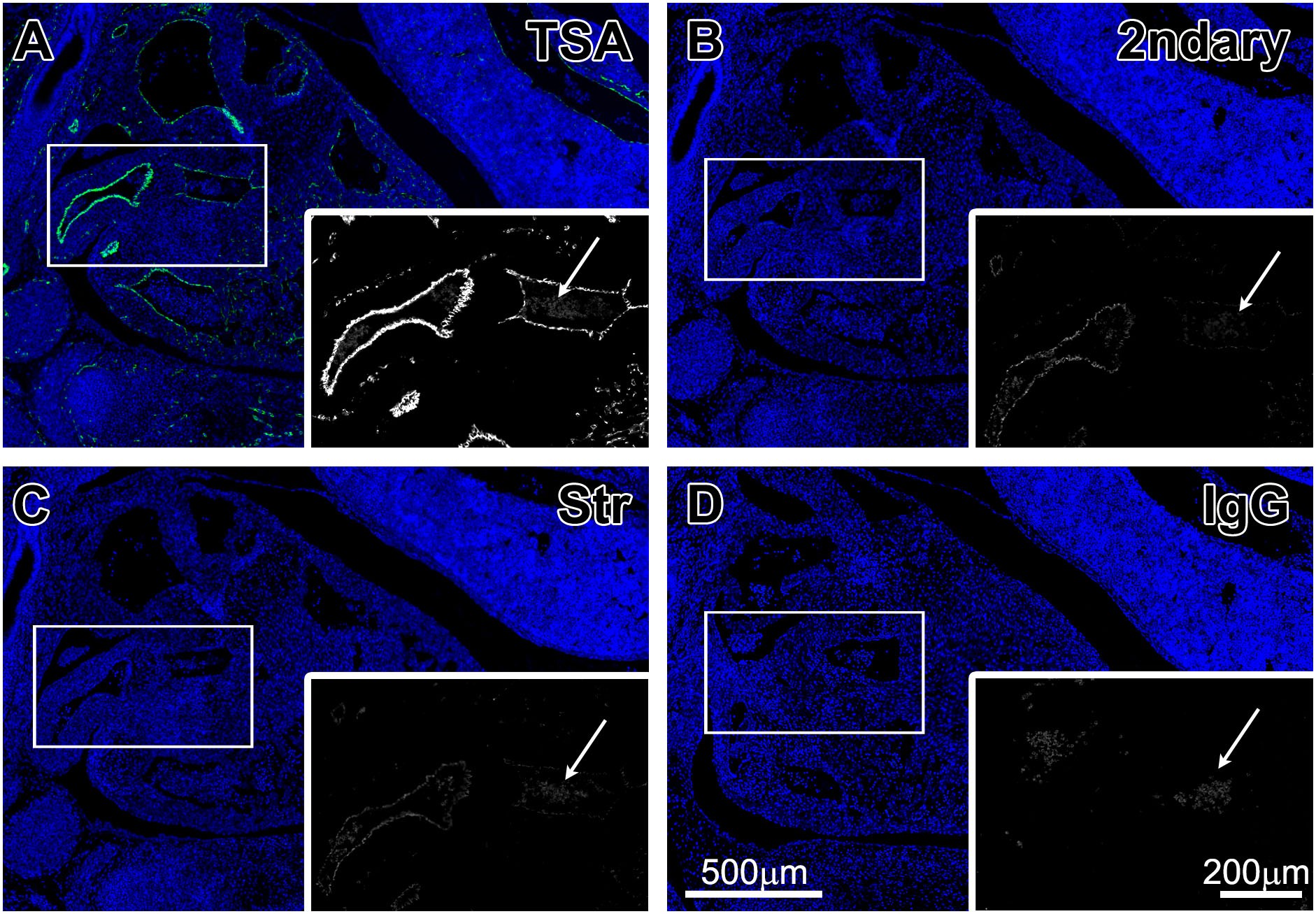
The study used the TSA tyramide amplification system, direct fluorescent secondary antibody conjugation system, and streptavidin direct-labeled fluorescence system to detect CD31, a mouse embryonic vascular endothelial marker. In large blood vessels with high CD31 expression, all three detection methods could detect clear signals, and the differences were not obvious. However, when facing microvessels and low-expression target protein regions that are common in organisms, the differences were completely revealed. The two traditional fluorescence detection methods could not capture weak specific signals at all; the microvessel structures were completely submerged by background fluorescence, and no effective positive signals could be identified. In contrast, the experimental group equipped with TSA signal amplification, relying on the enzymatic reaction to accumulate a large number of fluorescent tyramide molecules at antigen binding sites, efficiently amplified the originally invisible weak signals, completely outlining the fine microvascular network. At the same time, the autofluorescence levels of red blood cells in each group were completely consistent, indicating that TSA only specifically amplifies specific target signals without increasing non-specific background noise, perfectly balancing the two advantages of high sensitivity and low background, which is a technical breakthrough that all traditional fluorescence detection systems cannot achieve.

Figure 1

III. Multiplex Detection in Complex Immune Tissues: TSA Ensures No Crosstalk Between Multi-Channel Signals
Multiple rounds of sequential fluorescence staining are prone to signal attenuation and spectral crosstalk. To verify whether TSA can avoid this common experimental defect, the study selected human spleen tissue with complex cell subtypes and high staining difficulty for triple-label immunofluorescence detection, simultaneously distinguishing functionally different T cell subsets.

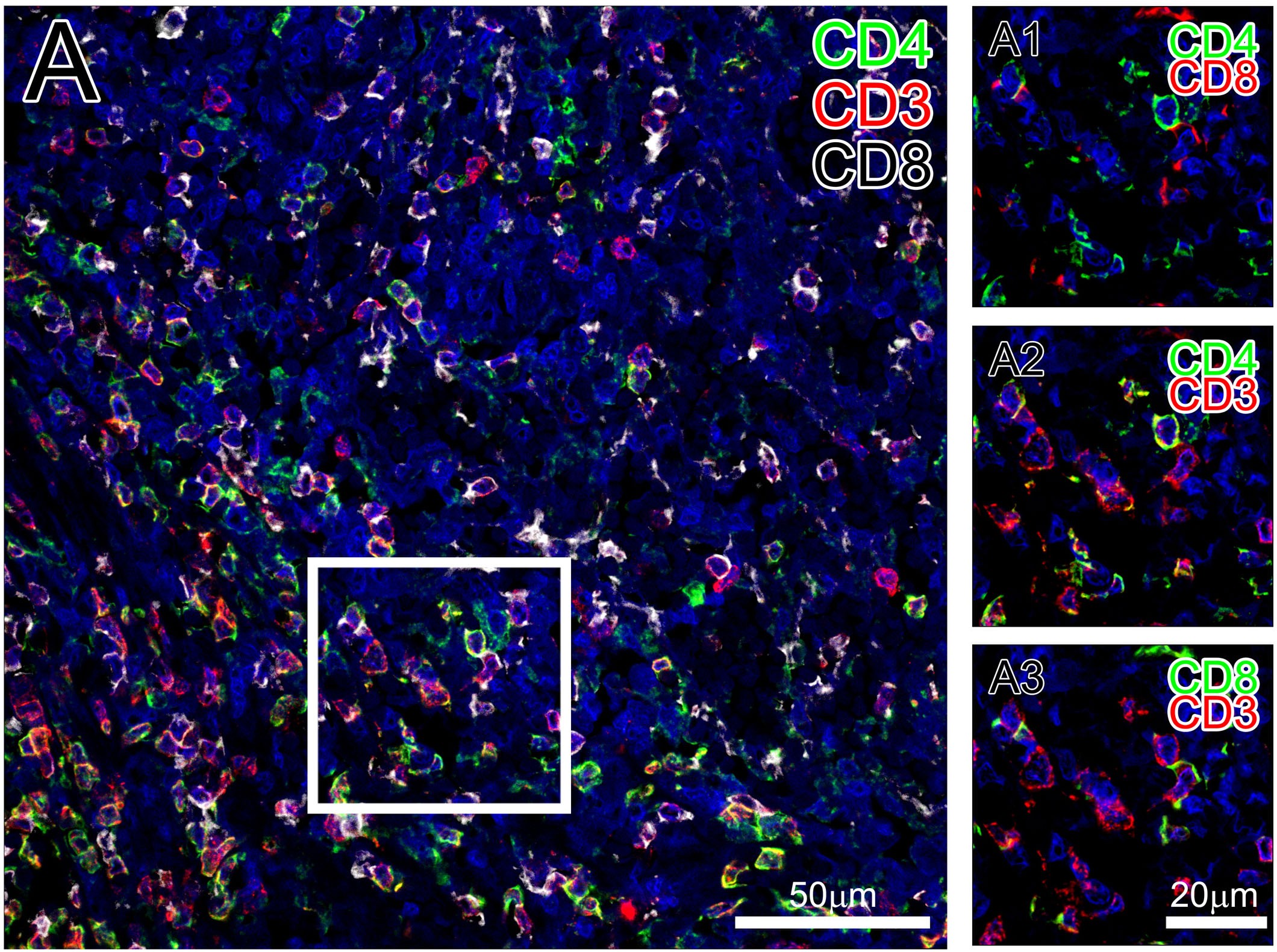
Spleen immune cell subtypes are complex, and multiplex staining is prone to signal overlap and non-specific antibody binding. The study used TSA technology to perform CD3, CD4, and CD8 triple-label fluorescence staining on human spleen sections, simultaneously distinguishing different T cell subsets. The results showed that relying on the TSA round-by-round signal amplification mode, the three fluorescence signals had clear boundaries without mutual interference. CD4 and CD8 positive cells had completely separate spatial distributions, allowing precise distinction between the two T cell subsets. At the same time, both cell types perfectly co-localized with the universal T cell marker CD3. Different from traditional multiplex fluorescence staining, where signals gradually attenuate and crosstalk becomes more severe as the number of markers increases, each round of TSA staining can independently complete signal amplification and fixation, ensuring that each target signal has sufficient fluorescence intensity. Even after three rounds of sequential staining, extremely high signal specificity can still be maintained, proving that TSA can stably support multi-target in-situ co-localization analysis in complex tissues.

Figure 2

IV. Endogenous Background Interference Control: TSA Specifically Amplifies Signals Without Amplifying Non-Specific Background
To further clarify the mechanism of TSA signal amplification and determine whether it non-selectively amplifies the overall fluorescence background of sections, the study conducted negative control verification relying on the inherent endogenous interference in mouse spleen, highlighting the uniqueness of TSA targeted amplification.

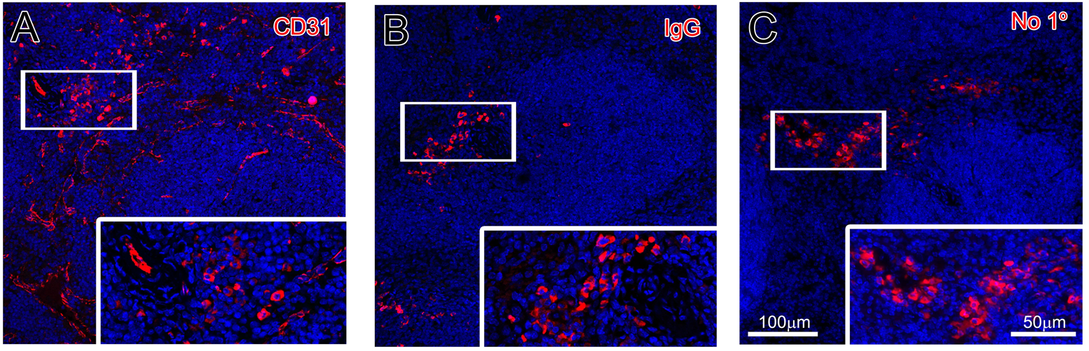
Plasma cells in mouse spleen naturally contain a large amount of endogenous immunoglobulins, which directly bind to secondary antibodies, producing inherent non-specific background. This type of background is an inherent interference of the tissue itself and cannot be eliminated by optimizing the staining process. The experimental results showed that even with this inherent background interference, TSA still did not amplify non-specific endogenous background signals, only specifically amplifying fluorescence signals at antigen-antibody specific binding sites. The specific vascular signals in the experimental group were clear and bright, while the non-specific plasma cell fluorescence signals always maintained their original weak levels and would not be synchronously amplified to aggravate background interference. This result proves that TSA has the core advantage of targeted site-specific amplification, does not non-selectively amplify all fluorescence signals on the entire section, and maximally retains the difference between effective signals and background signals, ensuring that the signal-to-noise ratio of staining results is always at a high level.

V. Ultra-High-Throughput Quadruple-Label Staining: TSA Breaks Through the Upper Limit of Multiplex Detection

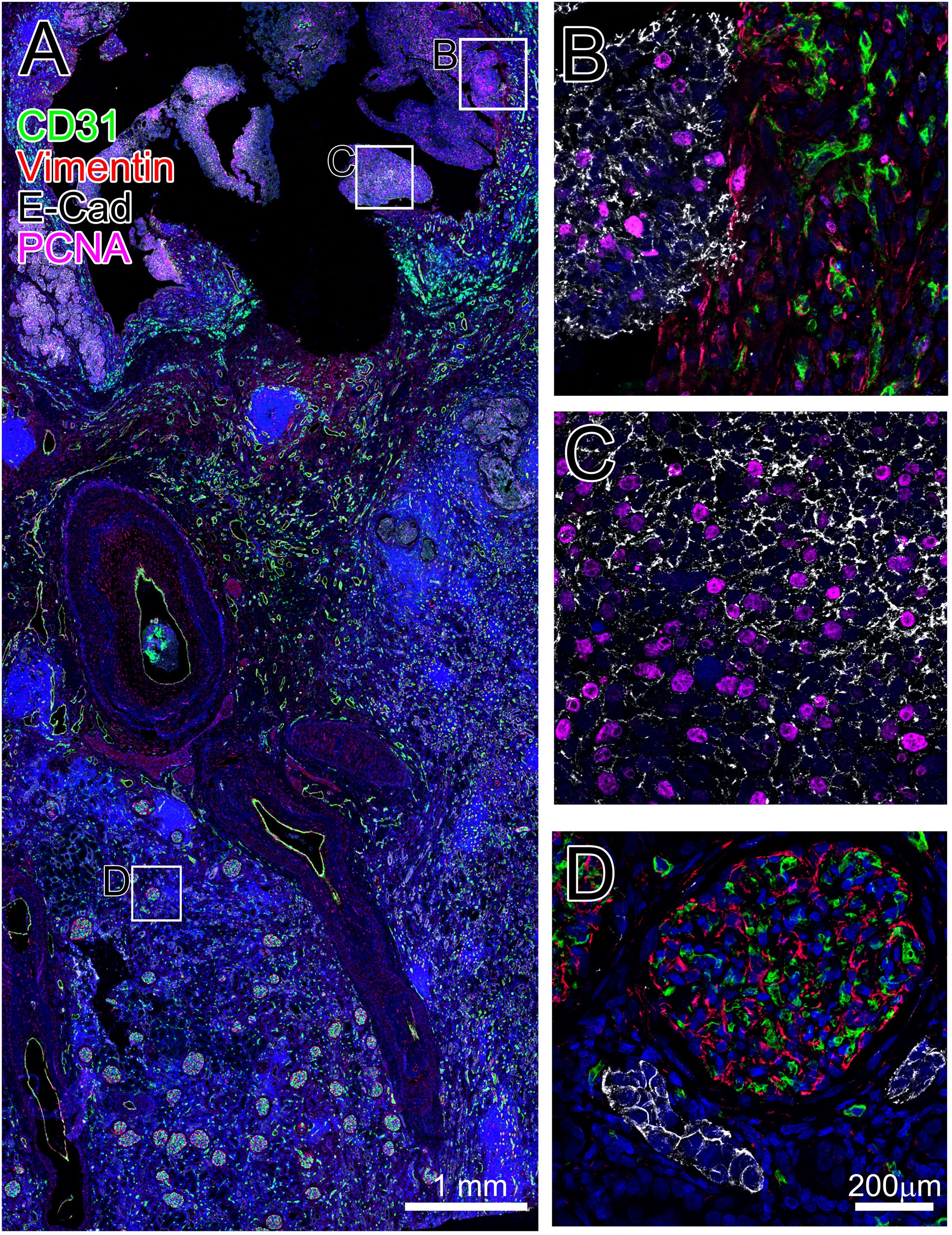
Due to antibody compatibility and signal interference issues, traditional immunofluorescence can achieve at most triple-label detection, and cannot use same-species primary antibodies, which greatly limits multi-dimensional molecular analysis of the tumor microenvironment. To break through this detection throughput bottleneck, this study relied on TSA technology to simultaneously detect four markers in human kidney tumor sections, including three mouse homologous primary antibodies. By completely inactivating the HRP enzyme activity of the previous round after each round of staining, TSA blocks cross-reactions between antibodies from different rounds. Finally, the four fluorescence signals did not interfere with each other, clearly distinguishing four cell populations in the tumor tissue: vascular endothelial cells, stromal cells, epithelial cells, and proliferating cells, and clearly delineating the junction area between tumor and normal tissue. It is sufficient to illustrate that TSA has completely broken two major constraints of traditional fluorescence staining: first, it breaks through antibody species restrictions, allowing free combination of homologous primary antibodies; second, it breaks through the limit of marker number, supporting quadruple-label and above ultra-high-throughput multiplex detection, significantly increasing the detection information content of a single tissue section.

Figure 4

VI. Homologous Primary Antibody Staining Verification: TSA Completely Eliminates Cross-Reactions Between Same-Species Antibodies
Cross-reactions between same-species primary antibodies have always been the biggest pain point in multiplex immunofluorescence experiments and a key problem limiting antibody selection. This part of the experiment specifically focuses on the scenario of homologous antibody combination to verify the anti-interference ability of the TSA combined blocking protocol.

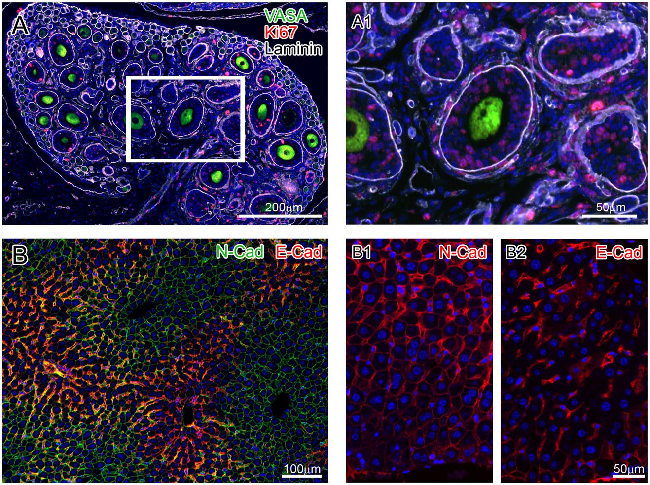
In previous multiplex fluorescence experiments, primary antibodies from different species must be used. Once same-species antibodies are used, subsequent secondary antibodies will non-selectively bind to all primary antibodies, causing comprehensive non-specific staining. This study used TSA combined with antigen saturation blocking treatment, using 3 rabbit primary antibodies to detect mouse ovary and 2 mouse primary antibodies to detect mouse liver, and finally no cross-binding phenomenon was observed in the staining results. The signals from single-channel independent staining completely matched those from multi-channel overlay, and the two homologous antibodies did not interfere with each other, with staining patterns identical to single-label staining. Relying on the enzymatic signal immobilization effect of TSA, antigen sites are completely blocked after each round of staining, preventing binding with homologous antibodies in the next round, completely liberating antibody selection restrictions. This is an advantage that cannot be achieved by all traditional multiplex staining methods without TSA support.

Figure 5

VII. Adaptability to Complex Embryonic Tissues: TSA Has Strong Versatility and Adapts to Various Difficult Tissue Samples
To comprehensively test the universality of the TSA automated staining system, avoiding conventional tissue samples with uniform structure, the study selected mouse embryonic tissues with fine structure and complex cell differentiation for multiple sets of gradient multi-label staining to test the stability of the technology in difficult samples.

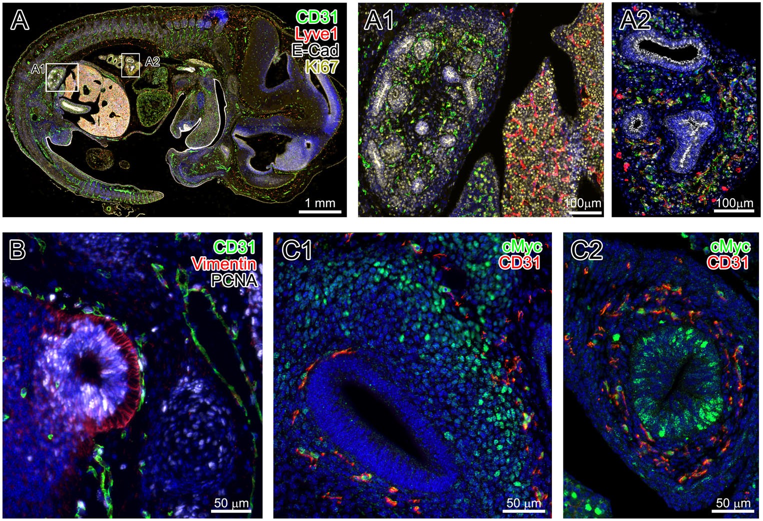
Mouse embryonic tissues have fast cell differentiation, fine tissue structure, and extremely high complexity. Conventional fluorescence staining is prone to problems such as signal loss and uneven staining. In contrast, the automated staining system based on TSA, whether for embryonic quadruple-label organ distribution detection, triple-label cell proliferation detection, or double-label metabolic activity detection, can output high signal-to-noise ratio and high-definition fluorescence images, enabling accurate identification of target molecules in the fine tissue structures of embryos. At the same time, this technology is compatible with various samples such as paraffin sections, frozen sections, and cultured cells, adapting to both clinical pathological samples and basic research samples. Compared with the strict sample requirements of traditional staining methods, TSA technology has a wider range of experimental applications and adapts to the needs of in-situ molecular detection of all types of tissues.

Figure 6

VIII. Summary of Core Advantages of TSA Technology
Based on the experimental design and six sets of imaging results in this article, Tyramide Signal Amplification (TSA) has effectively overcome multiple technical challenges in traditional immunofluorescence staining and is a key technology for in-situ multi-molecule detection in tissues. Combining this literature study, the five irreplaceable core advantages of TSA can be summarized:
• High detection sensitivity: It can effectively amplify weak fluorescence signals from low-abundance target proteins, solving the problem of easy missed detection of low-expression targets by traditional methods.
• Excellent signal targeting: It only amplifies signals at antigen-antibody specific binding sites without enhancing endogenous background fluorescence in tissues, ensuring high signal-to-noise ratio in imaging.
• Compatible with homologous primary antibodies: Combined with saturation blocking procedures, it can completely avoid cross-reactions between same-species antibodies, significantly expanding the range of antibody selection.
• Supports high-throughput multiplex detection: It breaks through the limit of traditional immunofluorescence with at most triple labels, enabling quadruple-label and above multi-target simultaneous staining, fully exploring the experimental value of a single tissue section.
• Good versatility and stability: It can seamlessly interface with fully automated staining equipment, improving experimental repeatability; at the same time, it is compatible with various experimental materials such as paraffin sections, frozen sections, embryonic tissues, and clinical pathological samples.

Compared with the immunohistochemistry system, TSA technology applied to immunofluorescence can also maintain a linear relationship between fluorescence signal intensity and target protein expression, with the ability for precise quantitative analysis. Overall, TSA technology has shown significant advantages in sensitivity, antibody compatibility, detection throughput, experimental repeatability, and quantitative ability, and has become an indispensable core technology in tissue multiplex immunofluorescence detection.

References

Yarilin D, Xu K, Turkekul M, Fan N, Romin Y, Fijisawa S, Barlas A, Manova-Todorova K. Machine-based method for multiplex in situ molecular characterization of tissues by immunofluorescence detection. Sci Rep. 2015 Mar 31; 5:9534. doi: 10.1038/srep09534. PMID: 25826597; PMCID: PMC4821037.


EnkiLife not only provides customers with a complete set of TSA multiplex labeling kits, but also offers various TSA specialty technical services, including IF fluorescence staining, fluorescence panoramic scanning, ultra-multiplex staining, and pathological analysis (5 markers and below).  

Product

Catalog Number

TSA Six-Label Seven-Color Multiplex Immunohistochemistry Kit

RA10012

TSA Five-Label Six-Color Multiplex Immunohistochemistry Kit

RA10011

TSA Four-Label Five-Color Multiplex Immunohistochemistry Kit

RA10010

TSA Three-Label Four-Color Multiplex Immunohistochemistry Kit

RA10009

TSA Two-Label Three-Color Multiplex Immunohistochemistry Kit

RA10008

For details, please check TSA mIHC Kit


   💬 WhatsApp