In modern life science research, the complexity of tissue microenvironments poses higher demands on scientific research. Multiplex immunofluorescence (mIHC) technology, with its advantages of simultaneously detecting multiple targets and preserving spatial information, has become an indispensable tool in fields such as tumor immunology, neuroscience, and stem cell research. However, traditional multiplex immunofluorescence often encounters several challenges:
- Complex operation: Each round of staining requires strict control of conditions, with tedious procedures.
- Signal interference: Fluorescence overlap and background signals affect result accuracy.
- Antibody species restrictions: Same-species antibodies are prone to cross-reactions, affecting multiplex analysis.
Faced with these problems, researchers urgently need a highly stable, highly sensitive, and easy-to-operate multiplex staining solution.
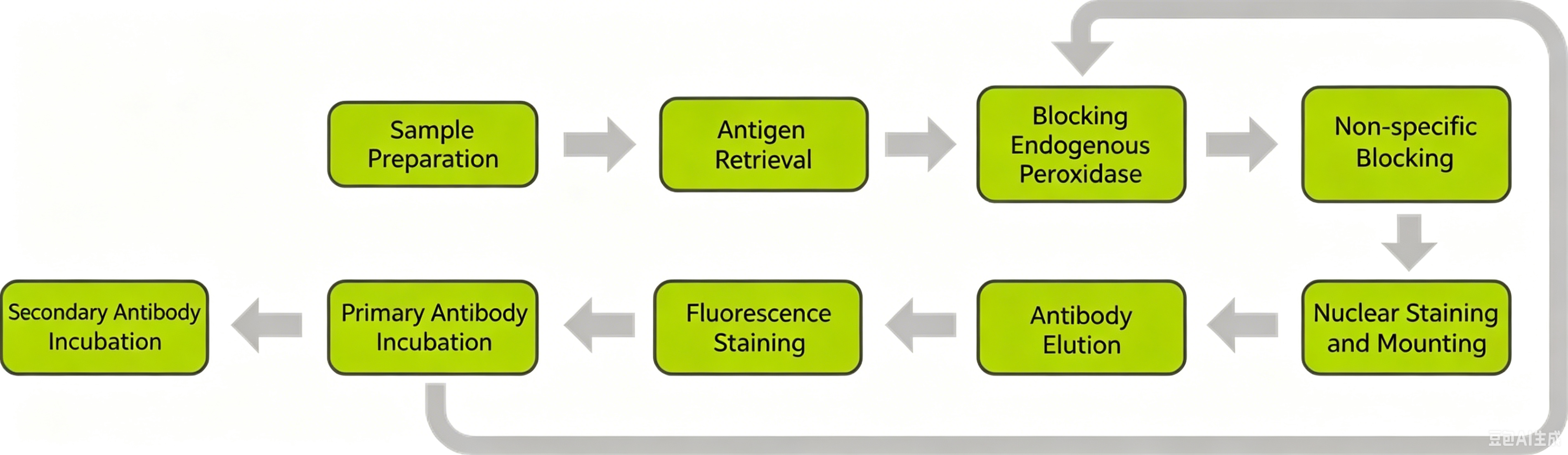
The TSA (Tyramide Signal Amplification) kit is a high-sensitivity immunoassay system developed based on the principle of HRP enzyme-catalyzed fluorescent tyramide molecule deposition. Its core mechanism utilizes the peroxidase reaction of tyramide. Under the action of HRP and H₂O₂, the tyramide fluorescein substrate is activated, and the activated fluorescent substrate can covalently bind to residues such as tyrosine on the target protein, resulting in massive deposition of fluorescein at the antigen-antibody binding site, achieving signal amplification. Through heat retrieval or antibody stripping solution, the non-covalently bound antibodies from the previous round are washed away, while the fluorescein remains stably bound to the protein for the next round of staining. This process continues until all antibody incubations are completed, followed by nuclear staining, mounting, and scanning.
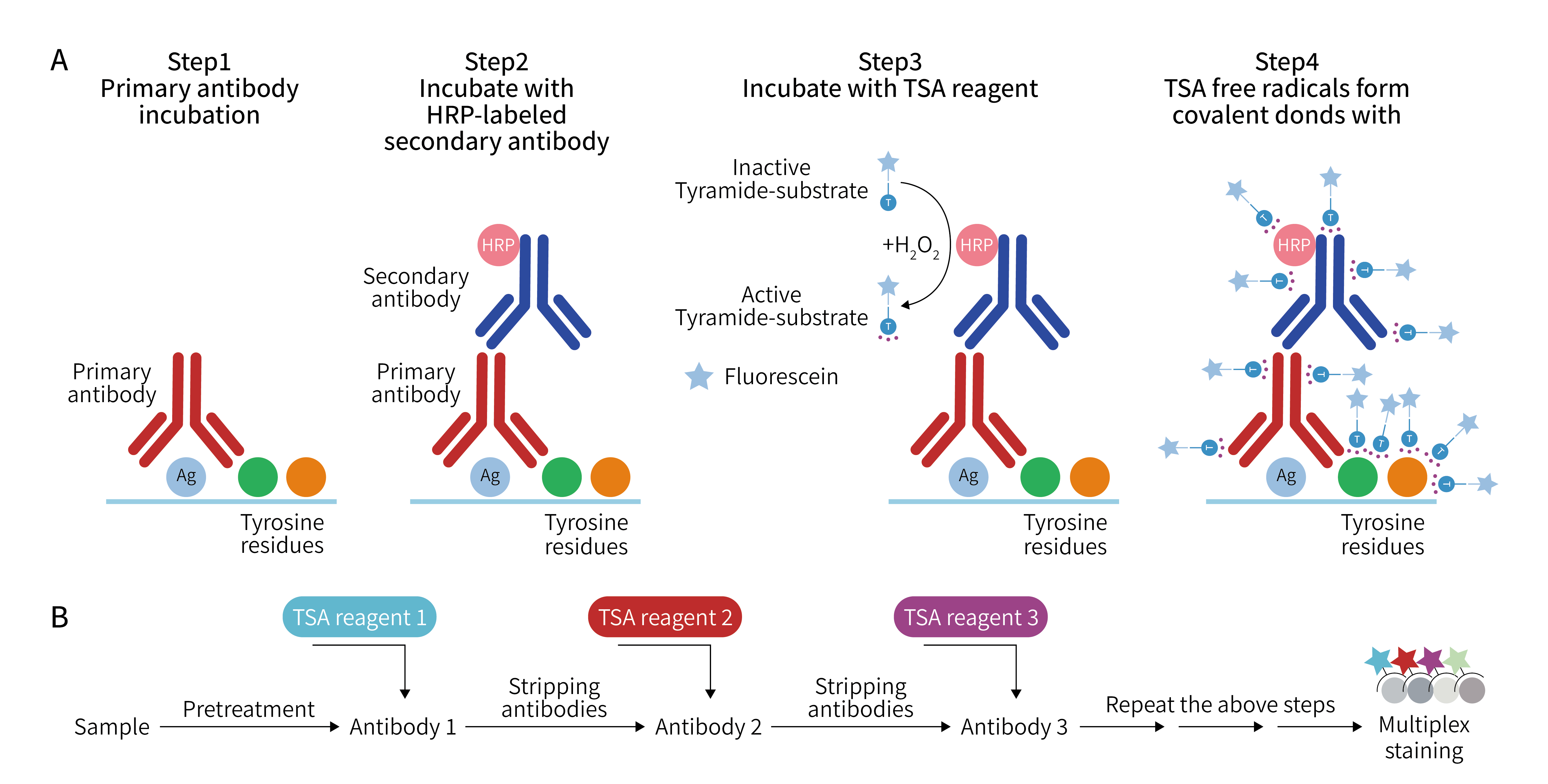
- TSA Signal Amplification System
The new kit adopts Tyramide Signal Amplification (TSA) technology, an enzyme-catalyzed signal amplification technique. Compared with ordinary fluorescent labeling, it can significantly amplify the fluorescence signal of each target while reducing background noise. Signal intensity is increased by 2-5 times, making low-abundance proteins clearly visible. - No Primary Antibody Species Restrictions
The kit is compatible with mouse and rabbit secondary antibodies. Researchers do not need to change antibodies to avoid species conflicts. They can directly select antibodies with sufficient literature validation, optimal performance, or those already in established databases to complete multiplex detection, significantly saving experimental time and costs. - Multiplex Staining Completed in One Run
The antibody stripping solution provided with the kit supports multiple fluorescent labels to be completed simultaneously in a single experiment, avoiding repeated high-temperature stripping processes, which improves efficiency and reduces sample loss. - High Reproducibility and Stability
The optimized fluorescent substrate and mounting medium formulations ensure stable signals between experiments with strong image comparability. Even on complex tissues or precious samples, reproducible high-quality data can be obtained. - Wide Application Range
It can be applied to multiple sample types including paraffin sections, frozen sections, cell slides, cell smears, tissue microarrays, organoids, etc. Whether for tumor microenvironment analysis, immune cell spatial distribution research, or stem cell or neuron labeling, it can easily handle the task.
- Sample pretreatment: Optimal section thickness is 4-5 μm. Antigen retrieval time should be adjusted according to tissue type.
- Antibody selection: Prioritize validated high-specificity primary antibodies to avoid background interference.
- Staining order: Stain targets from low to high expression levels sequentially to reduce fluorescence overlap effects.
- Fluorescence signal preservation: Store slides protected from light after mounting. Can be stored at room temperature for several weeks, or extended to several months at low temperature.
- Imaging considerations: Properly set excitation light intensity and exposure time to ensure balanced signals across all channels.
Combining these tips with the upgraded kit can double both experimental efficiency and data quality.
Stay Ahead in Research, Lead the Way in Experiments
On the "fast-track" of scientific research, high efficiency and high reliability are equally important. Choosing the new upgraded mIHC kit not only allows you to take the lead in experiments but also stay ahead in scientific output. Let multiplex immunofluorescence technology truly become your "accelerator" to explore the infinite possibilities of tissue microenvironments!
EnkiLife not only provides customers with a complete set of TSA multiplex labeling kits, but also offers various TSA specialty technical services, including IF fluorescence staining, fluorescence panoramic scanning, ultra-multiplex staining, and pathological analysis (5 markers and below).
Product | Catalog Number |
|---|---|
TSA Six-Label Seven-Color Multiplex Immunohistochemistry Kit | |
TSA Five-Label Six-Color Multiplex Immunohistochemistry Kit | |
TSA Four-Label Five-Color Multiplex Immunohistochemistry Kit | |
TSA Three-Label Four-Color Multiplex Immunohistochemistry Kit | |
TSA Two-Label Three-Color Multiplex Immunohistochemistry Kit |
For details, please check TSA mIHC Kit