What Exactly is "Spatial Information" that Reviewers are Increasingly Focused On? — Re-understanding Multiplex Fluorescence Immunohistochemistry
In recent years, across research fields such as tumor immunology, infection and inflammation, and neuroscience, an increasing number of reviewer comments have begun to feature a common keyword — "insufficient spatial information". Many researchers are puzzled: they have completed Western blot, qPCR, and even single-cell sequencing, yet are still asked to supplement spatial-level evidence. The core issue lies in an important transformation that modern life sciences are undergoing: the research focus is no longer just "whether something is expressed" or "how much is expressed", but "where it is expressed" and "who it coexists with". This cognitive leap from molecules to structures has gradually made spatial information a key dimension in the paper evaluation system, and TSA multiplex immunofluorescence technology has emerged as an important tool connecting molecular and tissue structures in this context.
I. What Exactly is "Spatial Information"?
Spatial information is essentially not a single indicator, but a systematic description of structural relationships within biological tissues. In traditional experimental systems, we typically focus on molecular expression levels, such as whether a gene is upregulated in tumor tissue or whether a protein changes significantly under disease conditions. However, this information is an "average", derived from tissue homogenates or mixed cell populations, thus naturally losing a crucial dimension of information — spatial structure.
In real biological tissues, cells are not uniformly distributed, but exist in a highly organized manner. For example, in the tumor microenvironment, CD8-positive T cells may concentrate at the tumor edge forming an "immune wall", while the tumor core region exhibits immune exclusion; macrophages may form barrier structures around blood vessels, affecting immune cell infiltration; even the same biomarker may have completely different functions in different spatial regions. Therefore, spatial information essentially answers three levels of questions: where cells are located, who cells are adjacent to, and whether these spatial structures have biological significance.
With the development of tumor immunotherapy, the importance of this spatial dimension has been further amplified. Increasing research finds that even with similar PD-L1 or CD8 expression levels, treatment responses still vary significantly among different patients, and these differences often stem from different cellular spatial organization structures. Therefore, reviewers' focus on spatial information essentially requires authors to move from "expression description" to "structural interpretation".
II. Why Can Traditional IHC and Molecular Experiments Not Answer Spatial Questions?
Although traditional immunohistochemistry can provide some tissue localization information, it is essentially a low-dimensional spatial observation. Single-marker IHC can only detect one indicator at a time. Even with serial sectioning or multiple staining, it can only make inferences through "visual stitching", and cannot establish true multi-dimensional spatial relationships in the same tissue section. This approach not only introduces tissue variation errors, but also cannot perform cell-level co-localization analysis, thus having obvious limitations in complex microenvironment research.
On the other hand, methods like Western blot and qPCR, while highly sensitive, completely lose spatial information. They grind the entire tissue and obtain a "mixed average signal". This signal cannot distinguish differences between tumor core and edge, nor can it reflect local interaction relationships between immune cells. In immune microenvironment research, this information loss is particularly fatal, because what truly determines function is often not overall expression level, but local spatial structure.
Therefore, when research questions enter the realms of "cell-cell interaction", "microenvironment structure", "immune exclusion zones", etc., traditional technologies can no longer provide sufficient explanatory power, which is the fundamental reason why reviewers increasingly tend to require spatial experimental verification.
III. Core Mechanism of TSA Multiplex Immunofluorescence Technology
The core idea of TSA multiplex immunofluorescence technology (Tyramide Signal Amplification) can be summarized as an amplification mechanism of "local signal deposition and permanent marking". In traditional immunofluorescence, fluorescence signals usually rely on direct or indirect antibody labeling. Such signals are easily affected by washing conditions and have limited intensity. In contrast, the TSA system uses HRP enzyme to catalyze the activation of tyramide substrate, causing it to undergo covalent deposition near the antigen, thereby "fixing" the signal around the target protein.
This mechanism brings about two key changes. First, signals no longer depend on the continuous presence of antibodies, but are permanently retained in the tissue in the form of chemical deposition, thus having extremely high stability. Second, due to the local diffusion characteristic of deposition, signals form an amplification effect around the antigen, enabling detection of low-abundance proteins. This high-sensitivity feature gives TSA technology obvious advantages in complex tissue analysis.

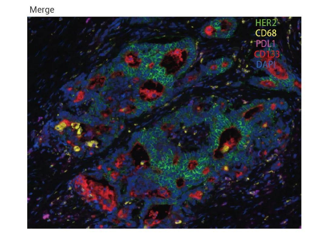
More importantly, this "signal fixation" characteristic makes multi-round staining possible. After completing one round of antibody reaction and signal deposition, the previous round of antibodies can be removed through antibody stripping without affecting the already deposited fluorescence signals. By repeating this process, multiple biomarkers can be sequentially detected on the same tissue section, thereby constructing a high-dimensional spatial information map.

IV. How Does TSA Technology Truly Generate "Spatial Information"?
The value of TSA multiplex immunofluorescence lies not merely in "multi-marker labeling", but in its ability to transform tissue from a two-dimensional image into a computable spatial system. After multiplex staining is completed, each cell not only carries expression information but also has explicit spatial coordinates, making the tissue no longer a static image but a spatial network composed of cells.
Based on this, further spatial neighborhood analysis can be performed. For example, by setting a certain spatial distance threshold, the contact probability between CD8-positive T cells and tumor cells can be calculated to determine whether immune attack truly occurs; it is also possible to analyze whether PD-1-positive T cells are concentrated at the tumor edge to identify immune exclusion structures; even the spatial enrichment zones of macrophages around blood vessels can be studied to infer the existence of immunosuppressive barriers.
Furthermore, TSA data can also be used to construct cell interaction network models. In this model, each cell is not only an expression unit but also a network node. The spatial distance and contact frequency between different cells constitute the weight of edges, thus forming a "tissue ecosystem". This analytical approach is gradually changing our understanding of tissue function, moving research from "average expression differences" to the level of "structure-driven mechanisms".
V. Typical Value of TSA in Tumor Microenvironment Research
In tumor research, one of the most important applications of TSA technology is to resolve the sources of immunotherapy response differences. Increasing research finds that even with similar PD-L1 expression levels, patients' responses to immune checkpoint inhibitors still vary significantly, and these differences are often closely related to spatial structures. For example, in some patients, although CD8-positive T cells exist in tumor tissue, they are confined to the tumor edge and cannot enter the core region, thus forming so-called "immune-excluded tumors". In other patients, immune cells can infiltrate deep into the tumor and make direct contact with tumor cells, showing better treatment responses.
In addition, TSA technology can also be used to analyze the spatial distribution patterns of tumor-associated macrophages. Studies find that M2-type macrophages tend not to be randomly distributed, but rather form structural enrichment around blood vessels or necrotic areas. This spatial structure may participate in immune escape processes by blocking T cell entry into the tumor core region. Such findings cannot be obtained through expression level analysis alone, and must rely on technical systems with higher spatial resolution.
VI. Why are Reviewers Increasingly Thinking "Spatially"?
Reviewers' focus on spatial information is not simply a change in technical preference, but the result of a paradigm shift in the entire life sciences. On one hand, the development of single-cell sequencing technology allows us to characterize cellular heterogeneity with unprecedented precision, but it always lacks a key dimension — spatial location information. On the other hand, the development of spatial transcriptomics and multiplex imaging technologies has led researchers to realize that cell function is not only determined by their own state, but also deeply influenced by the structure of their microenvironment.
At the same time, clinical translational research is also driving this trend. Compared to simple expression level indicators, spatial structure parameters are often more stable, more reproducible, and closer to the morphological judgment logic that pathologists have long relied on. Therefore, reviewers' requirement for spatial information essentially pushes research from "molecular description" to "structural interpretation".
Conclusion: From Expression Analysis to Spatial Understanding
When we re-examine the reviewer comment "insufficient spatial information", we find that it is not an additional technical requirement, but an implicit standard for research depth. Truly explanatory biological research not only needs to know whether a molecule changes, but also needs to understand the spatial context in which this change occurs, and how it affects real relationships between cells.
In this process, TSA multiplex immunofluorescence technology is playing a key role. It transforms traditional "staining results" into computable spatial structure data, enabling researchers to truly enter the micro-world inside tissues, see how cells are arranged, how they interact, and how these structures collectively determine biological functions.
In other words, modern pathology is undergoing a fundamental transformation: we are no longer just "seeing molecules", but trying to "read space".

Detection TechnologySpatial Information CompletenessMulti-biomarker Co-localizationCellular Spatial Interaction Analysis
WB/qPCRNoneNot supportedImpossible
Single-marker IHCLow-dimensional local localizationHigh error, unreliableOnly rough observation
TSA Multiplex FluorescenceComplete cell-level spatial coordinatesPrecise co-localization with multi-marker on same sectionQuantifiable neighborhood/cell interaction network


To meet the research needs of tumor microenvironment and spatial biology, Enkilife provides TSA-mIHC kits and technical services, supporting multi-biomarker detection, experimental optimization, and spatial analysis, helping researchers obtain more valuable information from a single tissue section.

Product

Catalog Number

TSA Six-Label Seven-Color Multiplex Immunohistochemistry Kit

RA10012

TSA Five-Label Six-Color Multiplex Immunohistochemistry Kit

RA10011

TSA Four-Label Five-Color Multiplex Immunohistochemistry Kit

RA10010

TSA Three-Label Four-Color Multiplex Immunohistochemistry Kit

RA10009

TSA Two-Label Three-Color Multiplex Immunohistochemistry Kit

RA10008


   💬 WhatsApp