
In the world of biomedical research, clinical diagnostics, and drug development, antibodies are the "exclusive detectors" that precisely identify specific antigens. However, antibodies themselves cannot be directly captured by the naked eye or instruments. To make their recognition process "visualizable, quantifiable, and traceable," antibodies must be bound with special "signal tags" — this is the core principle of antibody labeling technology.
Simply put, antibody labeling is the process of connecting functional markers to antibody molecules through chemical conjugation, electrostatic adsorption, or other methods. After antibodies bind to target antigens, they release detectable signals, enabling localization, qualitative, and quantitative analysis of antigens. Different markers vary greatly in characteristics, sensitivity, and applicable scenarios. Choosing the right marker is the key to the success of experiments such as immunohistochemistry, Western Blot, ELISA, and flow cytometry. This article will comprehensively break down mainstream antibody labeling markers, clearly explain their principles, advantages/disadvantages, and applicable scenarios, helping you thoroughly understand the core knowledge of antibody labeling.
Enzyme markers are currently the most widely used and technically mature labeling system in laboratories, and are the preferred choice for routine immunoassays. The core principle is to covalently conjugate catalytic enzymes with antibodies, producing chromogenic or chemiluminescent signals through enzymatic substrate reactions, converting invisible immune binding reactions into visualizable and quantifiable results. This labeling method does not destroy the antigen-binding specificity of antibodies, the system is stable and reproducible, and can be operated in ordinary laboratories.
The most popular enzyme marker, with small molecular weight, strong penetration ability, easy to penetrate tissue cells, high labeling efficiency, and low cost. The substrate reaction signal is stable, compatible with Western Blot, ELISA, immunohistochemistry IHC, and most basic experiments. In chemiluminescent detection mode, the sensitivity is extremely high, making it the core marker for protein detection.
Mild catalytic activity, lower background interference, stronger contrast in chromogenic reactions, especially suitable for tissue section and in-situ cell staining experiments. It has significant advantages in fine immunohistochemistry and in-situ hybridization detection, effectively avoiding the non-specific chromogenic problems that HRP is prone to.
Fluorescent markers are the core tools for cellular and molecular localization research. The principle is to conjugate fluorescent functional molecules to antibodies. Under specific wavelength excitation light, fluorescent substances can emit exclusive fluorescent signals, accurately marking the in-situ location of antigens in cells and tissues. This labeling technology breaks through the limitation of single detection and supports simultaneous detection of multiple targets, making it the core method for advanced scientific research. Based on the molecular properties and sources of fluorescent substances, commonly used antibody fluorescent markers can be divided into two categories: small-molecule fluorescent dyes and fluorescent proteins, which differ significantly in structure, characteristics, and applicable scenarios.
Currently the most commonly used organic fluorescent reagents for antibody labeling. They have small molecular weight, easy chemical conjugation, high labeling efficiency, and rich spectral types. They can flexibly adapt to various antibody labeling needs and are compatible with most immunofluorescence experiments and flow cytometry detection. The main small-molecule fluorescent dyes commonly used in laboratories include:
A classic green organic small-molecule dye with stable excitation and emission wavelengths, high brightness, low background, and excellent cost-performance ratio. It is the basic first-choice marker for flow cytometry and ordinary immunofluorescence staining, with an extremely wide range of applications.
A classic red organic small-molecule dye with significantly better fluorescence stability than FITC and stronger anti-quenching ability. It is not easily inactivated under light. It is often paired with FITC in red-green combinations to achieve dual-antigen co-localization analysis in cell and tissue samples, making it the basic combination for multicolor fluorescence staining.
A new generation of high-end modified small-molecule fluorescent dyes covering the full spectral range from blue to far-red light. They perfectly solve the problems of rapid quenching and low signal-to-noise ratio of traditional dyes. With high brightness, extremely low background interference, and excellent photobleaching resistance, they are specifically designed for fine imaging and subcellular localization research using high-resolution fluorescence microscopy and confocal microscopy, making them the first-choice dyes for high-precision fluorescence experiments.
Classic high-brightness cyanine small-molecule fluorescent dyes, the mainstream commercial dye series for biological imaging and spectral detection. Cy3 is orange-red fluorescence with high brightness, low background, and excellent signal-to-noise ratio, suitable for routine immunofluorescence and tissue section staining; Cy5 is far-red fluorescence with strong penetration and minimal autofluorescence interference, suitable for deep tissue imaging and trace sample detection; Cy7 belongs to near-infrared fluorescent dyes, mainly used for in-vivo imaging and non-invasive detection. Overall, the Cy series has high molar extinction coefficient, outstanding luminescence intensity, and wide spectral span, making it very suitable for multicolor fluorescence overlay detection. Widely used in immunofluorescence imaging, flow cytometry, and fluorescence quantitative experiments, it is the second most popular high-end small-molecule dye system after Alexa Fluor in scientific research.
These are natural or genetically modified macromolecular fluorescent proteins with excellent biocompatibility, strong specificity, no chemical dye toxicity, and luminescence brightness far exceeding most small-molecule dyes. They are especially suitable for low-abundance antigen detection and high-sensitivity flow cytometry analysis. Commonly used fluorescent proteins for antibody labeling include:
An ultra-high brightness natural fluorescent protein whose luminescence intensity and sensitivity far exceed ordinary organic small-molecule dyes. It has outstanding advantages in the identification and detection of low-expression and trace antigens with extremely high signal recognition. Widely used in flow cytometry multicolor sorting and high-sensitivity immunofluorescence quantitative analysis, it is the core fluorescent marker for flow multicolor panel design.
A classic long-wavelength phycobiliprotein with emission light in the far-red band, extremely low fluorescence background, minimal spectral cross-color interference, and excellent fluorescence brightness and signal-to-noise ratio. Compared to PE, APC has better photostability and slower fluorescence quenching rate, making it very suitable for flow cytometry multicolor matching and weak signal detection. It is also a commonly used high-end labeling fluorescent protein for clinical flow cytometry detection and rare cell sorting, often paired with PE to form a classic dual-color flow cytometry detection system.
A genetically encoded fluorescent protein distinct from phycobiliproteins, it is an iconic fluorescent labeling tool for life science imaging. Wild-type GFP has stable luminescence, and various variants modified through artificial mutations have significantly improved performance. Common mainstream variants include EGFP (Enhanced Green Fluorescent Protein), mCherry, YFP, CFP, etc., covering green, yellow, red, and blue multicolor spectra. These fluorescent proteins have extremely high specificity, no autofluorescence interference, and excellent biocompatibility, without affecting the biological activity of antibodies and cells. They are mainly used in fusion protein tracing, cell in-vivo dynamic imaging, cell lineage tracing, immunofluorescence localization labeling, and other scenarios. Compared to PE and APC, GFP variants have smaller molecular weight, stronger tissue penetration, and are suitable for in-vivo dynamic observation, making them the core fluorescent protein markers for cell biology and in-vivo imaging experiments.
Biotin labeling does not belong to the direct chromogenic labeling system. Instead, it relies on the ultra-high specificity and ultra-high affinity binding characteristics of biotin-avidin (streptavidin) to build a signal amplification labeling system, which is the key technology to solve the detection challenges of trace and low-abundance samples.
Its core advantage lies in the cascade amplification effect: first label biotin on the primary antibody, then perform secondary binding through avidin conjugated with enzymes or fluorescein. One avidin can bind multiple biotin molecules, greatly amplifying the detection signal and significantly lowering the detection limit. At the same time, the biotin labeling reaction is mild and almost does not damage antibody activity. The labeled antibodies can be stored for a long time without losing activity. Suitable for high-precision scientific research scenarios such as low-abundance protein detection, trace sample analysis, high-sensitivity immunoblotting, fine immunohistochemistry, and multi-channel fluorescence detection.
Colloidal gold labeling is an exclusive technology for applied rapid detection. The principle relies on the electrostatic adsorption of colloidal gold particles to directly and stably bind with antibodies without complex chemical conjugation processes. When colloidal gold particles aggregate, they can present visible red or purple-red bands to the naked eye, allowing direct result interpretation without instrument assistance.
The greatest features of this labeling system are simplicity, speed, stability, and long shelf life. It does not require complex experimental procedures or professional operators, making it perfectly suitable for batch rapid screening scenarios. It is also the core labeling technology for various in-vitro diagnostic test strips, such as COVID-19 antigen test strips, pregnancy test strips, and tumor marker rapid detection test strips.
Isotope labeling was the core technology of early immunoassays, with radioactive iodine isotope labeling as the main application form. The principle is to conjugate radioactive nuclides to antibodies and capture radioactive signals through γ counters or X-ray films to achieve ultra-high sensitivity quantitative detection. This technology has extremely high sensitivity, capable of capturing extremely trace antigen-antibody binding signals with excellent accuracy. However, with the iteration of biological detection technology, its shortcomings have become increasingly prominent. Currently, it has basically withdrawn from routine scientific research scenarios, with only a very few specialized scientific research applications remaining. Core disadvantages include: radiation safety hazards, strict requirements for experimental protection and operating qualifications; short isotope half-life, short validity period of labeled products, inability to preserve for a long time; complicated experimental procedures and long detection cycles.
In addition to the five major mainstream classic markers, several novel markers have become popular choices in cutting-edge research in recent years due to their unique advantages, complementing the shortcomings of traditional labeling systems.
High luminescence intensity, extremely strong anti-quenching ability, good spectral discrimination, suitable for multi-index combined detection and high-throughput screening, widely used in liquid chip detection technology.
Nano-scale fluorescent labeling materials with narrow emission, wide excitation, high brightness, and photobleaching resistance characteristics. Imaging resolution far exceeds traditional fluorescent dyes, making them the core tool for ultra-high-resolution cellular imaging and single-molecule detection.
Directly label antibodies to achieve luminescent signal output without enzymatic reactions. Extremely low background noise and wide linear range, compatible with high-end automated chemiluminescent immunoanalyzers, widely used in clinical precision quantitative diagnosis.
Antibody labeling markers are the "signal core" of immunoassay technology. From classic enzyme, fluorescence, and biotin labeling to novel nano and chemiluminescent labeling, different technologies complement each other, building a complete biological detection system. Mastering the core principles and applicable scenarios of various markers and accurately matching experimental needs can not only avoid common problems such as background interference, weak signals, and data distortion but also maximize experimental efficiency and detection accuracy, providing reliable technical support for scientific research exploration and clinical diagnosis.
EnkiLife has a comprehensive antibody labeling scientific research product and technical service system. The self-developed full series of kits covers mainstream categories including enzyme labeling, small-molecule fluorescent dye labeling, fluorescent protein labeling, and biotin labeling. They can be adapted to various experimental scenarios such as WB, ELISA, immunohistochemistry, confocal imaging, flow multicolor detection, and high-precision analysis of low-abundance samples. All kits have undergone strict process optimization, with advantages of high labeling efficiency, strong specificity, low background, and good stability, effectively improving experimental success rate and data reproducibility. At the same time, the brand can provide one-stop antibody labeling customized technical services, offering full-process exclusive solutions from experimental design, precise labeling, sample detection to data analysis for special antibody samples, personalized, and high-difficulty high-precision labeling needs.
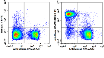
Flow cytometry detection results using APC labeling kit (RE80006p) labeled mouse CD3 antibody and R-PE labeling kit (RE80005p) labeled rat anti-Mouse CD45R and Rat IgM
| Label Type | Representative Markers | Core Advantages | Main Application Scenarios |
| Enzyme Labeling | HRP, AP | Low cost, stable signal, simple operation | WB, ELISA, routine IHC |
| Small-Molecule Fluorescent Dyes | FITC, Alexa, Cy Series | Multicolor imaging, clear localization | Immunofluorescence, confocal, flow cytometry |
| Fluorescent Proteins | PE, APC, GFP | High brightness, dynamic observation | Flow multicolor sorting, in-vivo imaging |
| Biotin | Biotin-Streptavidin System | Signal amplification, extremely low detection limit | Low-abundance proteins, high-sensitivity IHC |
| Colloidal Gold | Colloidal Gold Particles | Naked-eye visible, rapid detection | Rapid antigen detection test strips |
| Isotope | Radioactive Iodine | Ultra-high sensitivity | Very few specialized research, now obsolete |