
Ferroptosis: Mechanisms, Detection, and Interpretation
A research guide to iron-dependent lipid peroxidation, the SLC7A11–GSH–GPX4 axis, ferroptosis regulators, and multiparametric validation.
Ferroptosis differs from apoptosis and necroptosis in its dominant biochemical failure: membrane lipid hydroperoxides accumulate faster than the cell can remove them. The same treatment can engage more than one death pathway, so experimental conclusions should be based on matched perturbations, rescue controls, and a time-resolved panel rather than on a protein abundance change alone.
What ferroptosis is
Ferroptosis was defined as an iron-dependent, non-apoptotic form of regulated cell death distinguished by oxidative damage to membrane lipids. The defining event is not simply an increase in total reactive oxygen species (ROS); it is the accumulation of phospholipid hydroperoxides in a membrane environment that cannot be adequately protected or repaired.[1]
Ferroptotic cells often show shrunken mitochondria, increased mitochondrial membrane density, and reduced cristae, while the nucleus lacks the chromatin condensation typical of apoptosis. These ultrastructural features support a conclusion but are not sufficient on their own. A cell can show oxidative or mitochondrial stress without undergoing ferroptosis.
| Layer | Ferroptosis-associated feature | Interpretive boundary |
|---|---|---|
| Biochemistry | Iron-dependent phospholipid peroxidation and loss of antioxidant capacity | Total ROS or 4-HNE alone is not specific for ferroptosis |
| Regulation | Failure of GPX4/GSH protection, cystine limitation, iron loading, or altered PUFA-phospholipid remodeling | Expression changes can indicate susceptibility rather than execution |
| Cell state | Loss of viability that is blocked by a compatible ferroptosis inhibitor | Inhibitor rescue must be tested at the same dose and time point as the death signal |
| Morphology | Mitochondrial shrinkage and dense membranes with relatively preserved nuclear morphology | Electron microscopy is supportive but not a pathway-specific assay |
The SLC7A11–GSH–GPX4 defense axis
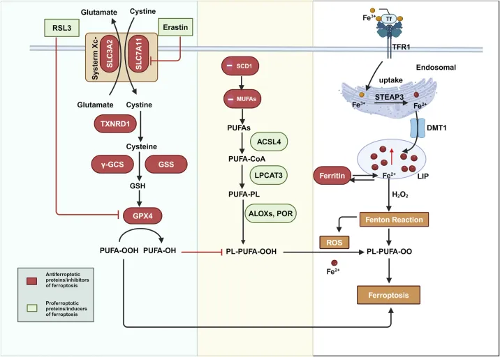
System xc− is a plasma-membrane cystine/glutamate antiporter composed primarily of SLC7A11 and SLC3A2. Cystine imported through this system is reduced to cysteine and used for glutathione (GSH) synthesis. GSH supplies the reducing capacity required by glutathione peroxidase 4 (GPX4), which converts membrane lipid hydroperoxides into less-reactive lipid alcohols.
When cystine uptake, GSH synthesis, or GPX4 activity is compromised, lipid peroxidation can become self-amplifying. However, the axis is plastic. FSP1/AIFM2, DHODH, GCH1, MGST1, and other systems can suppress lipid peroxidation in specific compartments or cell states. A fall in GPX4 protein is therefore neither necessary nor sufficient to define ferroptosis in every model.[2][3]
Iron, lipid remodeling, and execution
Iron can promote lipid-peroxide formation through redox chemistry and iron-dependent enzymes. Transferrin receptor 1 (TFRC), ferritin storage, labile iron, heme metabolism, and iron export all influence the size and availability of the intracellular iron pool. An iron increase may sensitize a cell without being the immediate execution event, and iron chelation can alter many processes besides ferroptosis.
ACSL4 shapes ferroptosis sensitivity by loading long-chain polyunsaturated fatty acids (PUFAs) into phospholipids that are vulnerable to oxidation. ALOX enzymes, POR, and other lipid-oxidizing systems can contribute to peroxidation, while phospholipase and aldehyde-detoxifying systems remove or repair damaged lipids. ACSL4 is therefore a context-dependent susceptibility determinant rather than a universal “on” marker.[4]
Once peroxidized membrane lipids exceed the cell’s repair and detoxification capacity, membrane integrity fails and the cell loses viability. The precise transition from reversible oxidative stress to irreversible ferroptotic death remains an active research question.
Common perturbations and rescue logic
| Perturbation | Primary action | What it does not prove alone |
|---|---|---|
| Erastin or related system xc− inhibitors | Reduce cystine import and weaken GSH/GPX4 protection | That all downstream death is ferroptosis; off-target and cell-line-specific effects must be tested |
| RSL3 or other GPX4 inhibitors | Directly compromise GPX4 activity and accelerate lipid-peroxide accumulation | That the observed response is independent of lipid peroxidation or rescue-sensitive |
| Iron loading or altered iron handling | Increase the available redox-active iron pool | That iron is the only causal driver or that a high-dose oxidative insult is pathway-specific |
| Ferrostatin-1 or liproxstatin-1 | Trap lipid radicals and suppress lipid-peroxidation-driven death | That rescue is specific if the inhibitor also changes the upstream stress or is used at an incompatible dose |
| Genetic GPX4, SLC7A11, ACSL4, or FSP1 manipulation | Test pathway dependence or susceptibility | That a gene-expression phenotype is equivalent to cell-death execution |
A practical ferroptosis claim is strongest when an inducer increases lipid peroxidation and cell death, a chemically distinct ferroptosis inhibitor rescues both readouts, and an orthogonal perturbation or genetic result supports the same pathway. Compare the timing of rescue with the timing of lipid-peroxide accumulation; a late rescue may not reverse an already irreversible membrane injury.
How to detect ferroptosis
Detection should be multiparametric. The international consensus literature emphasizes that lipid peroxidation, pathway perturbation, rescue, and cell-death measurement should be interpreted together rather than relying on a single marker or morphology.[5]
| Readout | What it measures | Key control or limitation |
|---|---|---|
| Cell viability or membrane integrity | Loss of metabolic activity, ATP, membrane exclusion, or clonogenic potential | Use a readout appropriate to the time point; viability loss alone does not identify the pathway |
| C11-BODIPY 581/591 or Liperfluo | Live-cell lipid oxidation or lipid hydroperoxide-associated fluorescence | Requires calibration, matched acquisition, and awareness of probe specificity and autofluorescence |
| Oxidized lipidomics | Specific oxidized phospholipid species and lipid-peroxidation products | Powerful but technically demanding; sample handling can introduce oxidation |
| GPX4, SLC7A11, ACSL4, TFRC, FSP1/AIFM2 | Pathway state or susceptibility | Protein or transcript levels are indirect and context-dependent |
| 4-HNE, MDA, or related adducts | Downstream lipid-peroxidation products | General oxidative stress can produce these products; they are not ferroptosis-specific |
| Electron microscopy | Ultrastructural support such as shrunken mitochondria and dense membranes | Supportive morphology does not replace biochemical and rescue evidence |
| Ferroptosis inhibitor rescue | Functional suppression of lipid-peroxidation-driven death | Use matched doses, timing, and vehicle controls; distinguish rescue from nonspecific cytoprotection |
A multiparametric validation workflow
- Define the model. Record species, cell type, culture conditions, tissue, treatment, time point, and whether the perturbation is genetic, pharmacological, or environmental.
- Measure viability and lipid peroxidation together. Use a live-cell lipid-oxidation probe or a validated lipidomics method alongside a viability or membrane-integrity readout.
- Run a rescue control. Add ferrostatin-1, liproxstatin-1, or another validated lipophilic radical-trapping antioxidant in a parallel condition. Confirm that the rescue reduces both lipid peroxidation and loss of viability where expected.
- Test pathway dependence. Measure or perturb GPX4, SLC7A11, ACSL4, FSP1/AIFM2, iron handling, or relevant lipid enzymes according to the question.
- Exclude common alternatives. Include apoptosis, necroptosis, or general oxidative-stress readouts when the treatment could engage several death modes. Do not infer ferroptosis from morphology alone.
- Report the full time course. A pathway marker can precede, coincide with, or follow loss of viability. Endpoint-only measurements can reverse the apparent order of events.
Limitations and troubleshooting
| Observation | Possible explanation | Next check |
|---|---|---|
| Cell death occurs but ferrostatin-1 does not rescue it | Death is not ferroptosis, inhibitor timing/dose is unsuitable, or the model uses a ferroptosis-suppressor pathway not covered by the test | Test a second rescue inhibitor, a different inducer, lipid peroxidation, and alternative death markers |
| C11-BODIPY signal increases without loss of viability | Reversible lipid stress, early ferroptotic signaling, probe oxidation unrelated to the target pathway, or excessive probe loading | Add a time course, viability readout, inhibitor rescue, and probe-only controls |
| GPX4 decreases but lipid peroxidation is unchanged | Residual GPX4 activity, compensatory FSP1/DHODH/GCH1 pathways, or antibody/normalization artefact | Measure enzyme activity or lipid peroxidation and test compensatory pathways |
| 4-HNE or MDA is elevated | General oxidative stress, inflammation, sample oxidation, or ferroptotic lipid damage | Use lipidomics or a calibrated probe plus ferroptosis inhibitor rescue |
| ACSL4 is high but cells resist ferroptosis | ACSL4 is a susceptibility factor, not a complete execution readout; antioxidant or iron pathways may dominate | Measure lipid composition, GPX4 activity, FSP1, iron handling, and response to matched inducers |
| Results differ between cell lines | Differences in PUFA composition, antioxidant reserve, iron metabolism, transporter expression, or drug uptake | Profile baseline pathway state and normalize the perturbation to the relevant response range |
Disease and research contexts
Ferroptosis research spans cancer biology, ischemia–reperfusion injury, neurodegeneration, kidney disease, infection, and metabolic stress. The pathway may be harmful in one context and therapeutically exploitable in another. In tumors, for example, ferroptosis sensitivity can be shaped by oncogenic state, lipid composition, and antioxidant dependencies. In tissue injury, lipid-peroxidation inhibitors are being evaluated in preclinical models, but clinical translation requires careful control of exposure, tissue compartment, and off-target antioxidant effects.
For fixed tissue, chemical lipid-peroxidation probes may not preserve the relevant signal, while 4-HNE or other products can be detected with immunostaining but are not specific. For in vivo work, combine tissue-compatible readouts with genetic or pharmacological rescue and a time-matched assessment of injury and viability.
Common questions
Is GPX4 loss enough to prove ferroptosis?
No. GPX4 loss or inhibition can increase susceptibility, but ferroptosis should be supported by lipid-peroxidation and viability readouts plus rescue by a suitable ferroptosis inhibitor.
Is iron accumulation alone ferroptosis?
No. Iron availability is one component of the pathway. The relevant outcome is iron-dependent phospholipid peroxidation that drives loss of viability in the tested model.
Can I use 4-HNE as a ferroptosis marker?
Use it as supportive evidence only. 4-HNE can rise during many forms of oxidative stress. Pair it with a ferroptosis rescue experiment, a lipid-peroxidation assay, and pathway-specific controls.
Are erastin and RSL3 interchangeable?
No. Erastin-like compounds primarily perturb cystine import and the GSH/GPX4 axis, whereas RSL3-like compounds directly inhibit GPX4. Their timing, potency, and pathway dependence can differ substantially across models.
Does a ferroptosis inhibitor rescue prove the mechanism?
It is strong but not sufficient evidence. Rescue should be paired with lipid-peroxidation measurement, cell-death quantification, and an orthogonal pathway perturbation because some antioxidants and inhibitors have broader effects.
References
- Dixon SJ, Lemberg KM, Lamprecht MR, et al. Ferroptosis: an iron-dependent form of nonapoptotic cell death. Cell. 2012;149:1060–1072. doi:10.1016/j.cell.2012.03.042
- Conrad M, Friedmann Angeli JP. Glutathione peroxidase 4 (Gpx4) and ferroptosis: what’s so special about it? Molecular and Cellular Oncology. 2015;2:e995047. doi:10.4161/23723505.2014.995047
- Yang WS, Stockwell BR. Ferroptosis: death by lipid peroxidation. Trends in Cell Biology. 2016;26:165–176. doi:10.1016/j.tcb.2015.10.014
- Doll S, Proneth B, Tyurina YY, et al. ACSL4 dictates ferroptosis sensitivity by shaping cellular lipid composition. Nature Chemical Biology. 2017;13:91–98. doi:10.1038/nchembio.2239
- Galluzzi L, Vitale I, Aaronson SA, et al. Molecular mechanisms of cell death: recommendations of the Nomenclature Committee on Cell Death 2018. Cell Death & Differentiation. 2018;25:486–541. doi:10.1038/s41418-017-0012-4
- International consensus guidelines for the definition, detection, and interpretation of autophagy-dependent ferroptosis. Autophagy. 2024. doi:10.1080/15548627.2024.2319901
- Feng H, Schorpp K, Jin J, et al. A systematic protocol for the detection of ferroptosis in cultured cells. Nature Protocols. 2023. Open-access protocol
- Kagan VE, Mao G, Qu F, et al. Oxidized arachidonic and adrenic PEs navigate cells to ferroptosis. Nature Chemical Biology. 2017;13:81–90. doi:10.1038/nchembio.2238
Ferroptosis should be assigned from a convergent pattern of lipid peroxidation, pathway dependence, cell-death rescue, and appropriate controls. Marker abundance alone is not a diagnosis of ferroptosis.
 | Flora Flora is a technical support expert at EnkiLife, familiar with immunology and neuroscience, dedicated to providing customers with high-quality product combinations and technical support to help achieve research in neurodegenerative diseases and other neuroscience areas. |