Metabolic Enzyme UPP1 How Does it Cross-Regulate Tumor Immunity?
I. Research Background
Lung cancer remains the leading cause of cancer-related deaths globally. As the most prevalent histological subtype of lung cancer, lung adenocarcinoma (LUAD) continues to have high incidence and mortality rates, posing a severe threat to human health. Despite recent advancements in targeted therapy, immunotherapy, and other novel diagnostic and treatment approaches that have significantly improved the clinical management of LUAD patients, many patients still face challenges such as high tumor microenvironment heterogeneity, acquired therapeutic resistance, and significant individual differences in treatment response. Overall prognosis remains unsatisfactory. Therefore, exploring the molecular mechanisms underlying LUAD progression and identifying novel prognostic biomarkers and precise therapeutic targets are critical for overcoming current clinical treatment bottlenecks. The complex remodeling of the tumor microenvironment (TME) is a core factor driving LUAD occurrence, invasion, metastasis, and immune escape. Abnormal interactions between tumor cells and immune/stromal cells in the microenvironment continuously shape an immunosuppressive microenvironment that blocks the host's anti-tumor immune response. Uridine phosphorylase 1 (UPP1) is a key enzyme mediating uridine metabolism and maintaining the homeostasis of the pyrimidine salvage synthesis pathway. Previous studies have confirmed its role in regulating tumor glycolytic metabolism, supporting tumor cell survival and proliferation under nutrient-deprived conditions, and preliminary evidence suggests potential associations with tumor immune checkpoints and anti-tumor T cell infiltration. However, the specific role of UPP1 in TME remodeling, its molecular regulatory pathways, and clinical translational value in LUAD have not been systematically elucidated. Based on this, Li et al. published a study in 2024 in Nature Communications entitled "UPP1 promotes lung adenocarcinoma progression through the induction of an immunosuppressive microenvironment". Through integrated multi-omics analysis and multi-dimensional experimental validation, this study systematically resolved the core mechanisms by which UPP1 mediates the formation of an immunosuppressive microenvironment and drives tumor progression in LUAD, providing novel theoretical basis for LUAD prognosis assessment and personalized immunotherapy.
II. Research Methods
This study employed a comprehensive research system combining integrated multi-omics analysis with in vitro and in vivo functional validation. First, five independent LUAD single-cell RNA sequencing datasets were integrated, including 117 LUAD patient samples and a total of 377,574 cells. After quality control processes including data integration, normalization, and batch effect removal, cell clustering and tumor cell subpopulation identification were completed. Combined with single-sample gene set enrichment analysis and survival analysis, a UPP1-high expressing tumor cell subpopulation closely associated with poor patient prognosis was identified, and its biological functions were predicted through functional enrichment analysis. The study utilized a tissue microarray cohort of 205 LUAD patients from Zhongshan Hospital, Fudan University, and validated the association between UPP1 expression and patient overall survival, recurrence-free survival, and tumor differentiation using immunohistochemical staining and AI image analysis. Meanwhile, multiplex immunofluorescence staining and cell communication analysis were employed to clarify the spatial distribution and interaction relationships between UPP1-high expressing tumor cells and immune/stromal cells in the microenvironment. High-throughput cytokine array and ELISA experiments were used to screen for core immunosuppressive cytokines regulated by UPP1, and Transwell co-culture systems were used to validate the mechanisms by which UPP1 regulates various microenvironment cell phenotypes through target factors. The study further utilized flow cytometry and CyTOF mass cytometry to resolve the remodeling effects of UPP1 on immune lineages and stromal cells in the tumor microenvironment at the cellular level, and validated the in vivo immune escape effects mediated by UPP1 using immunocompetent and immunodeficient mouse models. Additionally, the study integrated three major drug genomics datasets (CTRP, GDSC, PRISM) for drug screening, and validated potential targeted drugs for UPP1-high expressing LUAD through cell experiments, patient-derived organoid (PDO) models, and in vivo mouse administration experiments. Finally, pathway intervention experiments were performed to clarify the molecular signaling pathways through which UPP1 regulates target genes.
III. Results Analysis
3.1 Identification of Prognostic Value and Functional Characteristics of UPP1-high Expressing Tumor Cell Subpopulation
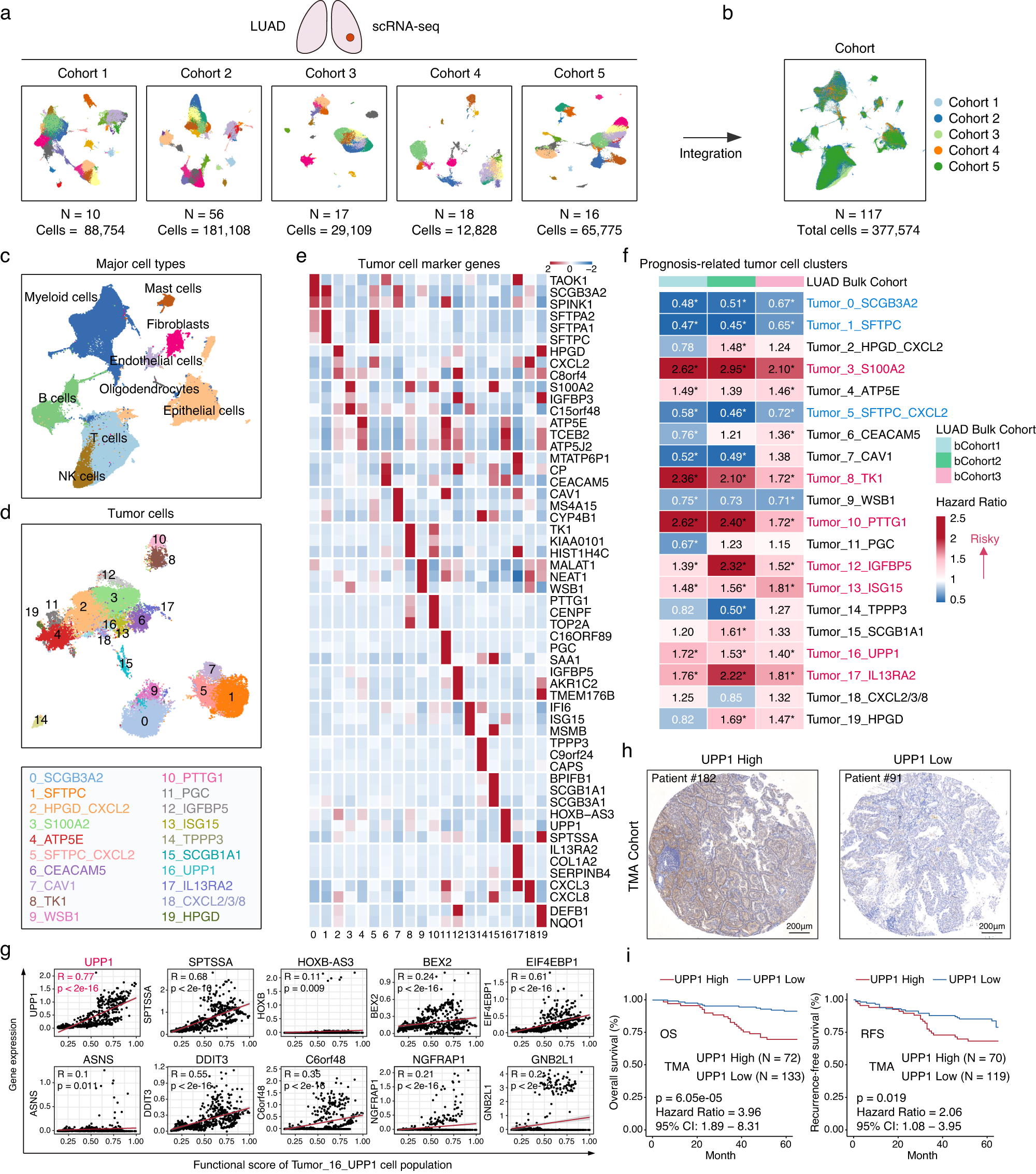
Through large-scale single-cell sequencing data integration analysis, this study accurately characterized the cellular atlas of the LUAD tumor microenvironment. After precise clustering of eight major cell populations including immune cells, stromal cells, and tumor cells, tumor cells were further subdivided into 20 subpopulations with unique molecular characteristics. Combined with survival prognosis data from large clinical samples, the association between each subpopulation and patient prognosis was verified one by one. Ultimately, seven tumor cell subpopulations that significantly drive poor prognosis in LUAD were identified, among which the UPP1-high expressing tumor cell subpopulation (Cluster 16) demonstrated strong prognostic value, and this subpopulation had been significantly understudied previously. Functional enrichment analysis revealed that UPP1-high expressing tumor cells simultaneously enriched two pro-cancer functional modules: on one hand, they significantly activated classical tumor-intrinsic malignant pathways including tumor invasion and metastasis, glycolytic metabolic reprogramming, MYC target genes, and MAPK, supporting tumor proliferation, invasion, and drug resistance; on the other hand, they significantly enriched microenvironment remodeling-related pathways such as TGF-β signaling, inflammatory response, and negative regulation of immune response. This fully demonstrates that UPP1 not only regulates tumor cell-intrinsic malignant phenotypes but also deeply participates in the immunosuppressive remodeling of the tumor immune microenvironment. More importantly, correlation analysis of the top 10 core signature genes of this subpopulation confirmed that UPP1 gene expression level showed the strongest positive correlation with the overall functional enrichment score of this tumor subpopulation, indicating it is the core driver gene that dominates both malignant proliferation and immunosuppressive phenotypes of this subpopulation. Large-scale cohort validation using clinical tissue samples further confirmed that high UPP1 expression in LUAD tissues was significantly negatively correlated with shortened patient overall survival and recurrence-free survival, and was closely associated with the malignant pathological feature of poor tumor differentiation. This clinically solidified UPP1's core value as a novel prognostic biomarker for LUAD and laid the foundation for subsequent exploration of its mechanisms in regulating the tumor microenvironment.
3.2 UPP1-high Expressing Tumor Cells Specifically Remodel Immunosuppressive Tumor Microenvironment
The spatial distribution characteristics of tumor cells often determine their malignant functions. Through multiplex immunofluorescence staining combined with AI visualization analysis, this study found that UPP1-high expressing tumor cells are not uniformly distributed within tumor tissues but specifically cluster at the invasive front of tumors. This special location enables them to directly interact with immune cells and stromal cells at the tumor boundary, making them key nodes for microenvironment remodeling. To systematically elucidate the microenvironment interaction network of UPP1-high expressing tumor cells, the study systematically analyzed their co-enrichment patterns, abundance correlations, and spatial distance relationships with various immune cells and stromal cells. The results confirmed significant spatial co-localization and close-distance distribution characteristics between UPP1-high expressing tumor cells and four types of typical pro-cancer, immunosuppressive cells: FOXP3+ regulatory T cells, MMP11+ cancer-associated fibroblasts, LAG3+PDCD1+ exhausted CD8+ T cells, and SPP1+ M2-type macrophages. Compared with UPP1-low expressing tumor cells, UPP1-high expressing tumor cells showed significantly shortened spatial distances to these four types of inhibitory cells, forming locally dense and stable immunosuppressive cell microecological niches at the tumor invasive front, providing a structural basis for tumor immune escape and invasive growth. Further CellphoneDB cell communication analysis precisely resolved the bidirectional signal interaction mechanisms at the molecular level. UPP1-high expressing tumor cells can construct complex inhibitory signal networks through multiple ligand-receptor pairs: through classical immune checkpoint pathways including PD-L1/PD-1, FGL1/LAG3, and NECTIN/TIGIT, they directly bind to and inhibit the anti-tumor killing function of T cells; through cytokine pathways including TGFB1-TGFBR and IL2-IL2R, they actively induce the activation and proliferation of immunosuppressive cells; meanwhile, through chemokine pathways including CCL2/CCR2 and CXCL8/CXCR1, as well as growth factor pathways, they continuously recruit macrophages and fibroblasts to infiltrate and accumulate at the tumor invasive front. These multi-level, multi-dimensional results collectively confirm that UPP1-high expressing tumor cells do not passively adapt to the tumor microenvironment but actively construct an immunosuppressive microenvironment with themselves as the core through spatial-specific colonization, inhibitory cell recruitment, and bidirectional signal interactions, creating optimal microenvironmental conditions for sustained tumor proliferation, local invasion, immune escape, and long-term recurrence and metastasis.

3.3 UPP1 Mediates Comprehensive Immunosuppressive Microenvironment Formation Through TGF-β1 Pathway
To unravel the core molecular mechanism by which UPP1 remodels the immunosuppressive microenvironment, the study used high-throughput cytokine array screening and found that UPP1-overexpressing LUAD tumor cells secrete large amounts of various immunosuppressive cytokines, with TGF-β1 showing the most significant upregulation, becoming the core effector factor for UPP1-regulated microenvironment. ELISA experiments also verified this result at the protein secretion level. To realistically simulate the physiological indirect interaction between tumor cells and microenvironment cells in vivo and avoid experimental biases from direct co-culture, the study constructed multiple Transwell indirect co-culture systems, separately co-culturing UPP1-overexpressing tumor cells with CD4+ T cells, CD8+ T cells, THP-1-derived macrophages, and lung fibroblasts, and set up TGF-β1 neutralizing antibody blocking experimental groups and IgG control groups to rigorously verify the core mediating role of TGF-β1 in UPP1-mediated microenvironment remodeling. The results showed that TGF-β1 secreted by UPP1-high expressing tumor cells can comprehensively regulate the phenotypic polarization of various immune and stromal cells. In adaptive immunity, TGF-β1 can induce CD4+ T cells to differentiate into immunosuppressive Treg cells, and simultaneously promote CD8+ T cells to highly express exhaustion markers such as PD-1 and LAG3, losing anti-tumor killing function; in innate immunity, TGF-β1 can drive THP-1-derived macrophages to polarize into SPP1+ M2-type pro-cancer macrophages, upregulating immunosuppressive markers such as PD-L1 and CD163; in stromal remodeling, TGF-β1 can induce fibroblasts to transform into cancer-associated fibroblasts highly expressing FAP and MMP11, activating pro-cancer programs such as extracellular matrix remodeling and epithelial-mesenchymal transition. After blocking TGF-β1 function with neutralizing antibodies, all the above immunosuppressive and pro-cancer phenotypes were significantly reversed, completely confirming the core mechanism by which UPP1 constructs an immunosuppressive microenvironment from both immune cell and stromal cell dimensions through activating TGF-β1 secretion, clearly linking the complete regulatory chain of UPP1, core cytokines, and microenvironment cell phenotype remodeling.

3.4 UPP1 Upregulates PD-L1 Through PI3K/AKT/mTOR Pathway to Promote Tumor Immune Escape
Abnormally high expression of the immune checkpoint PD-L1 is a core mechanism by which LUAD tumor cells achieve immune escape and develop clinical resistance to immunotherapy, and is also a core focus of current tumor immune regulation research. To comprehensively explore another key pathway by which UPP1 participates in tumor immune escape, this study broke through single-omics limitations and integrated single-cell sequencing, large clinical bulk transcriptome, CCLE tumor cell line mRNA, and protein multi-dimensional databases to cross-validate the expression correlation between UPP1 and various tumor immune checkpoint molecules. The results showed that UPP1 expression level was significantly positively correlated with PD-L1 expression in LUAD tumor cells at transcription, protein, and single-cell levels, suggesting a stable and close specific regulatory association between them. To deeply explore the upstream molecular mechanism by which UPP1 regulates PD-L1 expression, the study conducted enrichment screening of classical PD-L1 regulatory pathways in UPP1-high expressing tumor cells. Among the six known regulatory pathways, the PI3K/AKT/mTOR signaling pathway showed significant specific high activation and was identified as the core pathway mediating UPP1-regulated PD-L1. Subsequent molecular biology experiments verified this regulatory chain step by step: abnormal upregulation of UPP1 can significantly promote phosphorylation modification of PI3K, AKT, and mTOR proteins, continuously activating the PI3K/AKT/mTOR signaling pathway, thereby driving high PD-L1 expression at both transcriptional and protein levels; specific inhibition of UPP1 expression significantly downregulated PI3K/AKT/mTOR pathway activity, with PD-L1 expression decreasing accordingly. More importantly, pathway rescue experiments using the AKT-specific activator SC79 effectively reversed the PD-L1 downregulation phenotype caused by UPP1 inhibition, confirming the linear molecular mechanism by which UPP1 positively regulates PD-L1 expression through activating the PI3K/AKT/mTOR pathway from three-dimensional experimental perspectives of "inhibition-activation-rescue". Functional cell killing experiments further verified that PD-L1 upregulation caused by high UPP1 expression significantly inhibits the secretion of killing effector molecules such as perforin and granzyme B by CD8+ T cells, reducing the ability of CD8+ T cells to clear tumor cells, while blocking PD-L1 can effectively restore the anti-tumor activity of T cells. From both molecular mechanism and immune function perspectives, this completes another important pathway by which UPP1 mediates tumor immune escape, supplementing the multi-pathway mechanism by which UPP1 regulates the tumor microenvironment.

3.5 CyTOF Mass Cytometry Panoramically Validates UPP1's In Vivo Immunosuppressive Function
To avoid the limitations of in vitro cell experiments with single microenvironments and lack of in vivo immune homeostasis and stromal interactions, and to further validate UPP1's immunosuppressive function in a real in vivo environment, the study used CyTOF mass cytometry technology, selecting 21 core protein markers of the tumor microenvironment to perform panoramic immune atlas analysis on UPP1-overexpressing and control mouse tumor tissues, characterizing the remodeling effects of UPP1 on immune lineages and stromal components in the in vivo tumor microenvironment at the overall level. Immune cell subpopulation analysis results showed that the immune landscape of UPP1-high expressing tumor microenvironment underwent significant immunosuppressive remodeling: the infiltration proportion of immunosuppressive CD25+FOXP3+ Treg cells significantly increased, and the IL-10 immunosuppressive factor secretion capacity of these cells significantly enhanced, continuously suppressing local anti-tumor immunity; while the body's core anti-tumor effector cells, CD8+ T cells, showed typical severe exhaustion phenotypes, with multiple immune checkpoint molecules including PD-1, LAG3, TIM3, and CTLA4 simultaneously highly expressed, and the secretion of anti-tumor effector cytokines such as TNF-α and IFN-γ significantly decreased, completely losing tumor killing function. In myeloid immune cells, UPP1 overexpression significantly promoted the infiltration and polarization of CD163+PD-L1+ M2-type macrophages in the tumor microenvironment. These cells can secrete large amounts of anti-inflammatory and pro-cancer factors such as IL-4 and IL-10, further amplifying the overall immunosuppressive state of the microenvironment and forming a positive feedback loop of immunosuppression. In stromal remodeling, UPP1 significantly promoted the infiltration and activation of fibroblasts in the tumor microenvironment, inducing fibroblasts to highly express cancer-associated fibroblast characteristic markers such as MMP11 and FAP, accelerating tumor extracellular matrix remodeling, fibrosis, and invasion and metastasis processes. Through panoramic, unbiased in vivo immune mass spectrometry detection, this study intuitively and comprehensively confirmed that UPP1 can reshape the immunosuppressive tumor microenvironment in a real in vivo environment, consolidating the reliability of previous in vitro molecular mechanisms and cell function experiments at the animal level, and providing solid in vivo experimental evidence for UPP1 as a core target for promoting cancer and immune escape.

3.6 Inhibiting UPP1 Enhances CD8+ T Cell Function and Sensitizes Anti-PD-L1 Immunotherapy
Based on the core mechanism by which UPP1 dual-regulates TGF-β1 and PD-L1, mediates immunosuppressive microenvironment and tumor immune escape, the study further focused on clinical translational value, exploring the synergistic potential of UPP1-targeted intervention combined with immunotherapy to provide new strategies for solving the problem of immunotherapy resistance in LUAD. Through constructing LUAD mouse tumor-bearing models with stable UPP1 knockdown, four groups were set up: blank control, anti-PD-L1 immunotherapy alone, UPP1 inhibition alone, and UPP1 inhibition combined with anti-PD-L1 therapy. The anti-tumor effects and changes in immune microenvironment of different intervention methods were systematically compared. In vivo experimental results showed that anti-PD-L1 immunotherapy alone had limited inhibitory effect on LUAD tumor growth, with significant clinically common immunotherapy resistance. The reason is that there are multiple immunosuppressive barriers in the tumor microenvironment, such as Treg cells, exhausted T cells, and M2 macrophages. Blocking PD-L1 alone cannot relieve the overall immunosuppressive state. Specific knockdown of UPP1 expression in tumor cells alone can significantly inhibit tumor proliferation and growth in vivo, effectively reshape the tumor microenvironment, significantly increase the number and proportion of infiltrating CD8+ T cells in the microenvironment, significantly restore the secretion levels of anti-tumor effector molecules such as perforin, granzyme B, TNF-α, and IFN-γ in CD8+ T cells, effectively reverse the exhaustion state of CD8+ T cells, and relieve local immunosuppression. Most clinically valuable is that after combined intervention of UPP1 targeted inhibition and anti-PD-L1 immunotherapy, the two components showed strong synergistic anti-tumor effects, with tumor proliferation inhibition rate and tumor regression degree significantly better than all single treatment groups, maximizing the relief of multiple immunosuppressive barriers in the tumor microenvironment and completely awakening the body's systemic anti-tumor immune response. This part of the results fully confirmed that UPP1 is a key driver of immunotherapy resistance in LUAD. Targeted inhibition of UPP1 can effectively break through the immunosuppressive barriers of the tumor microenvironment, reverse immune resistance, and significantly sensitize anti-PD-L1 immunotherapy, providing a novel and reliable translational strategy for clinical personalized combined immunotherapy in LUAD.

3.7 Bioinformatics Screening and Validation of Targeted Drugs for UPP1-high Expressing LUAD
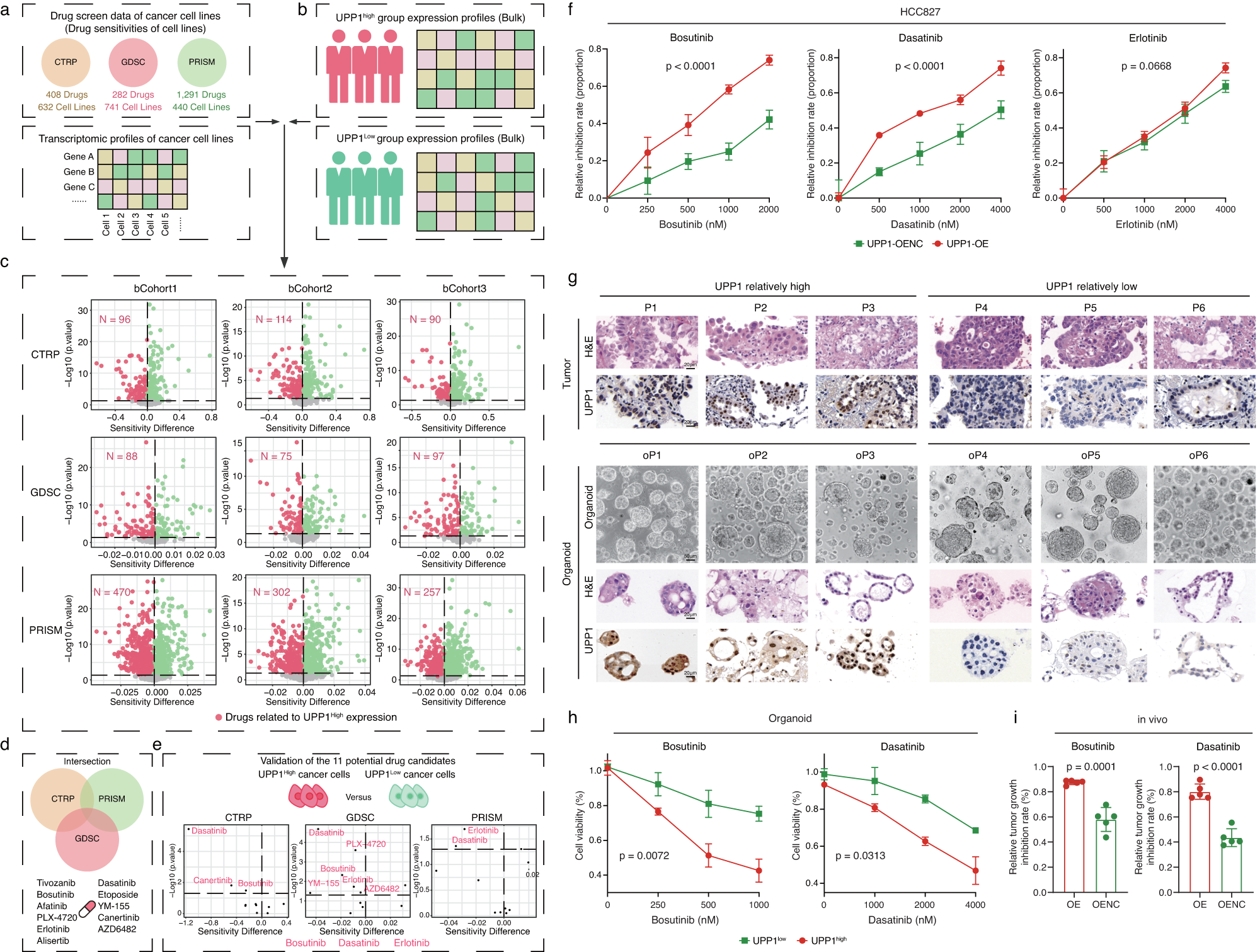
To achieve precise personalized treatment of LUAD based on UPP1 expression stratification and fill the clinical gap of lacking specific targeted drugs for UPP1-high expressing LUAD, the study relied on three major international authoritative drug genomics databases (CTRP, GDSC, PRISM), combined with gene expression and drug sensitivity data from large samples of LUAD patients, and performed differential drug sensitivity stratification analysis for UPP1-high and low expressing subgroups. Three small molecule targeted drugs with potential specific killing effects on UPP1-high expressing LUAD were initially screened: Bosutinib, Dasatinib, and Erlotinib. To verify the reliability of bioinformatics screening results, the study first conducted in vitro cell function validation. The results showed that compared with UPP1-low expressing LUAD tumor cells, UPP1-high expressing tumor cells had significantly increased drug sensitivity to Bosutinib and Dasatinib (two Src kinase inhibitors), with significantly higher cell proliferation inhibition rate and apoptosis level, while Erlotinib showed no significant efficacy difference on tumor cells with different UPP1 expression levels, excluding its specific targeting value. To maximize the approximation to the real tumor biological characteristics in clinical practice and avoid heterogeneity differences between ordinary cell lines and clinical tumors, the study constructed PDO organoid models from 6 clinical LUAD patients, preserving the original genetic characteristics, pathological phenotypes, and drug response characteristics of the patients' tumors. The PDO drug intervention results were highly consistent with cell experiments. Patient organoids with higher UPP1 expression showed more significant drug response to Bosutinib and Dasatinib, with more obvious organoid proliferation inhibition effects, fully verifying the targeting specificity of the two drugs. Finally, the study performed final efficacy verification through mouse in vivo tumor-bearing administration models. The results confirmed that Bosutinib and Dasatinib can specifically inhibit the in vivo growth of UPP1-high expressing LUAD, effectively block tumor proliferation and progression, with controllable toxic side effects. This part of the study completed drug effectiveness verification layer by layer and multi-dimensionally, from big data bioinformatics screening, in vitro cell verification, clinical organoid model simulation to animal in vivo efficacy verification, providing clear and reliable candidate drugs for stratified precise targeted therapy of UPP1-positive LUAD patients.
IV. Conclusion
Based on large-scale multi-omics integrated analysis and multi-level in vitro and in vivo experiments, this study systematically elucidated the dual roles of UPP1 as a novel prognostic biomarker and core pro-cancer factor in LUAD, completely revealing the molecular mechanisms by which UPP1 reshapes the immunosuppressive microenvironment and drives tumor progression through multiple pathways and dimensions, while exploring corresponding clinical treatment strategies, with extremely high basic research value and clinical translational significance. The study first successfully defined the UPP1-high expressing tumor cell subpopulation with poor prognosis through single-cell sequencing, confirming that UPP1-high expressing tumor cells are located at the tumor invasive front and can actively recruit immunosuppressive cells such as Treg cells, exhausted CD8+ T cells, M2-type macrophages, and cancer-associated fibroblasts through intercellular signal interactions to construct a pro-cancer microenvironment. At the mechanism level, two core regulatory pathways were identified: on one hand, UPP1 high expression can induce tumor cells to secrete large amounts of TGF-β1, comprehensively mediating the pro-cancer phenotypic polarization of immune cells and stromal cells; on the other hand, it upregulates PD-L1 expression through activating the PI3K/AKT/mTOR signaling pathway, directly inducing CD8+ T cell exhaustion and mediating tumor immune escape. These dual pathways jointly promote the malignant progression and immune resistance of LUAD. In vivo CyTOF panoramic immune atlas and mouse model experiments further consolidated UPP1's immunosuppressive function, confirming that targeted silencing of UPP1 can effectively restore the body's anti-tumor immunity and sensitize anti-PD-L1 immunotherapy. In addition, the study innovatively combined drug genomics screening, patient-derived organoids, and animal model validation to identify Bosutinib and Dasatinib as specific targeted drugs for UPP1-high expressing LUAD, providing a novel direction for stratified precise treatment of patients. Overall, this study improves the regulatory theory of the LUAD tumor microenvironment. However, the study did not further explore the cross-link between UPP1's metabolic function and immune regulatory function, and the long-term efficacy and safety of drug combined immunotherapy still need to be verified in large-scale clinical cohorts, leaving room for subsequent in-depth research and clinical application.
References
Li Y, Jiang M, Aye L, Luo L, Zhang Y, Xu F, Wei Y, Peng D, He X, Gu J, Yu X, Li G, Ge D, Lu C. UPP1 promotes lung adenocarcinoma progression through the induction of an immunosuppressive microenvironment. Nat Commun. 2024 Feb 8;15(1):1200. doi: 10.1038/s41467-024-45340-w. PMID: 38331898; PMCID: PMC10853547.