Antibody Preparation FAQs – Hybridoma Cell
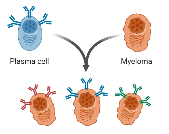
In the field of monoclonal antibody preparation, cell fusion is a key link in obtaining hybridoma cells. The purpose is to fuse B lymphocytes and myeloma cells together so that the genetic material of the two cells can be recombined with each other. The hybridoma cells obtained in this way can not only maintain the function of B cells secreting antibodies, but also obtain the characteristics of unlimited proliferation of myeloma cells.
Cell fusion steps
1. Myeloma cell culture: Myeloma cell lines such as SP2/0, NS0, or P3 - X63 - Ag8.653 from mouse. To culture, RPMI 1640 or DMEM medium containing 10% fetal bovine serum (FBS) was used at 37°C at 5% CO₂. Cells in the logarithmic growth stage should be selected, and the viability should reach more than 95%, and the cell morphology should be rounded and well-dispersed. Cells should be passaged 1-2 days before fusion to prevent cell aging, and cells should be washed with serum-free medium 3 times before fusion to remove components in the serum that may affect fusion.
2. Splenocyte preparation: 3-4 days after the last immunization, when the number of antigen-specific B cells in the spleen is the largest, the material is taken at this time. The specific operation is to remove the spleen of the immune mouse under sterile conditions, cut it with scissors, and grind it through a 200-mesh cell sieve to obtain a single-cell suspension. The cell suspension was then centrifuged at 1000 rpm for 5 minutes. Dump the supernatant, wash the cells twice with serum-free medium, and perform a cell count.
3. Cell mixing and centrifugation: Mix splenocytes and myeloma cells at a ratio of 5:1 to 10:1. Centrifuge the mixed cell suspension at 1000 rpm for 5 minutes, pour out the supernatant after centrifugation, and then gently flick the bottom of the tube to loosen the cell pellet and prevent cell agglomeration from affecting the subsequent fusion effect.
4. PEG-induced fusion: Preheat 50% PEG and serum-free medium to 37°C first. Slowly add 1 mL of 50% PEG to the cell pellet over 1 minute at 37°C, gently rotating the tube to allow the PEG to contact the cells evenly. After dropping is complete, let it sit at 37°C for 1 - 2 minutes, too long can increase cell mortality.
5. Termination of the fusion reaction: After the reaction is completed, add 10ml DMEM medium containing 10% FBS to terminate the reaction, gradually add from slow to fast, and add all the reactions in 6 minutes. Gently shake the tube as you add to gradually dilute the PEG to reduce its toxicity to cells. Centrifuge at 1000 rpm for 5 minutes, discard the supernatant, and resuspend the cells with complete medium containing HAT.
6. Plating and Screening: Inoculate the fused cell suspension into a 96-well cell culture plate, adding 105 cells per well to ensure an appropriate cell density for subsequent HAT screening. Place the culture plate in a constant-temperature incubator set at 37°C with 5% CO₂, and culture the cells in a complete medium containing HAT. After culturing for 3–7 days, aspirate and discard the culture supernatant, then replace the medium according to the cell growth status. After medium replacement, pay attention to observing the cell morphology and cell growth. Conduct ELISA to perform positive screening detection for fusion 2–3 days later.
Common Questions
1. Few Clones After Fusion
(1) Insufficient cell viability is the primary cause. The viability of immune B cells may decrease due to poor immune status of the animal or improper blood collection time; alternatively, myeloma cells may experience reduced proliferation capacity due to contamination during culture or excessive passage. Both scenarios directly affect the success rate of fusion.
(2) Imbalanced ratio of the two cell types can be a factor. Typically, the optimal ratio of B cells to myeloma cells ranges from 5:1 to 10:1. An excessively high or low ratio will lead to insufficient interaction between cells, failing to form an adequate number of hybridoma cells.
(3) Details in the fusion operation process require attention. In the commonly used PEG fusion method, the quality and concentration of PEG are core influencing factors: insufficient purity of PEG introduces impurity toxicity, excessively high concentration causes fatal damage to cells, and excessively low concentration fails to effectively induce cell membrane fusion. Meanwhile, improper temperature control during fusion can also cause problems—excessively high temperatures accelerate cell apoptosis, while excessively low temperatures inhibit the fluidity of cell membranes and hinder the fusion process.
(4) Issue with plating density: It is recommended that the total number of cells plated in each well of a 96-well plate be 105.
2. No Positive Clones Detected After Fusion
(1) Insufficient sensitivity of the detection method fails to identify positive clones with low antibody secretion. For example, in the ELISA experiment, this may result from excessively low concentration of coated antigen, poor activity of enzyme-labeled secondary antibody, or inactivated chromogenic substrate.
(2) Insufficient specificity of the detection method leads to interference from miscellaneous signals. The absence of properly designed negative and positive controls may cause misjudgment of negative results.
(3) Excessively small fused cell clusters result in insufficient antibody secretion (too little antibody produced to be detected).
(4) The growing cell clusters are either false fused cells or abnormal cell clusters (which do not secrete the target specific antibody, thus failing to be identified as positive clones).
3. Positive Signals in All Wells Detected After Fusion
(1) Insufficient specificity of the detection method and reagents. For instance, poor specificity of the secondary antibody, excessively high concentration or insufficient purity of the coated antigen, overly strong specificity of the chromogenic substrate, prolonged chromogenic time, or excessively high chromogenic temperature. Therefore, parallel negative and positive control wells should be set up simultaneously during detection.
(2) Nutrient enrichment of the medium leads to an excessively long survival time of unfused cells, which secrete a large amount of positive antibodies into the wells. The medium should be replaced before performing the detection again.
(3) Excessive cell colonies in each well of the cell plate. The cells should be diluted and transferred to a new plate for culture; once they grow, the detection should be conducted again.
4. Slow Growth of Fused Cells
(1) The culture medium, especially the serum, is not suitable for cell growth. It is essential to select high-quality serum that is suitable for hybridoma growth.
(2) The culture consumables are not cell-friendly.
(3) The cells are contaminated, such as by mycoplasma, which can sometimes even lead to cell death.
(4) Some individual cells have special nutritional requirements, making them difficult to grow or causing slow growth; however, they have a strong ability to secrete antibodies, and a strong signal can be detected even with only a few such cells in one well. This can be judged after determining the titer of the supernatant.
5. Positive Clones Turn Negative After Several Rounds of Subcloning
(1) The cell line is impure, and non-secreting cells dominate. During the subcloning process, positive cells may either fail to be seeded into the cell wells, or even if they are seeded in a timely manner, the clones fail to grow.
(2) The cell line is unstable and prone to chromosome loss. Use the limiting dilution method or single-cell sorting technology to perform at least 2–3 rounds of subcloning, and regularly detect the antibody secretion level and specificity. Eliminate clones with a rapid decline in antibody titer or altered specificity during passage, and retain cell lines with stable secretion function.
(3) The cell growth environment is unfavorable. For example, when attempting to rescue cells in poor condition, many cells may have already undergone degeneration.
(4) After cell contamination, the cells are also likely to stop secreting antibodies; the most typical case is mycoplasma contamination.
6. Hybridomas Injected into Mouse Peritoneum Fail to Produce Ascites, Only Form Solid Tumors, or the Produced Ascites Has No Titer
(1) An inappropriate mouse strain is selected. Abnormal immune function in the mouse itself can trigger an intraperitoneal inflammatory response, which hinders ascites production or leads to the formation of solid tumors.
(2) Insufficient dose of the sensitizer, incorrect injection method, or excessively long interval (more than 7–14 days) after sensitization will result in poor sensitization effect.
(3) The number of cells injected into the peritoneum is insufficient. Each mouse should be injected with 10⁵–10⁶ cells each time, suspended in sterilized PBS or incomplete medium.
(4) The reason for excessive solid tumor formation may be that during injection, the abdominal subcutaneous tissue, muscle, or intestine is punctured, and the cells are not fully injected into the peritoneum. Injection technique is crucial.
(5) The antibody produced by the hybridoma specifically binds to a certain molecule in the mouse body, resulting in neutralization of the produced antibody. The target protein against which such antibodies are directed has a high sequence homology with the mouse's own protein substances.
Regarding the potential issues that may arise during monoclonal antibody preparation, we will continue to collect relevant problems. We also welcome everyone to raise more issues encountered in practical work, so that we can discuss, learn and improve together. Our official website provides a variety of monoclonal antibodies for everyone to purchase. Visit our website to view Mouse Monoclonal Antibody.
 | Felicia Felicia is a technical support specialist at EnkiLife, with extensive professional experience in antibody development, optimization, and ELISA assay design and application. She is committed to assisting our clients in selecting suitable antibody products, optimizing ELISA experimental protocols, and resolving technical challenges encountered in the process, thereby supporting the smooth progress of their life science research projects. |