In pathology and tumor immunology research, tissue sections carry enormous amounts of information, but traditional immunohistochemistry (IHC) methods typically detect only 1-2 biomarkers at a time, which not only wastes samples but also limits the understanding of complex microenvironments. In recent years, multiplex immunohistochemistry/fluorescence (mIHC) technology, with its powerful capability of "one tissue section, multiple biomarkers detected simultaneously," is revolutionizing the landscape of tissue analysis. So, how powerful is mIHC exactly? Today, we will provide you with a comprehensive understanding of this revolutionary technology from the perspectives of technical principles, experimental workflow, application cases, and advantages analysis.
mIHC is a multiplex immunodetection technology based on fluorescent labeling and tyramide signal amplification (TSA). Through this technology, researchers can detect multiple protein biomarkers on the same tissue section while preserving spatial information, enabling "spatial omics" analysis.
Traditional IHC is limited by the number of staining channels and antibody species cross-reactivity issues, while mIHC breaks through these limitations through the following core principles:
TSA Signal Amplification
Enzymatic reaction generates fluorescent deposits that label the target protein location.
Signal intensity is 10-100 times higher than traditional fluorescence.
Even low-expression proteins can be clearly visible.
Cyclic Staining and Antibody Stripping
Each round detects one antigen, followed by removal of primary/secondary antibodies without affecting tissue structure.
The next round of staining can use different fluorescence channels, and after multiple rounds of accumulation, multi-biomarker co-localization is achieved.
Multi-channel Fluorescence Imaging
Through multispectral imaging technology, overlapping fluorescence signals can be deconvolved to resolve the spatial distribution of different biomarkers.
In short, mIHC allows you to "see the full picture with one tissue section" while maintaining tissue spatial information, which is unmatched by traditional IHC.
Tissue Pretreatment
Xylene I → Xylene II → 100% Ethanol → 95% Ethanol → 85% Ethanol → 75% Ethanol → Distilled Water Wash
Antigen Retrieval
High-temperature antigen retrieval → Cooling → Endogenous enzyme blocking
Cyclic Staining (1 biomarker per round)
Blocking → Primary antibody incubation → Secondary antibody incubation → TSA fluorescent substrate reaction → Washing → Antibody stripping
Repeat the above cycle for 6 rounds
Select different fluorescence channels for each round to accumulate biomarker fluorescence information
Nuclear Staining and Mounting
DAPI nuclear staining → Mounting → Multi-channel microscope imaging
Note: Strict antibody stripping is required after each round of cyclic staining, otherwise cross-signaling may occur.
Assuming we detect 6 immune-related biomarkers on a tumor tissue section: CD8, TIM-3, PD-1, CD68, LAG3, CK, we can obtain the following advantages:
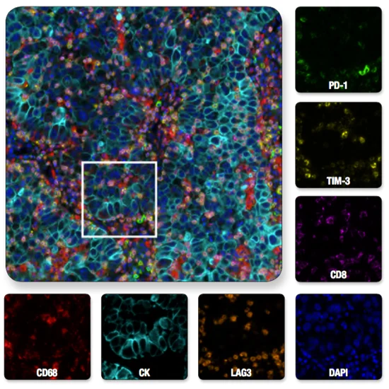
Through multi-channel overlay imaging, we can see:
- T cell infiltration distribution (CD8)
- Immune checkpoint expression (PD-1/ TIM-3/ LAG-3)
- Macrophage to tumor cell ratio (CD68/CK)
Adding immune cell proliferation status (Ki-67), a single tissue section can simultaneously obtain a panoramic view of the immune microenvironment, saving valuable samples while improving data comparability.
- Tumor Immune Microenvironment Analysis
In tumor immunotherapy research, mIHC can simultaneously detect T cell subsets, immune checkpoints, and tumor cell markers, helping to screen for responding patients.
- Rare Cell Population Identification
In inflammation or regeneration research, rare regulatory cells or stem cell subsets are difficult to detect through single-marker IHC. mIHC significantly improves detection accuracy through multi-biomarker combination identification.
- Drug Development and Efficacy Evaluation
Multiplex detection of drug targets can be completed on a single tissue section, providing intuitive and quantitative spatial data for pharmacodynamic studies.
Although mIHC has obvious advantages, there are still challenges:
- Antibody cross-interference → Use strict stripping steps and species/homology-optimized antibody combinations.
- Fluorescence photobleaching → Choose stable fluorescent dyes or rapid imaging, and avoid repeated light exposure.
- Complex imaging and analysis → Utilize multispectral microscopes and AI image analysis software to improve resolution and data reproducibility.
- Experimental cost → Although reagent costs are higher than traditional IHC, it saves tissue sections and experimental repetition costs, with significantly improved overall cost-effectiveness.
Detecting 8 biomarkers on a single tissue section is not science fiction, but a real capability of mIHC technology.
For scenarios such as tumor immune microenvironment, drug development, and rare cell population research, mIHC has become an indispensable "research tool."
If you are still troubled by limited samples, multiple experimental repetitions, and insufficient information, mIHC may be the key to breaking through your challenges.
- Multiple biomarker detection, easily covering complex tissue microenvironments
- High-sensitivity TSA signal amplification, low background, high stability
- Complete experimental protocol and technical support, saving time and effort
Compatible with multiple tissue types, no antibody species limitations
Product | Catalog Number |
|---|---|
TSA Six-Label Seven-Color Multiplex Immunohistochemistry Kit | |
TSA Five-Label Six-Color Multiplex Immunohistochemistry Kit | |
TSA Four-Label Five-Color Multiplex Immunohistochemistry Kit | |
TSA Three-Label Four-Color Multiplex Immunohistochemistry Kit | |
TSA Two-Label Three-Color Multiplex Immunohistochemistry Kit |