With the rapid development of tumor microenvironment research, spatial biology, and precision pathological diagnosis, Multiplex Immunofluorescence (mIHC) has become an important tool for tissue sample analysis. Compared with traditional immunohistochemistry (IHC) and conventional immunofluorescence (IF), mIHC enables simultaneous detection of multiple protein markers on the same tissue section, allowing cell phenotype identification, spatial localization analysis, and cell-cell interaction studies.
However, in actual experiments, many researchers encounter a common challenge: primary antibodies for multiple target proteins are from the same species, making conventional multiplex staining impossible. The emergence of TSA (Tyramide Signal Amplification) technology provides an ideal solution to this problem. Among them, TSA multiplex immunofluorescence staining technology without primary antibody species restriction has become an important technical approach in current spatial pathology research.
In conventional immunofluorescence experiments, different target proteins typically require primary antibodies from different species, for example:
• Mouse anti-CD8
• Rabbit anti-PD-L1
• Goat anti-FOXP3
Followed by detection using fluorescent secondary antibodies of corresponding species.
This approach has obvious limitations:
✓ Limited selection of primary antibody combinations
✓ Popular target antibodies often come from rabbit or mouse
✓ Signals cannot be distinguished when multiple targets use rabbit antibodies
✓ Multiplex labeling number is severely limited
If a researcher needs to detect four targets: CD3, CD8, PD-1, and PD-L1, but all available primary antibodies are rabbit-derived, simultaneous detection using traditional IF methods is nearly impossible.
The core principle of TSA technology is: using HRP (horseradish peroxidase) to catalyze fluorescent tyramide molecules, forming irreversible covalent deposition around the antigen.
Basic process: Antigen recognition → Primary antibody binding → HRP-labeled secondary antibody binding → TSA fluorescence deposition → Antibody stripping → Next round staining
Since the fluorescent signal is firmly deposited in the tissue, even if the previous round of antibodies is completely removed through heat repair or antibody elution steps, the fluorescent signal remains. Therefore, in the next round of detection, primary antibodies from the same species can be used again without cross-reactivity.
1. More Freedom in Antibody Selection
Researchers do not need to change antibodies to avoid species conflicts. They can directly select antibodies with sufficient literature validation, the best-performing antibodies, or antibodies with established databases, significantly reducing experimental development difficulty.
2. Achieve Higher Plexity Detection
Traditional IF can typically only perform 1-2 color detection. TSA technology can achieve 3-7 or even more plex detection, meeting the needs of complex tumor microenvironment research.
3. Significantly Improve Detection Sensitivity
TSA is an enzyme-catalyzed signal amplification technology. Compared with ordinary fluorescent labeling, signal intensity can be increased by 10-100 times. It has obvious advantages for low-expression proteins, rare cell populations, and trace biomarkers.
4. Wide Adaptability and Scene Compatibility
In terms of sample types, it can adapt to various biological samples such as paraffin sections, frozen sections, cell climbing slices, cell smears, tissue microarrays, and organoids. In terms of detection modes, it is compatible with commonly used equipment such as fluorescence microscopes and confocal microscopes, without requiring additional dedicated instruments.
5. Gentle Operation Protects Samples
The antibody eluent provided with TSA kits uses a gentle formula. Without relying on high-temperature microwave treatment, it can remove unbound antibodies while completely preserving target antigens and labeled fluorescent signals. This gentle elution method ensures that tissue morphology remains intact after each round of cyclic staining, significantly reducing the risk of sample damage. This advantage is particularly important for precious samples.
1. Scientific Research:
- Neuroscience: Detection of sparsely expressed synaptic proteins or neurotransmitters in neurons.
- Cancer: Localization of rare biomarkers in tumor microenvironment (e.g., PD-L1).
- Developmental Biology: Observation of low-abundance signaling molecules in embryonic tissues.
- Spatial Biology: Analysis of spatial relationships between tumor cells, immune cells, and stromal cells.
- Others
2. Clinical Diagnosis:
- Enhance detection rate of weakly expressed antigens in pathological sections (e.g., HER2/neu expression in breast cancer).
- Detection of circulating tumor cells (CTCs) or minimal residual disease (MRD).
3. Special Samples:
- Suitable for special samples such as needle biopsies, bone tissue, brain tissue, organoids, rare cells, and long-term preserved FFPE samples, enabling multi-target, high-quality spatial expression analysis in limited samples.
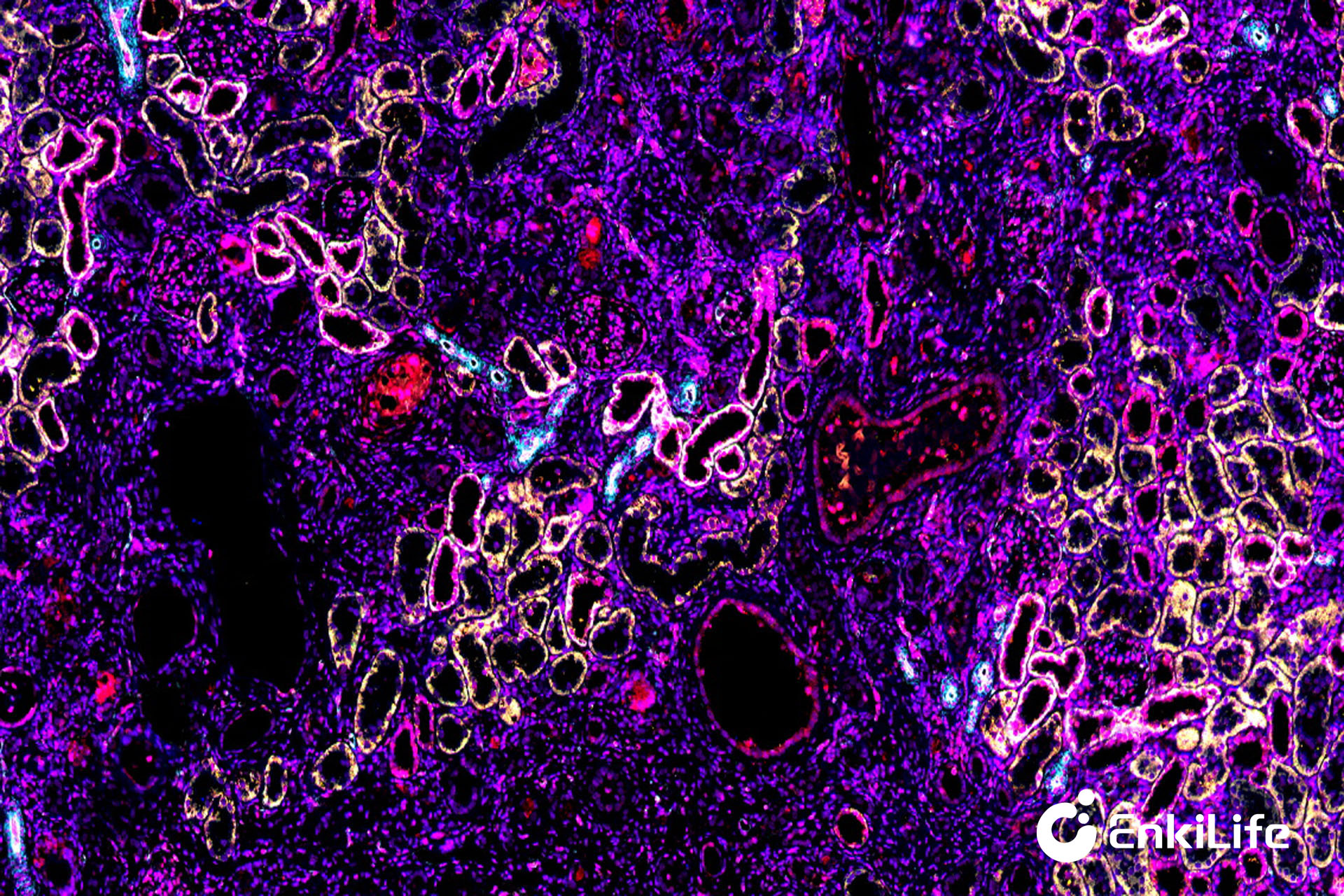
Rat Kidney (10×)
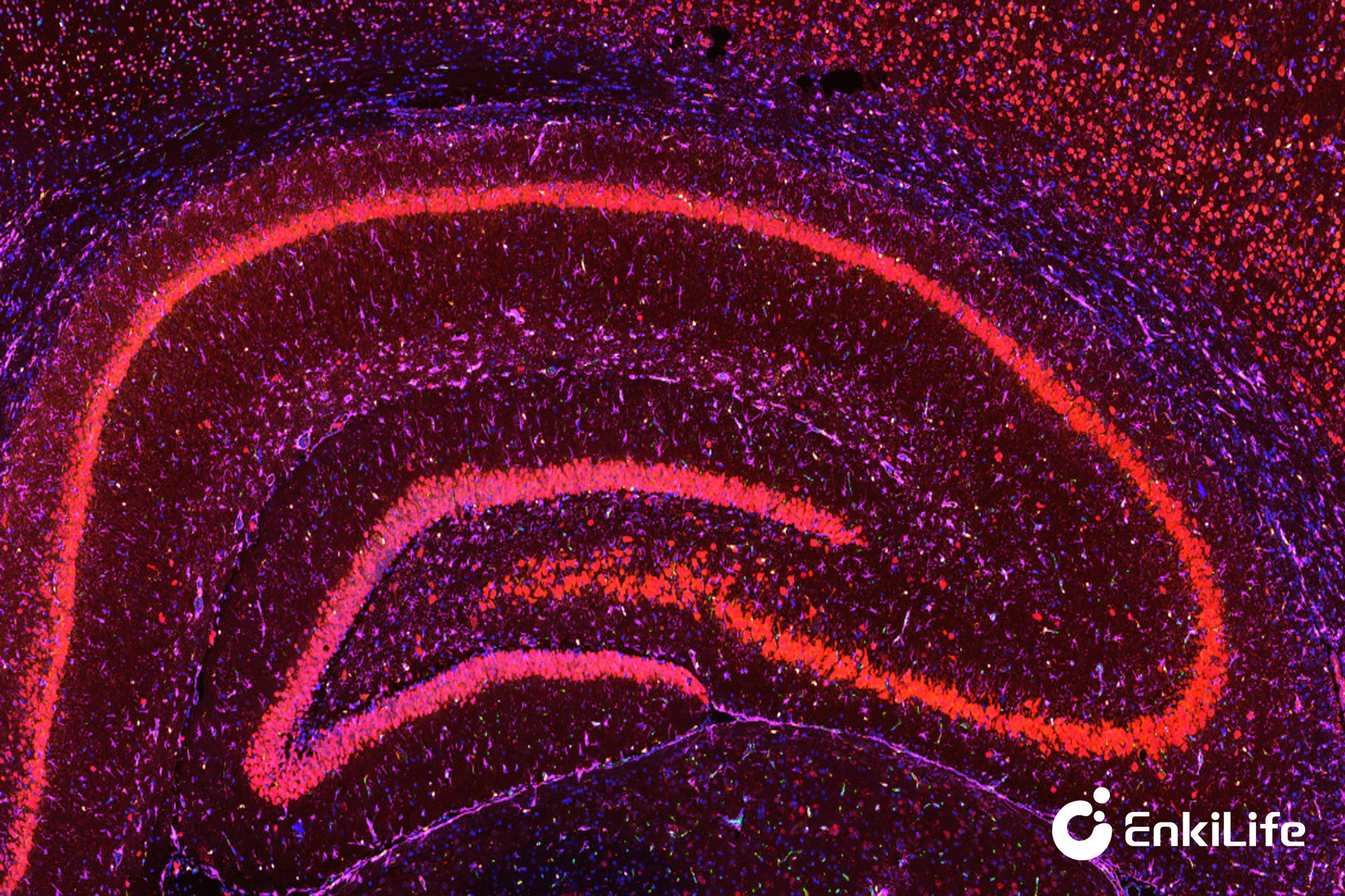
Rat Brain (5×)
Although TSA technology can break through species restrictions, experimental design still requires attention to:
✔ Antigen retrieval condition optimization
✔ Antibody staining sequence design
✔ TSA fluorescence channel matching
✔ Spectral overlap control
✔ Antibody stripping efficiency verification
✔ Multispectral imaging and spectral unmixing analysis
Reasonable panel design often determines the final experimental success rate.
With the development of spatial omics and digital pathology technologies, multiplex immunofluorescence has become an important tool for revealing tissue microenvironments. Through the innovative mechanism of "fluorescence deposition + antibody stripping", TSA signal amplification technology completely breaks through the restriction of traditional immunofluorescence on primary antibody species origin, allowing researchers to freely combine antibodies, achieve high-plex detection, and obtain higher sensitivity and richer tissue spatial information.
For tumor immunology research, spatial biology, drug development, and clinical translation research, TSA multiplex immunofluorescence technology without primary antibody species restriction is gradually becoming the standard protocol for high-quality spatial pathology analysis. Choosing a mature and stable TSA multiplex immunofluorescence reagent system and professional technical support platform will help improve experimental success rates, accelerate scientific output, and provide stronger technical support for precision medicine research.
EnkiLife not only provides customers with a complete set of TSA multiplex labeling kits, but also offers various TSA specialty technical services, including IF fluorescence staining, fluorescence panoramic scanning, ultra-multiplex staining, and pathological analysis (5 markers and below).
Product | Catalog Number |
|---|---|
TSA Six-Label Seven-Color Multiplex Immunohistochemistry Kit | |
TSA Five-Label Six-Color Multiplex Immunohistochemistry Kit | |
TSA Four-Label Five-Color Multiplex Immunohistochemistry Kit | |
TSA Three-Label Four-Color Multiplex Immunohistochemistry Kit | |
TSA Two-Label Three-Color Multiplex Immunohistochemistry Kit |
For details, please check TSA mIHC Kit