Podocyte GOLM1: The "Invisible Driver" of Diabetic Nephropathy Progression?——Analysis of the Latest Research Findings in Adv. Sci.
Diabetic nephropathy (DN), one of the most devastating complications of diabetes, has been increasing in prevalence globally. Approximately 40% of patients with type 2 diabetes and 30% of patients with type 1 diabetes will suffer from kidney damage, which may eventually progress to end-stage renal disease. However, its pathogenesis has not been fully elucidated, and there is still a lack of effective targeted therapies in clinical practice. Is there a key molecule that plays a central role in the development of diabetic nephropathy and could become a potential therapeutic target? In July 2025, researchers published a study titled "Inhibition of AMPKα Pathway by Podocyte GOLM1 Exacerbates Diabetic Nephrology in Mice" in Advanced Science. Through multi-model and multi-dimensional experiments in vivo and in vitro, the study for the first time revealed the pathogenic role and regulatory mechanism of GOLM1 in podocytes during the progression of diabetic nephropathy, providing novel insights for clinical treatment.
I. Research Background and Methods
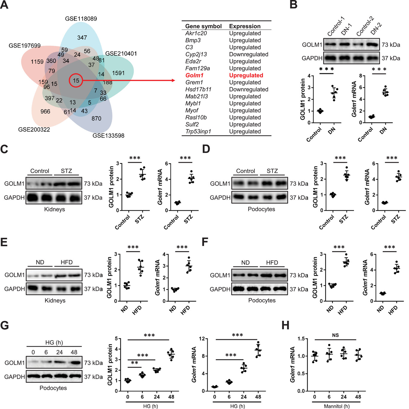
The core pathological features of diabetic nephropathy include renal inflammation, oxidative damage, podocyte apoptosis, and renal fibrosis. Podocyte injury is considered one of the initiating factors in the development of diabetic nephropathy, and its quantitative reduction and functional abnormalities often precede clinically detectable proteinuria. However, the key molecules and mechanisms regulating podocyte injury remain unclear. GOLM1, a known oncogene, is highly expressed in various malignancies and promotes tumor progression. Recent studies have found its association with inflammation and oxidative stress, but its role and regulatory mechanism in diabetic nephropathy have not been reported. Based on this, the research team integrated 5 GEO datasets to screen 15 overlapping differentially expressed genes (DEGs) in diabetic nephropathy mouse models, ultimately identifying GOLM1 as the research subject to clarify its expression characteristics, function, and molecular mechanism in diabetic nephropathy. In terms of research methods, the study combined in vivo and in vitro approaches. In vivo, type 1 diabetes (STZ injection) and type 2 diabetes (HFD feeding) mouse models were constructed, and podocyte-specific GOLM1 knockout and overexpression mouse models were established simultaneously. In vitro, mouse podocytes were cultured and treated with high glucose to simulate the diabetic environment. Various techniques including qRT-PCR, Western blot, immunofluorescence, immunohistochemistry, ELISA, and RNA sequencing were used to detect gene and protein expression, and pathological staining was performed to observe kidney tissue changes.
II. Results Analysis: Unlocking the Key Code of GOLM1 in Regulating Diabetic Nephropathy
1. GOLM1 is Abnormally Elevated in Diabetic Nephropathy and Closely Associated with Renal Function Impairment
In the first step of the study, the team outlined the "abnormal trajectory" of GOLM1 in diabetic nephropathy through multi-dimensional experiments. In clinical samples, both mRNA and protein levels of GOLM1 in kidney tissues of diabetic nephropathy patients were significantly higher than those in healthy controls. This phenomenon was perfectly replicated in two diabetic mouse models—both STZ-induced type 1 diabetic mice and HFD-induced type 2 diabetic mice showed significantly upregulated GOLM1 expression in kidney tissues and isolated primary podocytes. More importantly, this upregulation was not a universal phenomenon in all kidney cells. In vitro experiments confirmed that high glucose stimulation could only significantly increase GOLM1 expression in podocytes, but had no significant effect on other kidney cells such as mesangial cells and tubular epithelial cells, indicating that the upregulation of GOLM1 is podocyte-specific.
As a molecule defined as a novel gluconeogenic hormone, can serum GOLM1 levels serve as a potential biomarker for diabetic nephropathy? Further clinical and animal experiments provided a positive answer: serum GOLM1 levels were significantly elevated in diabetic nephropathy patients and positively correlated with chronic kidney disease (CKD) stages. Higher serum GOLM1 concentrations were associated with higher serum creatinine (Cr) levels and lower estimated glomerular filtration rate (eGFR), suggesting a close association with the degree of renal function impairment. In diabetic mouse models, serum GOLM1 levels were also significantly elevated, and high glucose stimulation in vitro could promote podocyte secretion of GOLM1 into the culture medium. This finding not only confirmed that GOLM1 can be secreted by podocytes but also suggested that serum GOLM1 may serve as a non-invasive biomarker for early diagnosis and progression monitoring of diabetic nephropathy.
2. The "Gain" and "Loss" of Podocyte GOLM1 Directly Determines the Outcome of Diabetic Nephropathy
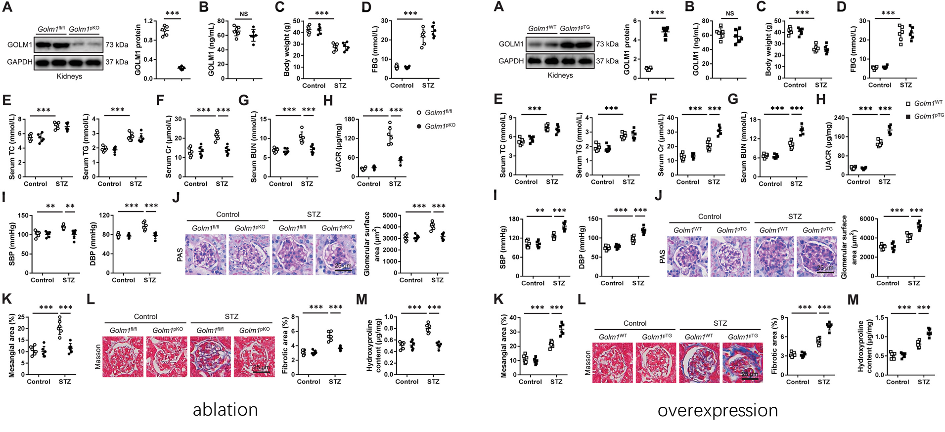
To clarify the specific function of GOLM1 in podocytes, the research team constructed podocyte-specific GOLM1 knockout and overexpression mouse models. By losing and gaining GOLM1 function, they observed its effects on diabetic nephropathy, which was also the core breakthrough of the entire study. In the STZ-induced type 1 diabetes model, after podocyte-specific knockout of GOLM1, the serum Cr, blood urea nitrogen (BUN), and urine albumin/creatinine ratio (UACR) in mice were significantly reduced. Diabetes-induced hypertension was also significantly alleviated. Renal pathological examination showed that glomerular hypertrophy, mesangial expansion, and renal fibrosis were significantly reduced, indicating that GOLM1 in podocytes plays a key role in promoting the progression of diabetic nephropathy.
In contrast, podocyte-specific overexpression of GOLM1 had negative effects. Overexpression mice showed further aggravated renal function impairment under STZ or HFD induction, with significantly elevated serum renal function indicators and more severe renal pathological damage, including significantly increased glomerular hypertrophy and renal fibrosis. More importantly, neither knockout nor overexpression of GOLM1 affected body weight, fasting blood glucose (FBG), total cholesterol (TC), triglycerides (TG), or other systemic metabolic indicators in mice, indicating that the regulatory effect of GOLM1 on diabetic nephropathy is specifically exerted on the kidney rather than through affecting systemic metabolism. This series of experiments constructed a clear logical chain: GOLM1 in podocytes is a key driver of diabetic nephropathy progression, and its specific inhibition may become a potential therapeutic strategy.
3. GOLM1 Promotes Podocyte Apoptosis and Renal Injury by Exacerbating Inflammation and Oxidative Damage
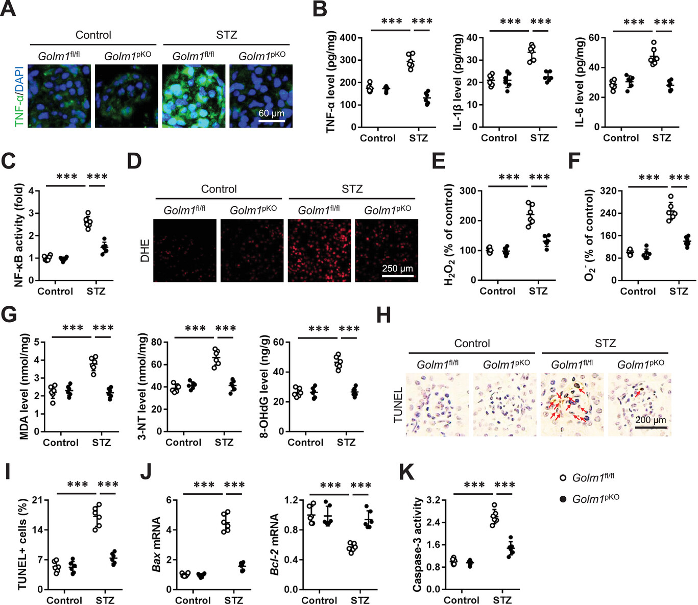
After clarifying the function of GOLM1, the research team further explored the specific pathways through which it regulates diabetic nephropathy. Inflammation and oxidative damage are two core pathological mechanisms of diabetic nephropathy, which synergistically promote podocyte apoptosis and renal fibrosis. Experiments showed that in STZ-induced diabetic mice, after podocyte-specific knockout of GOLM1, the levels of pro-inflammatory factors such as tumor necrosis factor-α (TNF-α), interleukin-1β (IL-1β), and interleukin-6 (IL-6) in kidney tissues were significantly reduced. The transcriptional activity of nuclear factor-κB (NF-κB) was also significantly inhibited, indicating that GOLM1 promotes renal inflammation by activating the NF-κB signaling pathway.
In terms of oxidative damage, knockout of GOLM1 significantly reduced the production of reactive oxygen species (ROS) in the kidneys of diabetic mice and decreased the levels of oxidative damage markers such as malondialdehyde (MDA), 3-nitrotyrosine (3-NT), and 8-hydroxydeoxyguanosine (8-OHdG). The reduction of these markers indicated that oxidative damage to kidney cells was effectively alleviated. At the same time, podocyte apoptosis was also significantly reduced, as evidenced by decreased TUNEL-positive cell count, decreased expression of pro-apoptotic gene Bax, and increased expression of anti-apoptotic gene Bcl-2. In vitro experiments confirmed that high glucose-induced podocyte apoptosis could be significantly inhibited by silencing GOLM1, while overexpression of GOLM1 had the opposite effect, further confirming that GOLM1 promotes podocyte apoptosis by exacerbating inflammation and oxidative damage.
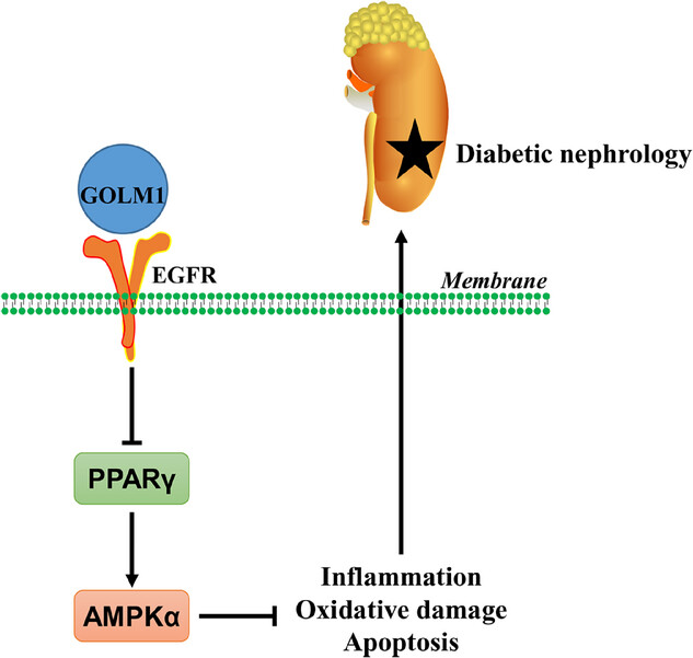
4. GOLM1 Regulates Diabetic Nephropathy Progression Through the EGFR/PPARγ/AMPKα Pathway
To completely unlock the molecular code of GOLM1 in regulating diabetic nephropathy, the research team screened differentially expressed genes in kidneys between GOLM1 knockout mice and wild-type mice through RNA sequencing analysis. They found that these genes were mainly enriched in the AMPK signaling pathway. As the core molecule of the AMPK pathway, the phosphorylation level of AMPKα was significantly reduced in the kidneys of diabetic mice, while podocyte-specific knockout of GOLM1 could restore its phosphorylation level, and overexpression of GOLM1 would further inhibit its phosphorylation. To verify the role of AMPKα, researchers used the AMPKα-specific inhibitor DMP to treat podocyte-specific GOLM1 knockout diabetic mice, and found that the protective effect of GOLM1 knockout on the kidneys was significantly reversed, confirming that AMPKα is a key downstream effector of GOLM1 in regulating diabetic nephropathy.
Further studies found that PPARγ, as an upstream regulator of the AMPKα pathway, had significantly increased expression in the kidneys of GOLM1 knockout mice, while overexpression of GOLM1 significantly decreased its expression. Silencing PPARγ could effectively block the activation of AMPKα and the renal protective effect induced by GOLM1 knockout, indicating that GOLM1 inhibits PPARγ, thereby suppressing the AMPKα pathway. Mechanistically, GOLM1 can directly bind to epidermal growth factor receptor (EGFR) on the podocyte membrane, activate EGFR and its downstream signaling pathways, thereby promoting the phosphorylation of STAT3, which in turn binds to the PPARγ promoter region and inhibits its transcription. This series of experiments constructed a complete regulatory pathway: GOLM1 activates EGFR/STAT3 signaling by binding to EGFR, inhibits PPARγ transcription, thereby suppressing the AMPKα pathway, and ultimately promotes inflammation, oxidative damage, and podocyte apoptosis, exacerbating the progression of diabetic nephropathy.
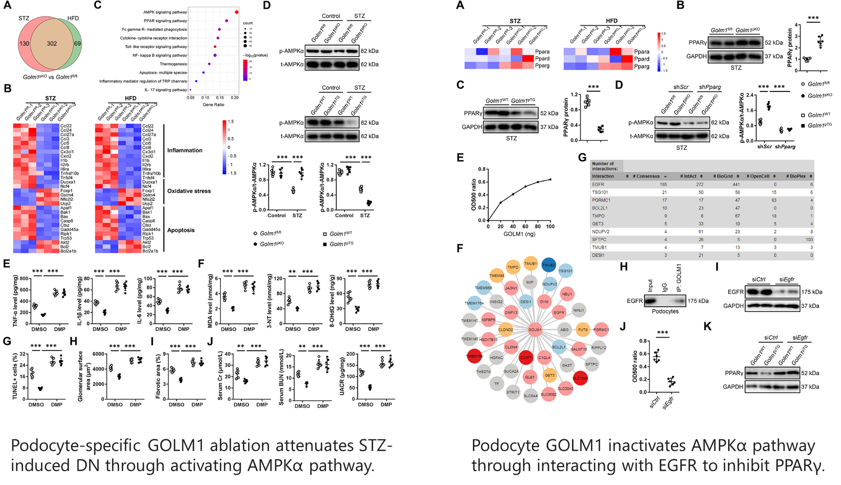
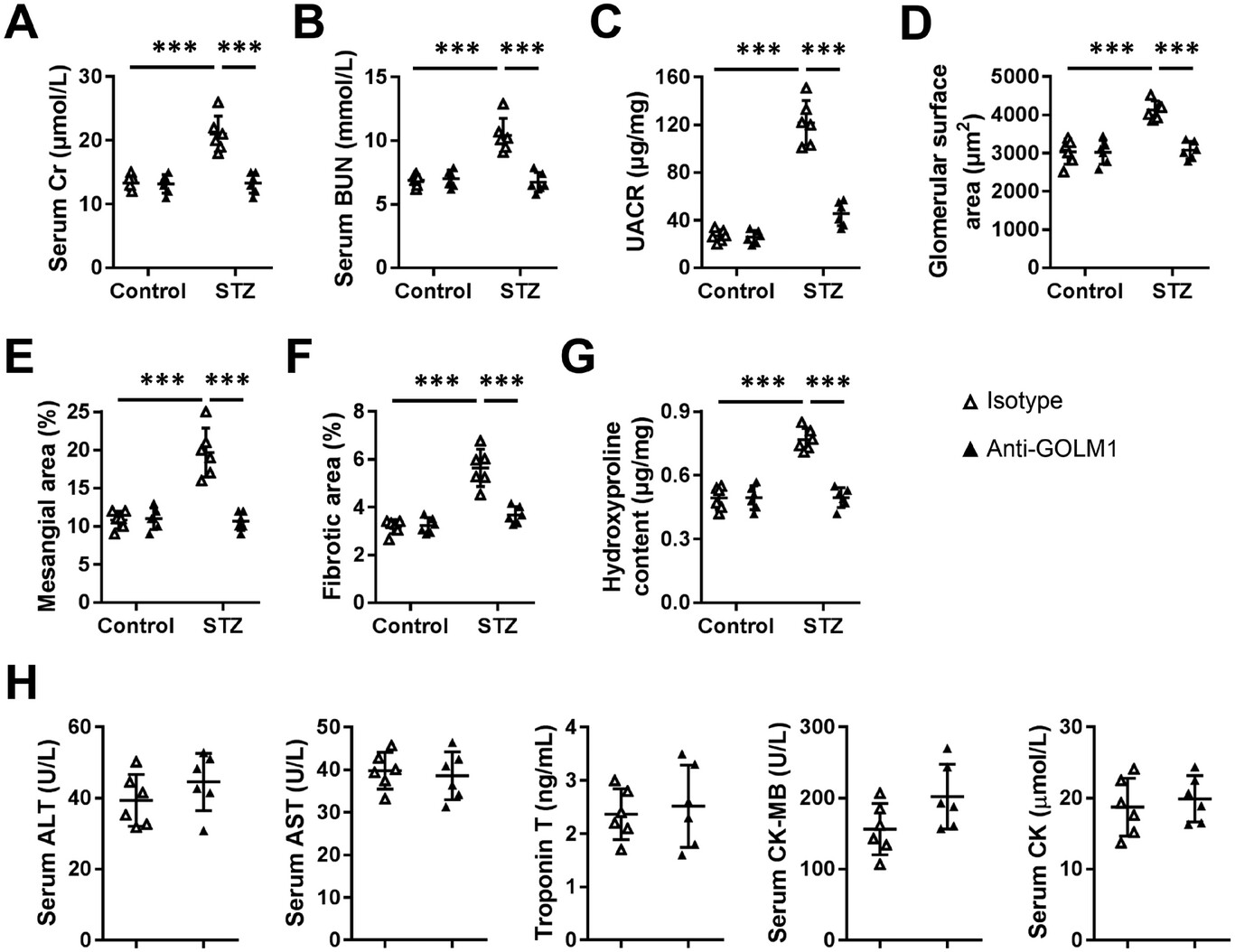
5. GOLM1 Neutralizing Antibody Provides a New Strategy for Diabetic Nephropathy Treatment
Based on the above findings, the research team further explored the therapeutic potential of targeting GOLM1. They treated diabetic mice with GOLM1 neutralizing antibody. The results showed that antibody treatment significantly reduced serum Cr, BUN, and UACR levels in STZ-induced diabetic mice, alleviated glomerular hypertrophy, mesangial expansion, and renal fibrosis. Its therapeutic effect was similar to that of podocyte-specific GOLM1 knockout. Mechanistically, the GOLM1 neutralizing antibody effectively blocked the binding of GOLM1 to EGFR, restored the activity of the PPARγ/AMPKα pathway, thereby inhibiting inflammation and oxidative damage, reducing podocyte apoptosis, and ultimately exerting a renal protective effect. This experiment not only verified the feasibility of targeting GOLM1 for the treatment of diabetic nephropathy but also provided a promising direction for the development of targeted drugs.
III. Research Implications and Prospects
Through multi-model and multi-technical approaches, this study for the first time clarified the pathogenic role of podocyte GOLM1 in diabetic nephropathy and constructed the regulatory pathway of "GOLM1-EGFR-PPARγ-AMPKα", which not only enriched the research on the pathogenesis of diabetic nephropathy but also provided novel biomarkers and therapeutic targets. For kidney disease researchers, the innovations of this study include: first, discovering the specific role of GOLM1 in podocytes, breaking the previous understanding of GOLM1's role in metabolic diseases; second, clarifying the autocrine regulatory mode of GOLM1, providing new ideas for the treatment targeting secreted molecules; third, verifying the therapeutic effect of GOLM1 neutralizing antibody, laying a foundation for clinical translation.
In the future, researchers can further explore the expression changes of GOLM1 in different stages of diabetic nephropathy to clarify its clinical diagnostic value as a biomarker. At the same time, they can optimize the administration method and dosage of GOLM1 neutralizing antibody to promote its clinical translation research. In addition, this study found that the GAP activity of GOLM1 is unrelated to its function in regulating diabetic nephropathy. The reason behind this phenomenon still needs further investigation, which is expected to provide a new direction for the functional research of GOLM1.
References
Xu P, Li K, Liu H, Xu A, Zhang Z, Yang Y, Lai X, Hao K, Fang K, Lai Z, Ou X, Cai Y, Wang Z, Lu K, Jiang W. Inhibition of AMPKα Pathway by Podocyte GOLM1 Exacerbates Diabetic Nephrology in Mice. Adv Sci (Weinh). 2025 Oct;12(37):e05695. doi: 10.1002/advs.202505695. Epub 2025 Jul 13. PMID: 40652516; PMCID: PMC12499477.