Over the past few decades, immunohistochemistry (IHC) has been the standard tool for tumor tissue research: from HER2, Ki-67 to PD-L1, it has allowed researchers and pathologists to clearly see the distribution of individual proteins in tissues. However, as tumor research enters the "spatial biology era," information from single markers is far from sufficient.
Today's tumor microenvironment (TME) research is not only concerned with how much protein is expressed, but more importantly:
- Have immune cells actually infiltrated the tumor core?
- What is the spatial relationship between tumor-associated macrophages, CAFs, and T cells?
- Which cell populations may affect immunotherapy efficacy?
These questions are difficult for traditional IHC to answer in one go. Multiple sections, repeated staining - not only consumes samples, but also easily loses spatial information.
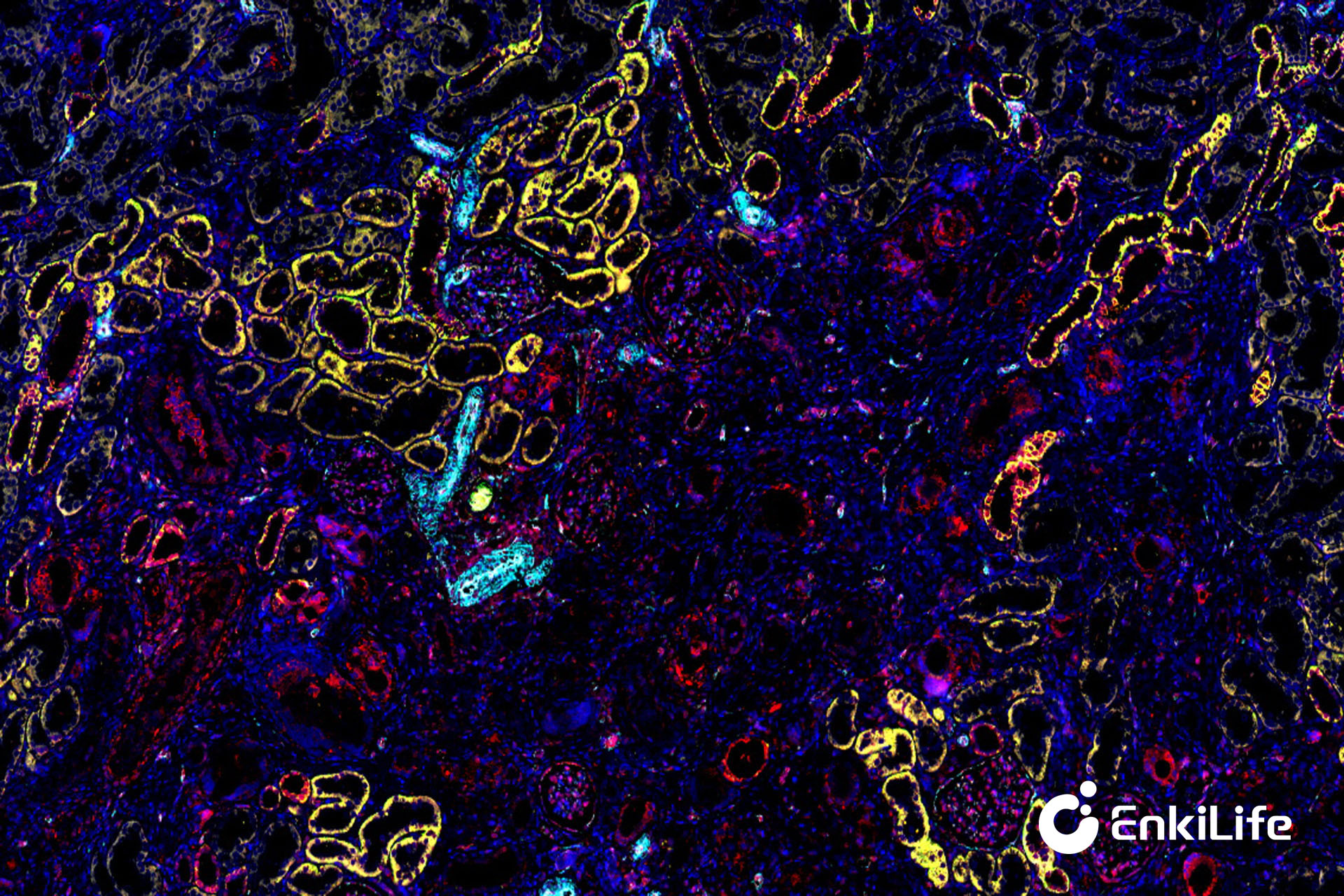
Multiplex immunofluorescence (mIHC) technology was developed precisely to solve this dilemma. It allows researchers to simultaneously detect multiple protein markers on a single tissue section, and obtain cell type, localization, and spatial relationship information through fluorescence imaging and multispectral analysis.
Compared with traditional IHC, the core advantages of mIHC are:
- Spatial information visualization: Understand the distribution and proximity relationships of immune cells within tumors.
- Multi-marker co-localization: Observe multiple cell types and protein expressions in a single experiment.
- High sample utilization: Precious needle biopsy or PDX samples can also maximize information output.
- More accurate quantitative analysis: Can generate publishable digital data charts.
For example, in PD-1/PD-L1 immunotherapy research, single IHC can only tell you whether the tumor expresses PD-L1, while mIHC can simultaneously show CD8+ T cell infiltration and the spatial relationship between tumor cells and immune cells - which is crucial for predicting therapeutic efficacy.
- Research Needs Drive Technological Upgrades
The complexity of TME means single-marker detection cannot meet research demands. Researchers need multi-dimensional data to support studies on tumor immunotherapy, drug resistance mechanisms, etc. - "Invisible Threshold" for High-Impact Papers
In recent years, high-impact journals have increasingly favored experimental data that provides spatial information. mIHC not only provides expression information, but also the spatial pattern of the immune microenvironment, significantly enhancing the persuasiveness of research. - Maximize Value of Precious Samples
Clinical needle biopsy samples or small animal model tissues are limited. Traditional IHC requires multiple sections, while mIHC can complete multi-marker analysis on a single section, saving time and samples. - Development of Digital Pathology and Spatial Analysis
The maturation of multispectral imaging and image analysis software makes mIHC data quantifiable, reproducible, and supports high-throughput analysis.
TSA (Tyramide Signal Amplification) technology combined with mIHC enables multiple rounds of staining and signal amplification, solving the problems of fluorescence quenching and weak signals. With professional kits and technical services, researchers can:
- Avoid tedious optimization and quickly obtain high-quality multi-marker staining results
- Obtain quantifiable spatial data directly for research analysis and paper publication
- Save precious tissue samples and improve experimental success rates
From traditional IHC to mIHC is not just a technological upgrade, but a conceptual upgrade in tumor microenvironment research. Single-marker observation can no longer meet the needs of complex TME research, while mIHC allows researchers to "see the entire tumor ecosystem" in a single section, providing a reliable tool for immunotherapy research and high-level scientific achievements.
From "seeing a single protein" to "deciphering the tumor microenvironment," mIHC is redefining the new standard for tumor research.
From sample to data, Enkilife helps researchers efficiently conduct multiplex immunofluorescence research.