Popular Spatial Pathology Technology: Overview of TSA Multiplex Fluorescence Results
The iterative advancement of spatial pathology technology has completely broken through the limitations of traditional pathological detection, including single markers, weak signals, and missing spatial information, becoming a core supporting technology for tumor microenvironment analysis, disease mechanism research, and precise pathological diagnosis. Among the numerous multiplex detection technologies, TSA (Tyramide Signal Amplification) multiplex fluorescence technology, with its core advantages of high sensitivity, multi-target co-staining, and precise in situ localization, has become the popular mainstream technology in the current spatial pathology field. Different from the detection modes of traditional immunofluorescence and conventional immunohistochemistry, TSA technology relies on the efficient enzymatic catalytic reaction of horseradish peroxidase (HRP) to drive the specific deposition of fluorescence-labeled tyramide molecules at target sites, achieving signal amplification by dozens of times, perfectly solving the pain points of difficult detection of low-abundance proteins and easy loss of weak signals. At the same time, this technology can complete multi-color target simultaneous labeling on a single tissue section through multiple cycles of staining and signal quenching, completely preserving the in situ spatial structure and cell interaction information of the tissue, providing reliable technical support for multi-dimensional and in-depth analysis of pathological samples.
TSA Multiplex Fluorescence Different Panel Results Display
2-Plex / 3 Colors
2-Plex / 3 Colors is a relatively basic combination form in TSA multiplex fluorescence, usually including 2 target markers and DAPI nuclear staining. This scheme is suitable for basic co-expression validation, positive cell localization, and simple tumor-immune cell relationship analysis. 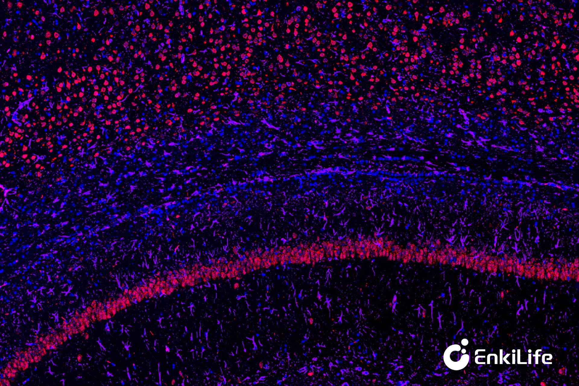
Rat Brain (10×)
Rat Kidney (10×)
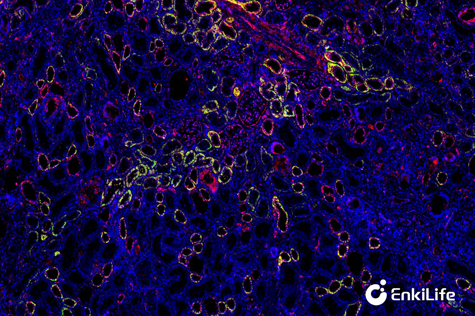
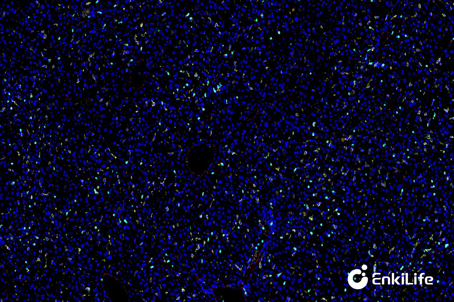
3-Plex / 4 Colors
3-Plex / 4 Colors can simultaneously analyze the expression of 3 target proteins in tissues and further demonstrate the spatial distribution relationships between different cells, widely used in tumor immune microenvironment research.
Rat Kidney (10×)
Mouse Liver (10×)
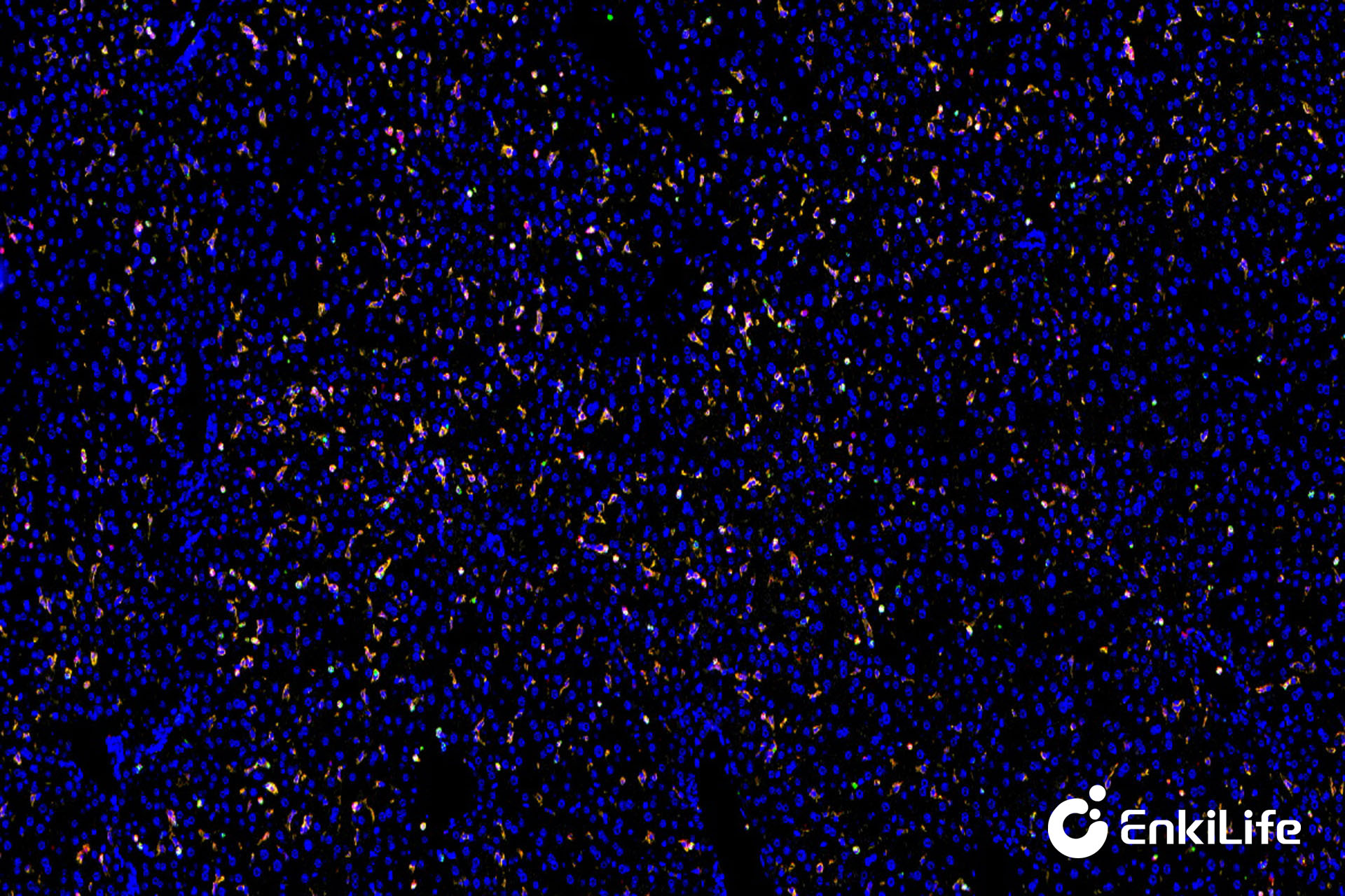
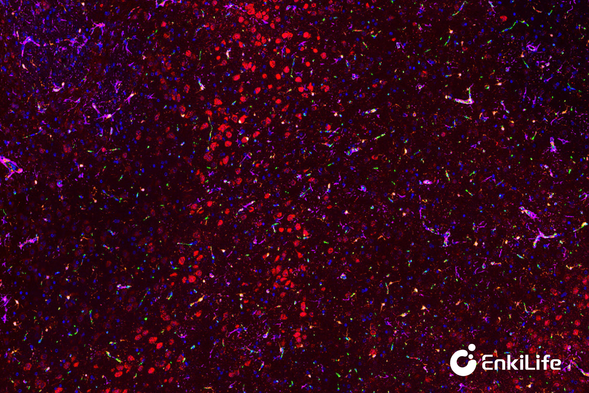
4-Plex / 5 Colors
4-Plex / 5 Colors can already meet the needs of most tumor immune research, and can be used for complex immune cell subpopulation analysis and multi-marker co-expression research.
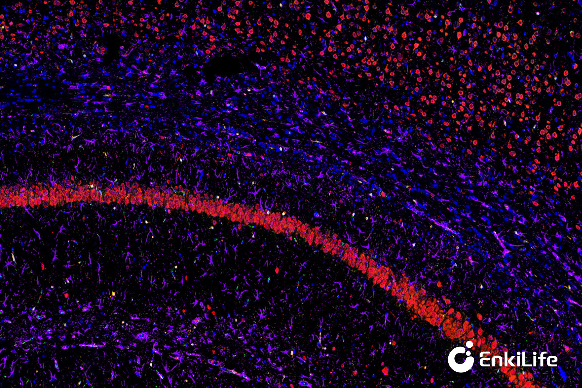
Rat Brain (10×)
Mouse Liver (10×)
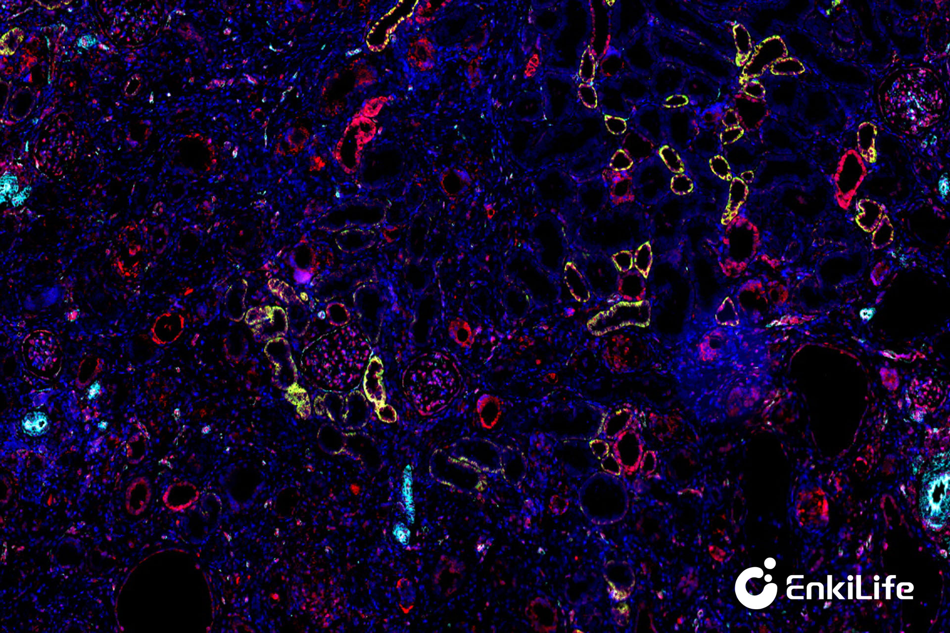
5-Plex / 6 Colors
5-Plex / 6 Colors belongs to a medium-to-high throughput TSA multiplex fluorescence scheme, which can more comprehensively analyze the cell composition and interactions in the tumor microenvironment.
Rat Brain (10×)
Rat Kidney (10×)
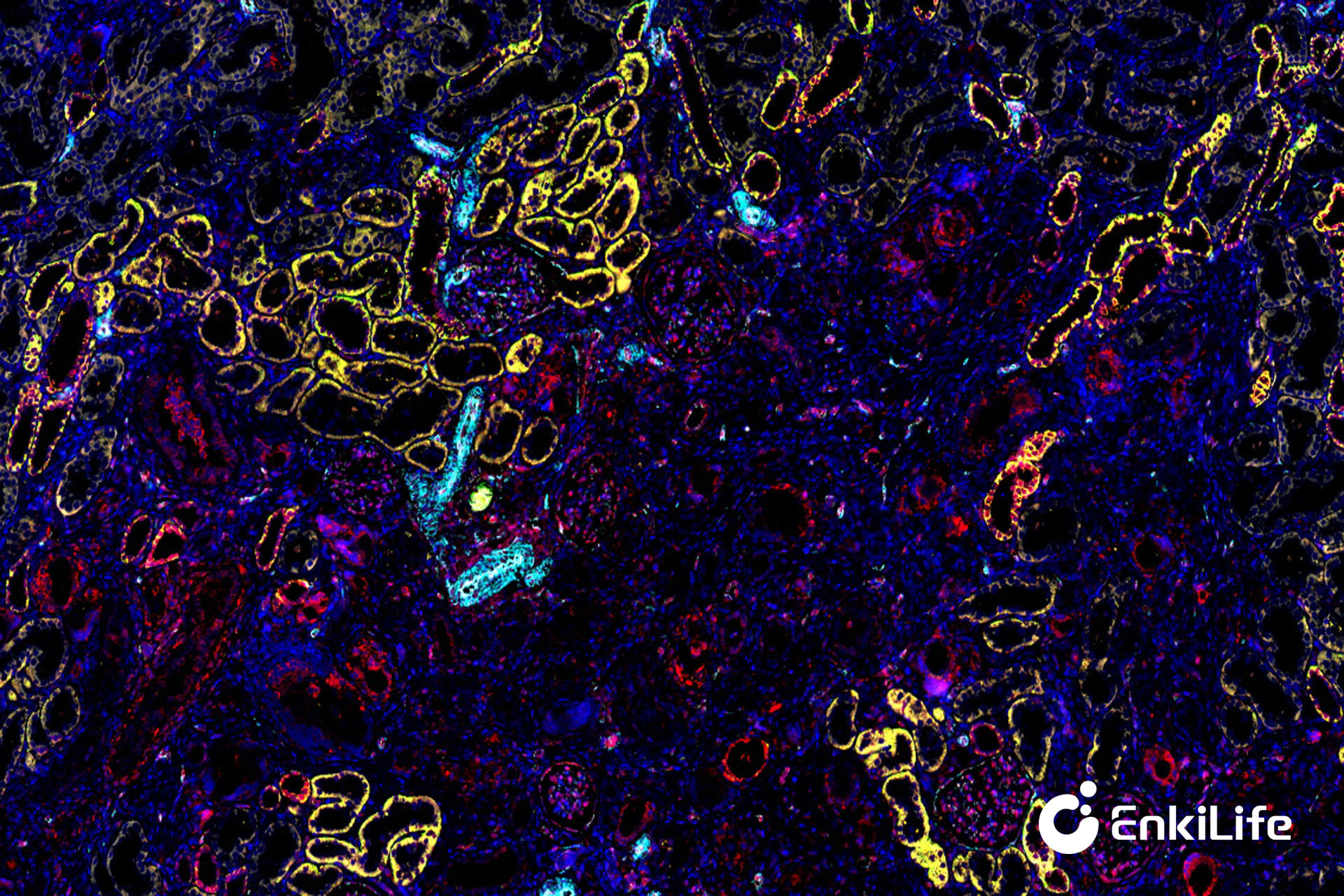
6-Plex / 7 Colors
6-Plex / 7 Colors is one of the more common high-throughput TSA-mIHC schemes currently, which can achieve high-dimensional spatial resolution of complex tumor microenvironment in a single tissue section.
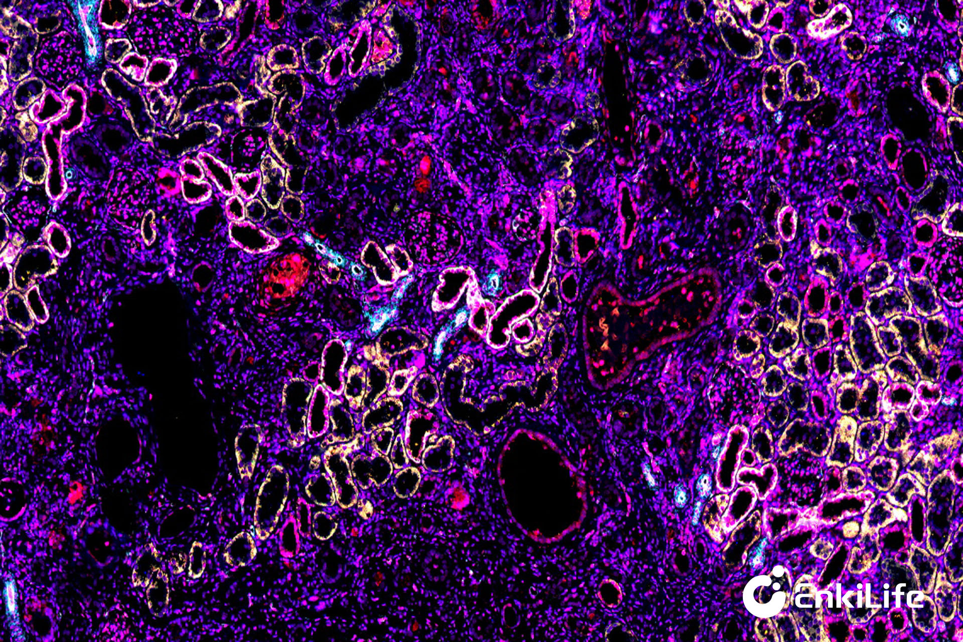
Rat Kidney (10×)
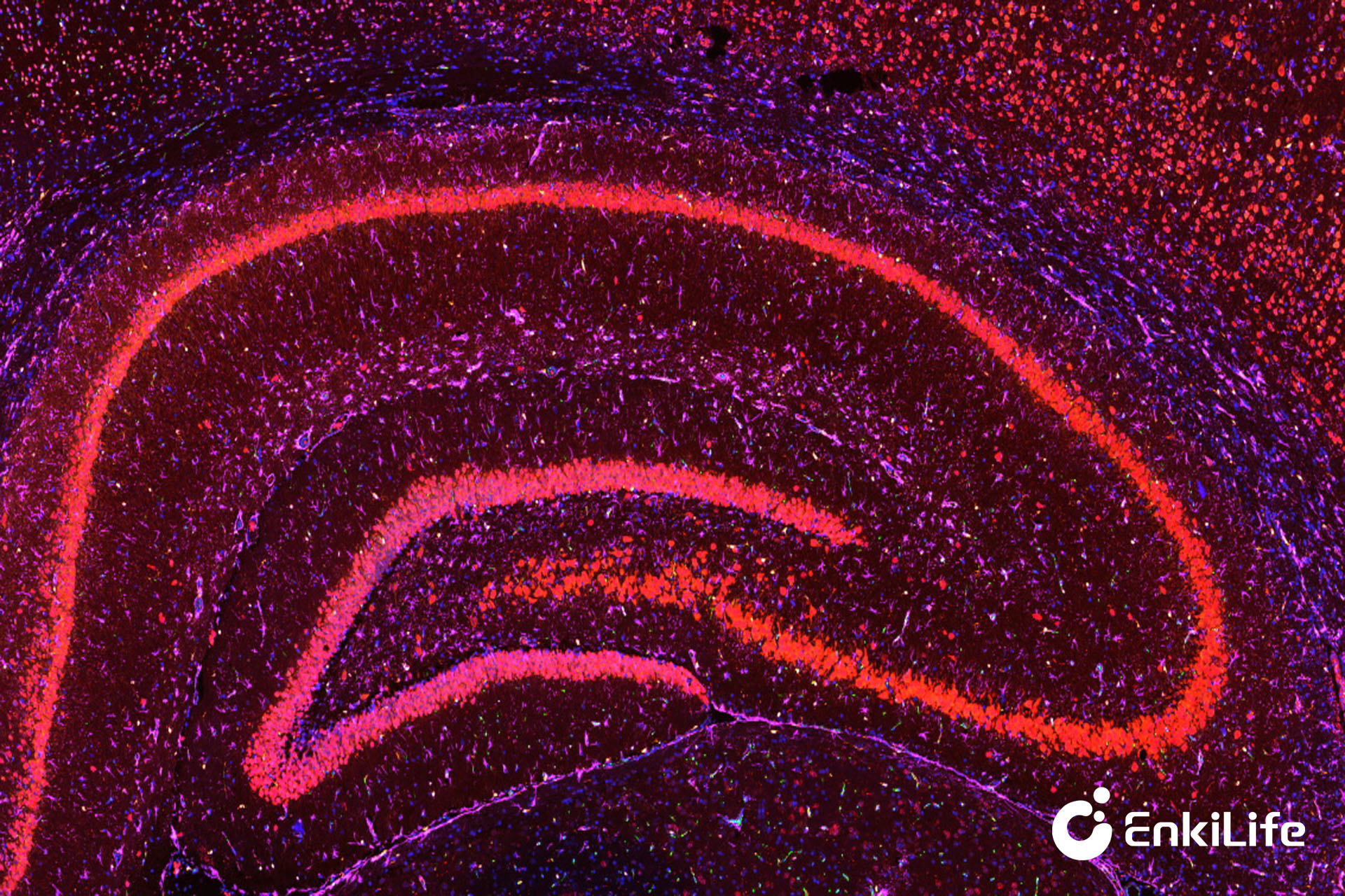
Rat Brain (5×)
EnkiLife not only provides customers with a complete set of TSA multiplex labeling kits, but also offers various TSA specialty technical services, including IF fluorescence staining, fluorescence panoramic scanning, ultra-multiplex staining, and pathological analysis (5 markers and below).
Product | Catalog Number |
|---|---|
TSA Six-Label Seven-Color Multiplex Immunohistochemistry Kit | |
TSA Five-Label Six-Color Multiplex Immunohistochemistry Kit | |
TSA Four-Label Five-Color Multiplex Immunohistochemistry Kit | |
TSA Three-Label Four-Color Multiplex Immunohistochemistry Kit | |
TSA Two-Label Three-Color Multiplex Immunohistochemistry Kit |
For details, please check TSA mIHC Kit