
Fluorescent labeling technology is one of the most important visualization tools in modern life science research. From intracellular protein localization and gene expression monitoring to immune cell typing, multi-parameter flow analysis, and spatial biology research, fluorescent signals are almost ubiquitous throughout the life science experimental workflow. With the development of fluorescent protein engineering and optical imaging technology, researchers can now simultaneously detect multiple target molecules in the same sample, achieving the leap from single labeling to high-dimensional, multi-parameter analysis.
However, although common fluorescent markers like GFP, mCherry, PE, and APC in experiments can all emit bright fluorescence, their sources, working principles, and application methods are completely different. GFP and mCherry can be continuously expressed in cells through genetic engineering, making them suitable for dynamic observation of living cells; while PE and APC need to be conjugated to biomolecules such as antibodies, making them more suitable for flow cytometry and immunoassays. Understanding the characteristics of different types of fluorescent proteins not only helps in correctly designing experimental protocols but also enables the selection of the most appropriate fluorescent labeling system based on the experimental purpose.
Genetically encoded fluorescent proteins refer to fluorescent proteins that can be directly expressed in cells through genetic engineering techniques. Their coding sequences can be fused with target protein genes, forming fluorescently active fusion proteins during cell translation, thereby enabling real-time visualization of target proteins. Unlike traditional labeling methods that require additional fluorescent dyes or antibodies, these fluorescent proteins can spontaneously form chromophores relying on their own amino acid residues and produce stable fluorescence without exogenous cofactors. Therefore, they are particularly suitable for live cell imaging, protein dynamic tracking, and long-term expression studies.
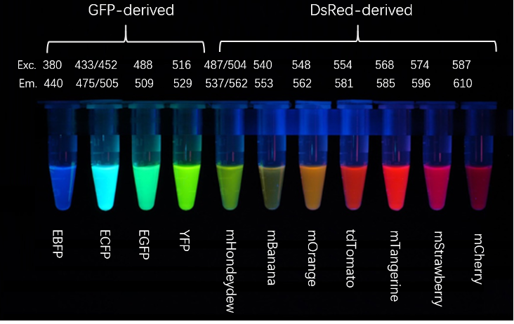
Green Fluorescent Protein (GFP) originates from the jellyfish Aequorea victoria and is the starting point for the development of modern fluorescent protein technology. Natural GFP possesses the unique property of autonomously forming chromophores, allowing it to emit green fluorescence without the need for exogenous substrates. To improve its expression efficiency and imaging performance in mammalian cells, researchers have developed multiple improved versions such as Enhanced Green Fluorescent Protein (EGFP), Superfolder GFP (sfGFP), and more recently, mNeonGreen with even better performance. Among these, EGFP has become the most commonly used tool for protein localization and gene expression reporting due to its high fluorescence brightness, good folding efficiency, and stable expression performance; sfGFP is more suitable for complex fusion proteins and difficult-to-fold protein research; while mNeonGreen, derived from amphioxus, has higher brightness and faster maturation speed, demonstrating significant advantages in low-abundance protein detection and super-resolution microscopy. Due to the characteristics of green channel including low background, high excitation efficiency, and mature microscope configuration, green fluorescent proteins remain the preferred marker for the vast majority of live cell experiments and transgenic model construction.
Yellow Fluorescent Protein (YFP) and Cyan Fluorescent Protein (CFP) are spectral variants obtained through site-directed mutagenesis based on GFP. Their greatest value lies not simply in adding another color, but in providing new technical means for protein-protein interaction studies. Since the emission spectrum of CFP overlaps well with the excitation spectrum of YFP, the two can form a classic Fluorescence Resonance Energy Transfer (FRET) system. When two proteins labeled with CFP and YFP respectively interact, the distance between them shortens, enabling energy transfer, thereby reflecting protein binding, enzymatic activity changes, or signal pathway activation processes in real-time through fluorescence signal changes. In recent years, improved YFP and CFP variants such as Venus and Cerulean have further improved brightness, maturation speed, and photostability, enabling FRET technology to more accurately study complex molecular events within cells. Therefore, compared to being used for ordinary protein localization, yellow and cyan fluorescent proteins are more commonly applied in molecular interaction analysis, functional sensor construction, and cell signal transduction research.
With the increasing demand for multicolor imaging and in vivo imaging, red fluorescent proteins have gradually become the most important fluorescent protein family after GFP. The early red fluorescent protein DsRed originated from coral, but due to its slow maturation speed and tendency to form tetramers, it had certain limitations in fusion protein research. Subsequently developed monomeric or improved red fluorescent proteins such as mCherry, tdTomato, and mScarlet effectively solved these problems. mCherry has the advantages of fast maturation, good photostability, and suitability for fusion expression, and has now become a classic red tag in multicolor fluorescence experiments; tdTomato has extremely high fluorescence brightness, suitable for neuronal tracing and low-expression protein detection; while mScarlet combines high brightness, excellent monomeric properties, and good photostability, and is considered one of the best-performing red fluorescent proteins currently available. Due to the longer wavelength of red light, stronger tissue penetration capability, and less autofluorescence interference, red fluorescent proteins are not only widely used in cell co-localization analysis but also become important tools for in vivo imaging and animal experiments.
In addition to genetically encoded fluorescent proteins, there is another widely used class of fluorescent proteins in life science research — phycobiliproteins. These proteins are mainly derived from red algae, cyanobacteria, and blue-green algae, and are natural light-harvesting proteins in photosynthesis systems. They have extremely high molar extinction coefficients and quantum yields, thus producing fluorescence intensity far higher than most organic fluorescent dyes and some fluorescent proteins. Due to the complex structure of phycobiliproteins, their chromophores require specific enzymatic modification and protein assembly processes, and are currently difficult to achieve functional expression through simple fusion like GFP. Therefore, in scientific research, they are usually used through chemical conjugation, linking them to antibodies, streptavidin, or other biomolecules. With high brightness and mature conjugation technology, phycobiliproteins have become important fluorescent labeling systems for flow cytometry, immunofluorescence, multiplex immunoassays, and immune analysis.
Phycoerythrin (PE) is one of the most widely used phycobiliproteins and one of the brightest natural fluorescent proteins in commercial fluorescent labels. PE has extremely high molar extinction coefficient and quantum yield, can be efficiently excited by 488 nm and 561 nm lasers, and emits orange-yellow fluorescence. Therefore, it can maintain excellent detection sensitivity even at low antigen expression levels. For this reason, PE is often used to detect low-expression immune checkpoint molecules in the tumor microenvironment, rare cell subsets, and other weakly expressed markers, and is one of the most commonly used high-sensitivity fluorophores in multicolor flow cytometry. It should be noted that due to the large molecular weight of PE and its broad emission spectrum, it is easy to have spectral overlap with PE tandem dyes or adjacent bands in multicolor experiments. Therefore, reasonable fluorescence compensation and channel planning are required during panel design.
Allophycocyanin (APC) also belongs to the phycobiliprotein family. Its excitation wavelength is approximately 650 nm, usually excited by 633 nm or 640 nm red lasers, and emits far-red fluorescence around 660 nm. Since the far-red region is less affected by cellular autofluorescence, APC can achieve higher signal-to-noise ratio, especially suitable for the detection of tissue-derived cells, primary cells, and samples with complex backgrounds. In modern multicolor flow cytometry, APC is often combined with FITC, PE, and other fluorophores to form a basic detection system, and is also widely used in the detection of classic immune cell markers such as CD3, CD4, CD8, and CD45. In addition, APC is the basic fluorescent donor for tandem dyes such as APC-Cy7 and APC-R700, providing important support for high-parameter flow analysis of 20 colors, 30 colors, or even 40+ colors.
| Comparison Dimension | GFP and Its Derivatives (Genetically Encoded Fluorescent Proteins) | PE/APC (Phycobiliproteins) |
|---|---|---|
| Spectral and Brightness Characteristics | Cover blue, cyan, green, yellow spectra; overall brightness lower than R-PE | Longer excitation/emission wavelengths, stronger penetration, less cellular autofluorescence interference; R-PE has extremely high brightness, APC has excellent signal-to-noise ratio |
| Core Application Scenarios | Live cell dynamic imaging, protein localization, gene tracing, long-term observation in living animals | Flow cytometry, cell surface antigen detection, rare cells, multi-parameter immunophenotyping; cannot be used for live cell endogenous labeling |
| Fluorescence Generation Principle | Spontaneously forms chromophore from its own tripeptide core, can be expressed as gene fusion | Phycobilin-binding protein backbone, complex structure, cannot be simply gene-fused, needs extraction from algae and chemical conjugation to antibodies |