Immunohistochemistry (IHC) is one of the most classic tissue detection techniques in pathological diagnosis and life science research. Whether for tumor marker detection, disease mechanism research, or drug development, IHC plays an important role.
However, with the rapid development of tumor microenvironment (TME), immunotherapy, and spatial biology research, researchers increasingly hope to simultaneously observe multiple cell types and their interactions in the same tissue section. The limitations of traditional IHC single-marker detection are gradually emerging, while multiplex immunohistochemistry (mIHC) has become a new technology that has attracted much attention in recent years.
So, what are the differences between traditional IHC and mIHC? Why are more and more studies choosing mIHC?
The core principle of IHC is to use the specific binding between antigens and antibodies to localize and detect target proteins in tissues.
During the experiment, specific antibodies first recognize the target antigen in the tissue, then react with the chromogenic substrate through enzyme-labeled secondary antibodies to form colored precipitates (most commonly DAB staining produces a brown-yellow signal). Finally, the expression and tissue distribution of the target protein are observed under a microscope. Due to its mature operation process, intuitive results, and low cost, IHC has become an important technique for clinical pathological diagnosis. For example, tumor marker detection such as ER, PR, HER2, and Ki-67 widely uses IHC methods.
However, traditional IHC also has obvious limitations. Due to the easy overlap of chromogenic signals, one section can usually detect only one marker. Even for double or triple staining experiments, it is limited by color differentiation and analysis difficulty. When researchers need to observe multiple cell populations simultaneously, they often need to cut multiple consecutive tissue sections and stain them separately. This approach not only increases experimental costs but also leads to the loss of tissue spatial information. For puncture samples, mouse model tissues, or precious clinical samples, the limited tissue volume is often insufficient to support a large number of consecutive detections.
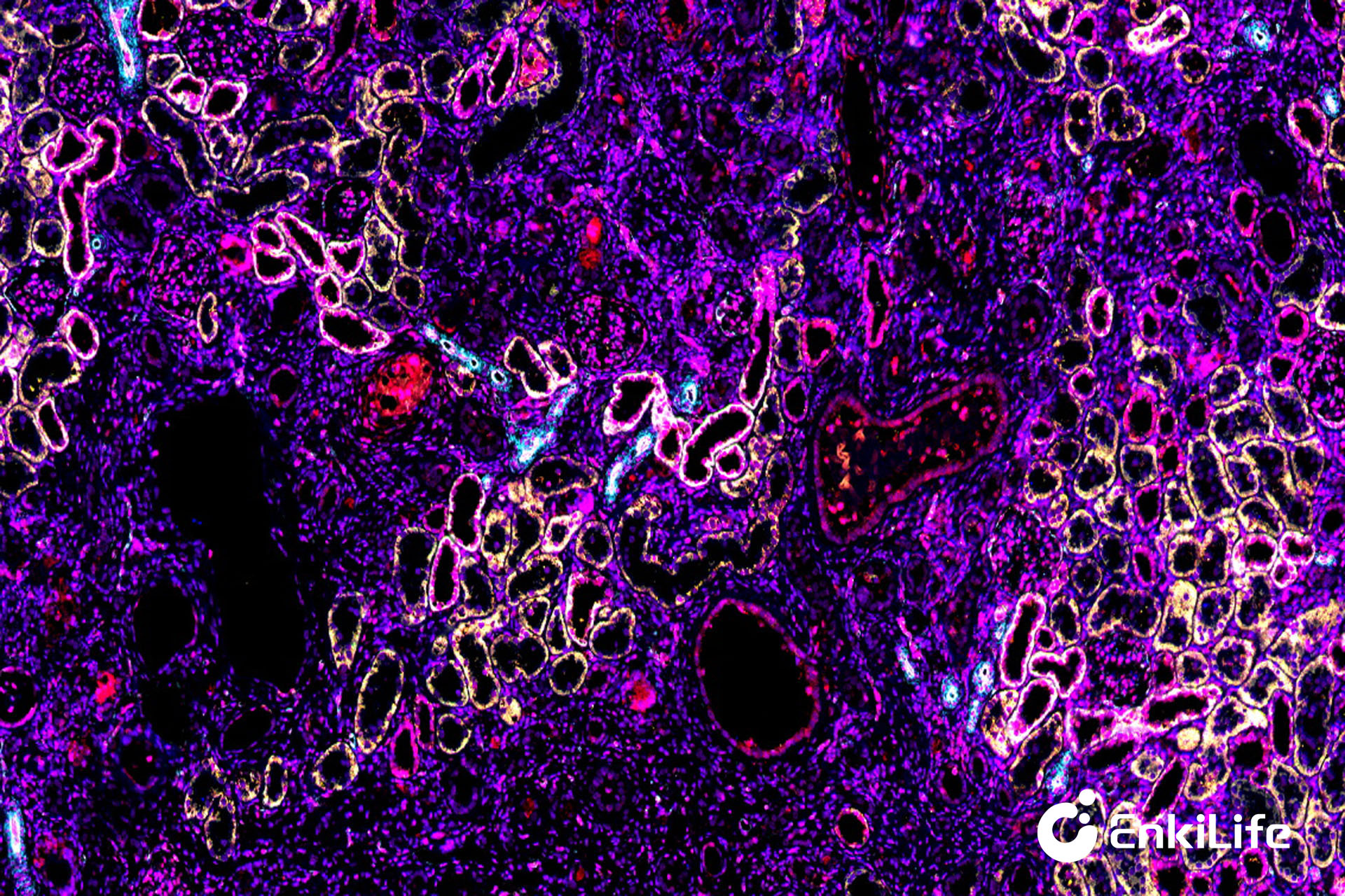
Multiplex Immunohistochemistry (mIHC) is an important upgrade based on traditional IHC. Its core idea is to simultaneously detect multiple protein markers in the same tissue section and use fluorescent dyes of different wavelengths for differentiation, thereby achieving multi-marker co-localization analysis. One of the most widely used protocols is the mIHC system based on TSA (Tyramide Signal Amplification) technology.
In the TSA system, HRP enzyme can catalyze the deposition of fluorescently labeled tyramide molecules near the antigen, achieving covalent immobilization of the signal. Subsequently, the previous round of antibodies is removed through an antibody stripping step, and the next round of staining is performed. After multiple cycles, the fluorescent signals of multiple markers are retained on the same tissue section. Finally, images are collected using a multispectral scanning system, and spectral unmixing, cell segmentation, phenotype recognition, and spatial analysis are completed through professional analysis software. Compared with traditional IHC, mIHC can simultaneously detect 4-9 or even more markers while maintaining tissue structure integrity.
From a technical perspective, the biggest difference between the two is not just the increase in the number of detected markers, but the improvement in research dimensions.
Traditional IHC mainly answers the question: "Is this protein expressed?" while mIHC further answers: "Which cells express this protein?" "Where are these cells located?" "How close are these cells to each other?" "Do they form specific spatial structures?" In other words, mIHC not only provides expression information but also cell composition information and spatial relationship information.
Therefore, mIHC is essentially not only a staining technique but also a spatial biology research tool.
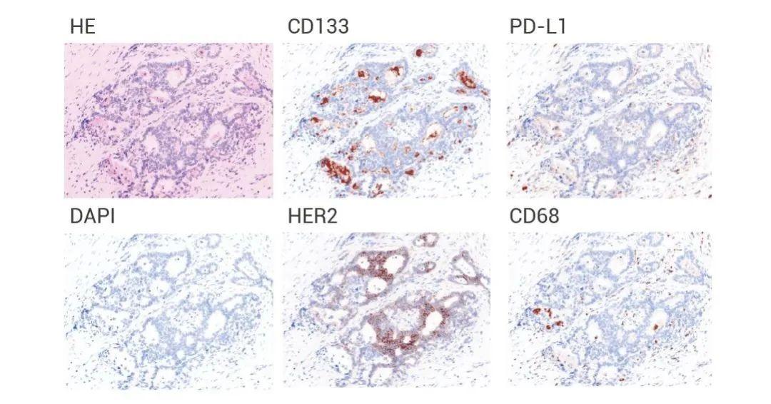
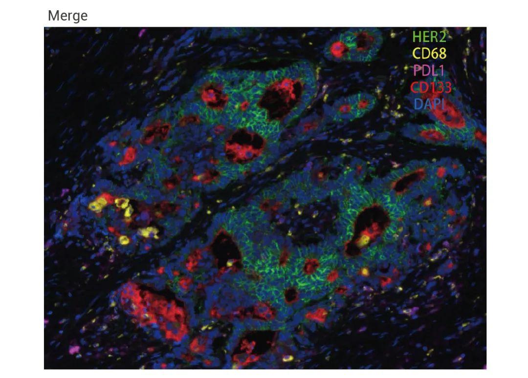
Figure. Monochromatic IHC (top) and Multispectral mIHC (bottom) Validation of Cancer Tissue
In recent years, one of the hottest topics in tumor research is the tumor microenvironment.
Studies have found that tumor development depends not only on tumor cells themselves but also on the combined influence of surrounding immune cells, fibroblasts, vascular cells, and other cell types.
For example, in immunotherapy research, detecting PD-L1 expression levels alone is often insufficient to fully predict patient response.
Researchers are more concerned about:
• Whether CD8+ T cells have infiltrated the tumor area;
• Whether regulatory T cells are abundantly aggregated;
• Whether macrophages are in M1 or M2 state;
• The spatial distance between immune cells and tumor cells.
These questions require combined detection of multiple markers and spatial localization analysis, which is exactly where mIHC excels.
By simultaneously detecting markers such as CD3, CD8, FoxP3, PD-1, PD-L1, and CK in one experiment, researchers can reconstruct the cell distribution map of the entire immune microenvironment on a single section.
Traditional IHC is an important basic technique in pathology and histology research, and still has irreplaceable value in clinical diagnosis and routine scientific research. mIHC is a further development based on IHC. Through its ability to simultaneously detect multiple markers and perform spatial analysis, it provides richer information for tumor microenvironment research, immunotherapy research, and spatial biology research.
From "observing single protein expression" to "analyzing tissue microenvironment ecology", mIHC is driving tissue pathology research into a more refined and digital new stage.
mIHC Product Recommendation - Multiplex Fluorescence Detection Kit
Product | Catalog Number |
|---|---|
TSA Six-Label Seven-Color Multiplex Immunohistochemistry Kit | |
TSA Five-Label Six-Color Multiplex Immunohistochemistry Kit | |
TSA Four-Label Five-Color Multiplex Immunohistochemistry Kit | |
TSA Three-Label Four-Color Multiplex Immunohistochemistry Kit | |
TSA Two-Label Three-Color Multiplex Immunohistochemistry Kit |
For details, please check TSA mIHC Kit