Multiplex Immunofluorescence Technology: In-depth Analysis and Practical Guide Based on Tyramide Signal Amplification
Against the backdrop of the rising interest in spatial biology and tumor microenvironment research, multiplex immunofluorescence (mIHC/mIF) has evolved from a "upgraded tool" of traditional immunohistochemistry to a core technical platform for analyzing tissue spatial information. Among them, the multiplex immunofluorescence system based on Tyramide Signal Amplification (TSA), with its high sensitivity, high stability, and cyclic staining capability, has become one of the mainstream technical routes for current spatial phenotyping analysis.
Unlike traditional fluorescent immunostaining, the TSA system does not simply enhance fluorescence intensity, but achieves "tissue deposition" of signals through enzymatic catalysis, fundamentally changing the signal existence form and detection logic, thereby supporting tissue in-situ detection systems with up to 5-8 colors or even higher dimensions.
Unlike traditional fluorescent immunostaining, the TSA system does not simply enhance fluorescence intensity, but achieves "tissue deposition" of signals through enzymatic catalysis, fundamentally changing the signal existence form and detection logic, thereby supporting tissue in-situ detection systems with up to 5-8 colors or even higher dimensions.
I. The Essence of TSA Technology: Mechanism Transition from "Antibody Labeling" to "Tissue Deposition"
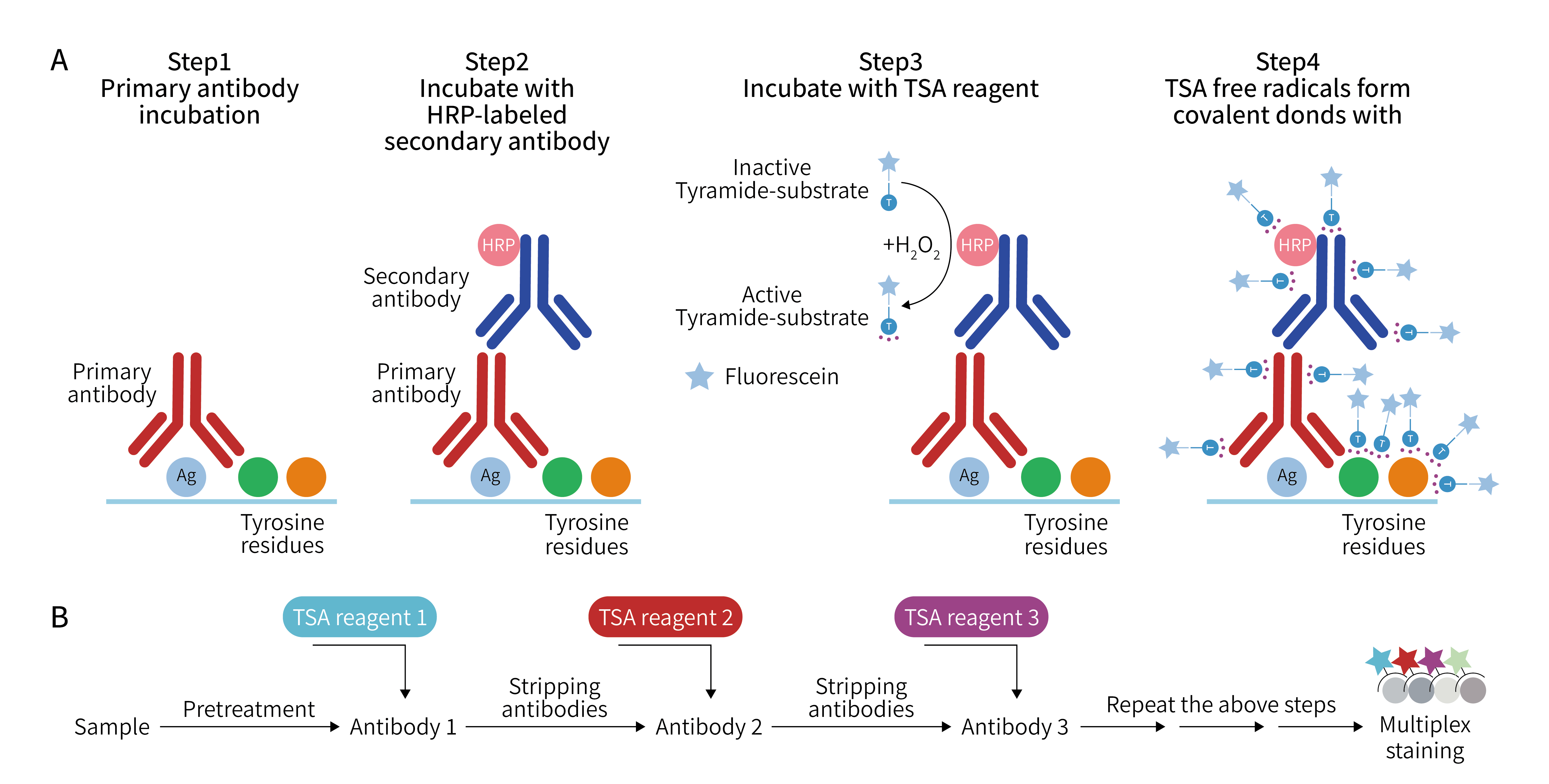
The core mechanism of TSA (Tyramide Signal Amplification) is based on the HRP (Horseradish Peroxidase)-catalyzed tyramide reaction system, which can be summarized as the following process:
Therefore, the TSA system possesses three distinct technical characteristics:
A. Primary antibody recognizes target antigen
B. HRP-labeled system binds to antibody complex
C. Add fluorescent-labeled tyramide substrate (Tyramide-fluorophore)
D. HRP catalyzes tyramide oxidation reaction
E. Activated tyramide rapidly forms covalent bonds with tyrosine residues in tissue
F. Fluorescence signal is "fixed" in local tissue structure
Therefore, the TSA system possesses three distinct technical characteristics:
Signal Permanence: Covalent bond structure ensures fluorescence signals remain stable during subsequent antibody stripping or multi-round staining processes.
Local Amplification Effect: A single HRP molecule can catalyze the deposition of multiple tyramide molecules, achieving exponential signal enhancement.
Spatial Locking Ability: Deposition reaction is confined within nanoscale range, avoiding spatial blurring caused by traditional fluorescence diffusion.
II. Technical Architecture of Multiplex Immunofluorescence System: Cyclic Staining Logic
TSA-based mIF technology is essentially a "cyclic iterative staining system", whose standard workflow is as follows:
1. Antigen Exposure and Retrieval: Restore antigen epitopes through heat-induced epitope retrieval (HIER), typically using EDTA (pH 8.0) or citrate buffer system.
2. Primary Antibody Incubation: Specific antibody recognizes target protein, this stage determines the upper limit of detection specificity.
3. HRP Signal System Construction: Use HRP-secondary antibody, which can significantly reduce background signal and improve stability.
4. TSA Fluorescence Deposition Reaction: Add fluorescent-labeled tyramide substrate, forming irreversible covalent deposition signals under HRP and H₂O₂ catalysis.
5. Antibody Stripping: Remove antibody complexes through high-temperature or low-pH treatment, while preserving deposited fluorescence signals.
6. Multi-round Cyclic Staining: Repeat the above steps to achieve layer-by-layer labeling of multiple targets.
III. Key Control Points in Experimental Workflow
1. Selection and Control of Antigen Retrieval Conditions
Antigen retrieval is a core step determining overall signal intensity and tissue structural integrity. Typically, high-temperature and high-pressure retrieval using 1× citrate buffer (pH 6.0) is performed, and basic antigen exposure can be completed within approximately 2 minutes after steaming while ensuring tissue structure stability; for weakly expressed targets, switching to EDTA (pH 9.0) can enhance antigen exposure efficiency, while for easily detached samples such as brain tissue and bone tissue, gentle microwave retrieval at around 80℃ is recommended to reduce mechanical stress, and the buffer concentration can be appropriately increased to 2× citrate to compensate for insufficient retrieval strength, thereby achieving a balance between "signal exposure" and "tissue integrity".
2. Optimization of Endogenous Peroxidase Blocking System
Endogenous enzyme blocking is mainly used to eliminate interference from tissue's own peroxidase activity on the TSA signal system. Conventionally, incubation with 3% H₂O₂ at room temperature for 20 minutes can effectively suppress background signals, but for hemoglobin-rich tissues such as liver and spleen, the blocking time can be appropriately extended to avoid autofluorescence or false positive enhancement; for easily detached tissues, the H₂O₂ concentration should be reduced to 1-2% to minimize tissue damage, thereby achieving dynamic balance between background control and tissue stability.
3. Antibody System Selection and Concentration Optimization Strategy
Antibody system directly determines detection specificity and signal-to-noise ratio. Monoclonal antibodies are preferentially recommended to reduce cross-reaction risk, while mouse-derived primary antibodies should be avoided as much as possible in mouse samples to reduce endogenous IgG interference; primary antibody working concentration must be determined through gradient pre-experiments. Excessively high concentration tends to increase non-specific binding background, while excessively low concentration may cause signal loss of weakly expressed targets. Therefore, it is recommended to determine optimal working conditions through high, medium, and low three-gradient screening to ensure signal stability and reproducibility.
4. TSA Dye Usage and Signal Amplification Control
TSA fluorescent substrates belong to highly sensitive enzyme-catalyzed deposition systems, which must be freshly prepared and used with strict light-shielding operations to avoid photodegradation affecting signal quality. In target allocation, the principle of "low expression with high gain, high expression with anti-saturation" should be followed, i.e., low-abundance proteins should preferentially select high signal intensity dyes such as Cy5 or AF594 to improve detection rate, while high-expression targets should use medium-intensity dyes such as FITC or Cy3 to avoid signal oversaturation, thereby ensuring balanced distribution of dynamic range.
5. Spectral Design and Crosstalk Control Principles
The success of multiplex fluorescence experiments highly depends on the rationality of spectral design. Dye selection must ensure that emission wavelengths are separated by at least 50 nm. For example, DAPI (461 nm), AF488 (519 nm), Cy3 (570 nm), and Cy5 (670 nm) form a classic four-channel combination, which can effectively reduce spectral overlap; at the same time, highly overlapping combinations such as FITC with AF488 and Cy3 with AF594 should be avoided, as they are likely to produce irreversible crosstalk interference, affecting the accuracy of subsequent quantitative analysis.
6. Matching Optimization of Imaging System and Dye System
Different microscopic imaging systems have direct constraints on dye selection. Due to fixed filters, ordinary fluorescence microscopes are recommended to prioritize visible light range dyes such as DAPI, FITC, and Cy3 to ensure imaging clarity, while for confocal or multispectral imaging systems, far-infrared dyes such as Cy5 or AF647 can be introduced, and spectral unmixing technology can further reduce channel crosstalk. Therefore, experimental design must be based on equipment capabilities, rather than simply pursuing dye quantity expansion.
7. Antibody Elution Efficiency and Cyclic Staining Stability Control
The core of multi-round TSA staining system lies in the balance between antibody stripping efficiency and tissue stability. If antibody affinity is high or binding is strong, removal efficiency can be improved by increasing elution times or optimizing heat retrieval intensity, but over-treatment leading to antigen damage must be avoided; during operation, slices must be kept horizontal to prevent reagent loss affecting local elution effect. For easily detached samples, elution temperature should be appropriately reduced or incubation time shortened, thereby achieving optimal control between "elution completeness" and "tissue integrity".
IV. Technology Development Trends: From "Multiplex Staining" to "Spatial Quantitative Analysis"
The development of multiplex immunofluorescence (TSA-mIHC) technology is progressing from the traditional "biomarker quantity expansion" stage to a quantification stage centered on "spatial structure analysis". Its essential change is the transition of data dimension from two-dimensional expression information to three-dimensional spatial relationship modeling. Due to its stable covalent deposition characteristics, the TSA system makes fluorescence signals no longer dependent on antibody presence, thereby providing a stable data foundation for subsequent digital image analysis. Against this technical background, research focus is shifting from "whether a certain protein is expressed" to "spatial distribution relationships between different cell types in tissues and their functional neighborhood structures". For example, distance gradients between CD8+ T cells and tumor cells, spatial co-localization probability of PD-1/PD-L1 axis, and aggregation patterns of macrophages in immunosuppressive regions. These indicators are gradually being converted into computable spatial parameters and further input into artificial intelligence image analysis and spatial statistical models, transforming mIF technology from a traditional histology tool into an important component of spatial omics analysis.
Conclusion
TSA-based multiplex immunofluorescence technology is essentially not a simple staining enhancement method, but a "tissue spatial information encoding technology". Its core advantage lies in converting molecular expression information into stable, iterable, and analyzable spatial structure signals through enzyme-catalyzed deposition mechanisms. With the development of spatial omics and digital pathology, TSA-mIF is evolving from a laboratory tool into a key bridge connecting "molecular expression - spatial structure - disease mechanism".
To meet the research needs of tumor microenvironment and spatial biology, Enkilife provides TSA-mIHC kits and technical services, supporting multi-biomarker detection, experimental optimization, and spatial analysis, helping researchers obtain more valuable information from a single tissue section.
Product | Catalog Number |
|---|---|
TSA Six-Label Seven-Color Multiplex Immunohistochemistry Kit | |
TSA Five-Label Six-Color Multiplex Immunohistochemistry Kit | |
TSA Four-Label Five-Color Multiplex Immunohistochemistry Kit | |
TSA Three-Label Four-Color Multiplex Immunohistochemistry Kit | |
TSA Two-Label Three-Color Multiplex Immunohistochemistry Kit |