Frontier Technologies And News

TUNEL Apoptosis Staining: Complete Experimental Protocol

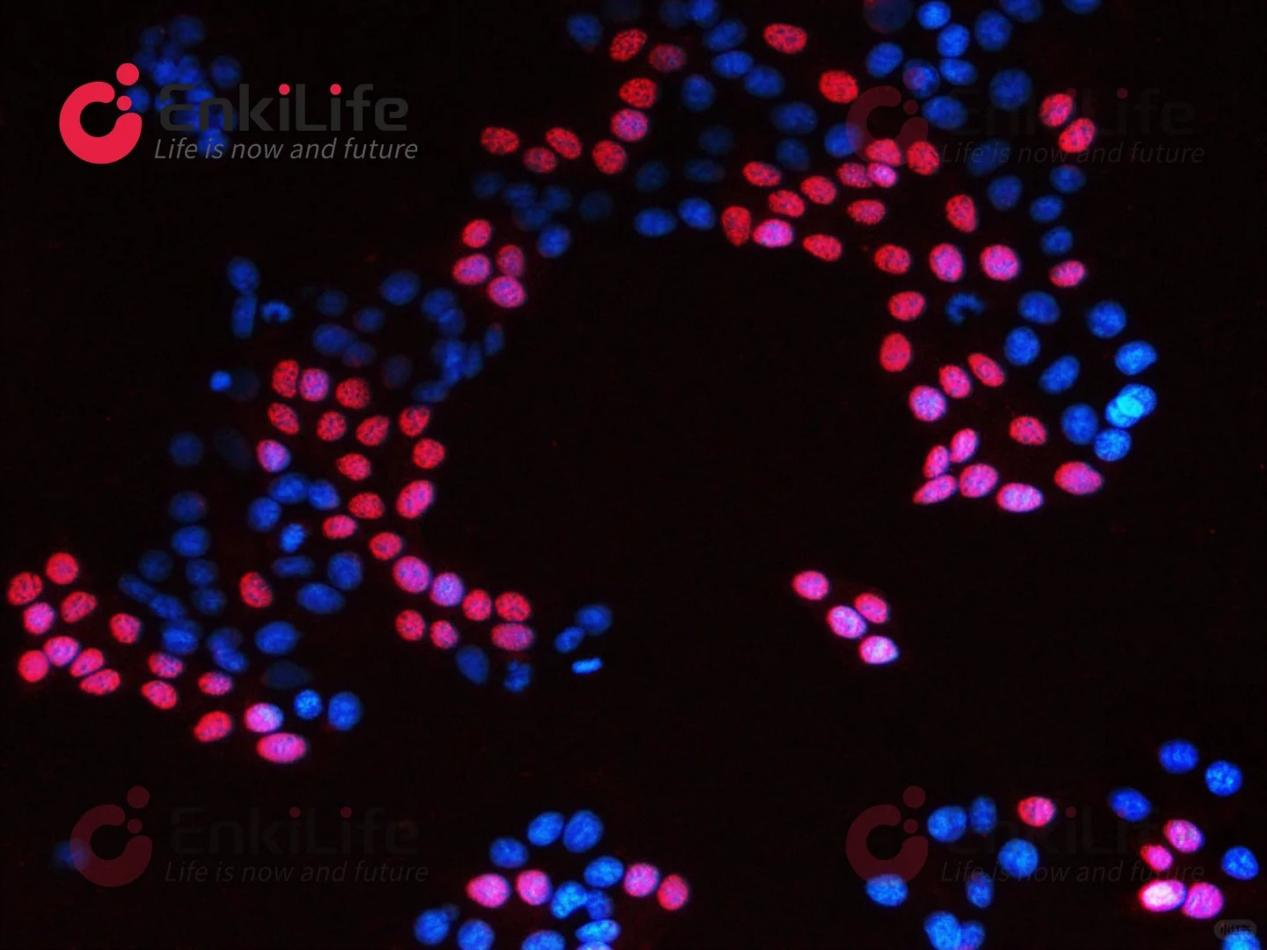
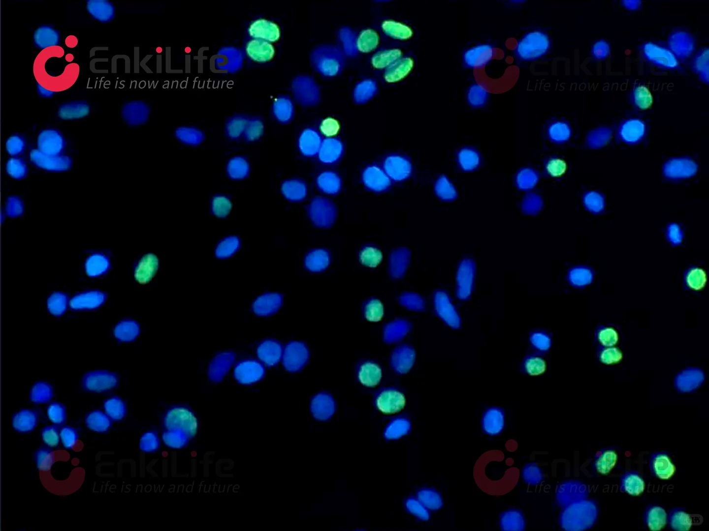
TUNEL labeling is the standard method for detecting apoptotic DNA fragmentation in situ. This protocol describes its rationale — apoptotic endonucleases cleave DNA between nucleosomal units to generate 180–200 bp fragments that expose abundant 3'-OH ends, which terminal deoxynucleotidyl transferase (TdT) labels with fluorophore- or biotin-conjugated dUTP in a template-independent manner — and provides a complete, sample-specific workflow for five specimen types: adherent cells and cell smears, suspension cells, paraffin sections and frozen sections, plus DNase I–treated positive controls. Key control points include fixation with 4% paraformaldehyde (30 min at 4 °C for cells, 30 min at room temperature for frozen sections), permeabilization with 0.4% Triton X-100 or Proteinase K (2 mg/mL diluted 1:50 to 40 μg/mL, 30 min at 37 °C for paraffin sections and 20 min for frozen sections), thorough removal of Proteinase K before labeling, and reaction with 50 μL TUNEL mixture at 37 °C in the dark (15–30 min for cells, 1 h for tissue), followed by optional DAPI counterstaining and anti-fade mounting. Results are quantified as the apoptotic index, AI = TUNEL-positive nuclei / total DAPI-stained nuclei × 100%, using at least five randomly selected fields under identical exposure settings and consistent counting criteria; fluorescence intensity (IOD) alone is not recommended as the primary readout. Troubleshooting tables address weak or absent signal, non-specific staining with high false positives, and excessive background, linking each artifact to fixation, dewaxing, permeabilization, photobleaching and washing parameters and to the corresponding corrective measures, thereby supporting reproducible, semi-quantitative assessment of apoptosis across cell and tissue samples.

Annexin V/PI Flow Cytometry Apoptosis Detection: Principle, Standard SOP and Quadrant Analysis

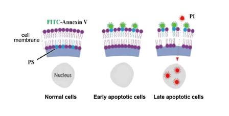
Annexin V/propidium iodide (PI) double staining is a widely used flow cytometric method for quantifying apoptosis and distinguishing it from necrosis. The assay exploits two independent markers: translocation of phosphatidylserine (PS) from the inner to the outer leaflet of the plasma membrane, which is bound selectively by the 35–36 kDa Ca²⁺-dependent phospholipid-binding protein Annexin V, and loss of membrane integrity, which permits the membrane-impermeant DNA dye PI to stain nuclei. Because PS externalization precedes membrane breakdown, the two probes resolve four populations in a quadrant plot: Annexin V⁻PI⁻ live cells (Q4), Annexin V⁺PI⁻ early apoptotic cells (Q3), Annexin V⁺PI⁺ late apoptotic or secondarily necrotic cells (Q2), and Annexin V⁻PI⁺ cells representing mechanical damage, primary necrosis or debris (Q1). This protocol details cell collection for suspension and adherent cultures (EDTA-free trypsinization to preserve Ca²⁺-dependent binding), washing and counting, resuspension in 100 μL 1× binding buffer, sequential addition of Annexin V and 5 μL PI, incubation at 20–25 °C in the dark for 10–15 min with intermittent resuspension, and immediate acquisition after adding 400 μL binding buffer without further washing. Total apoptosis is calculated as Q3 + Q2, and Q1 is recommended to remain below 5% as an internal index of technical quality. Common failure modes — an unexpectedly high Q1, results discordant with expectations, and absent positive signal due to residual EDTA, omission of Ca²⁺-containing buffer or post-staining washing — are analyzed together with optimization of stimulus dose and duration, dissociation intensity and initial cell viability, providing a practical basis for reproducible apoptotic-rate determination.

   💬 WhatsApp