

Product Introduction
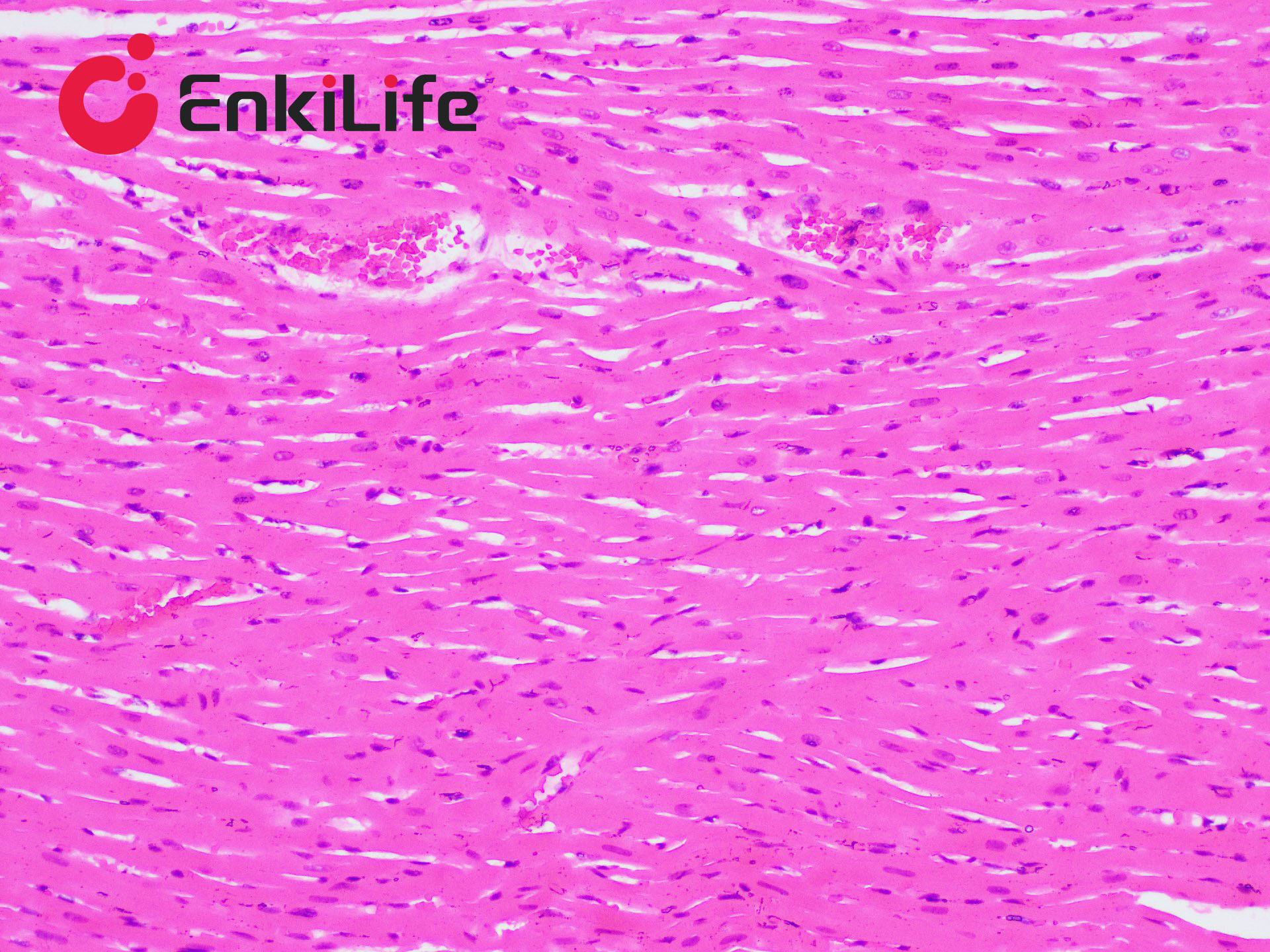
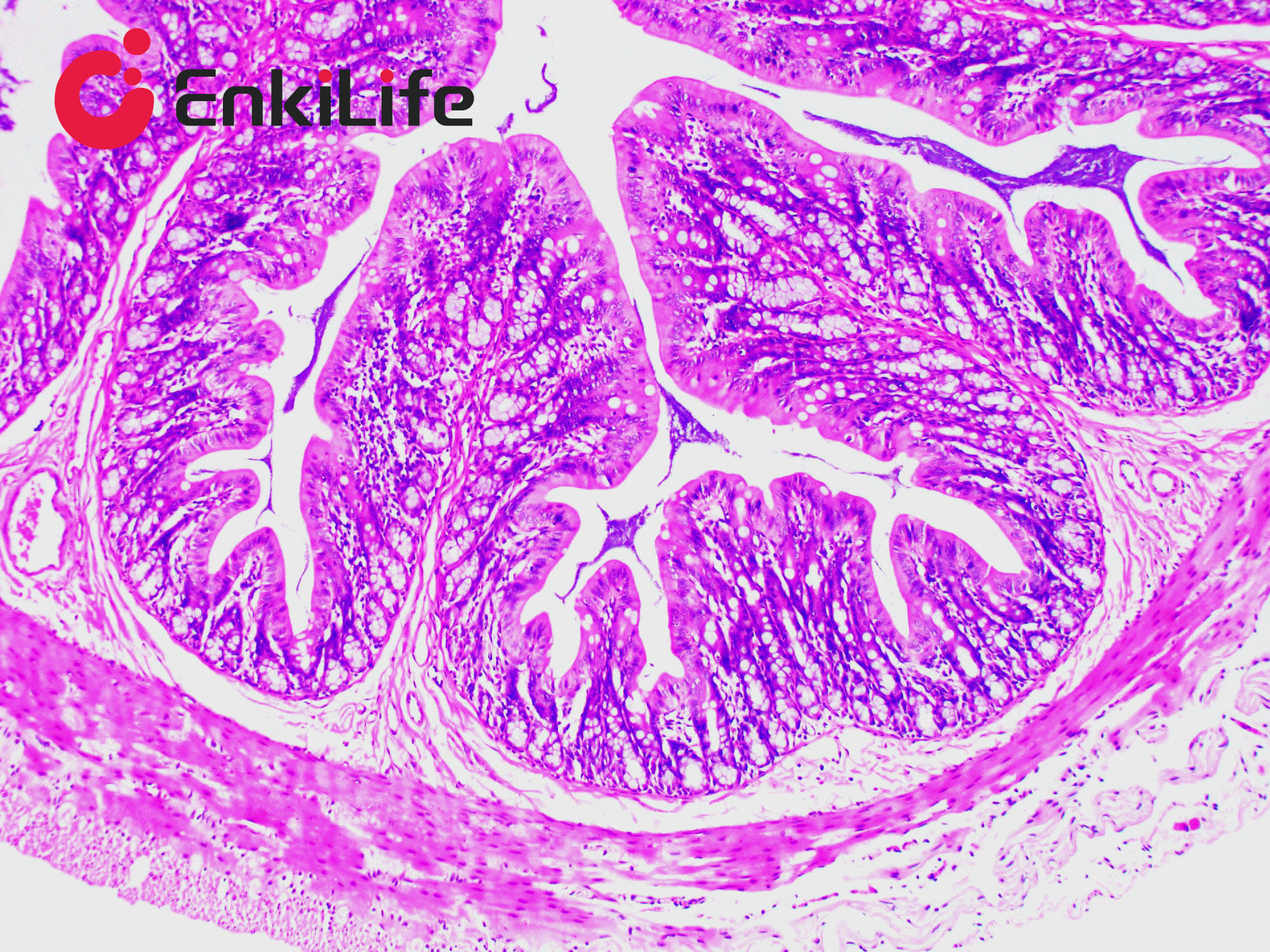
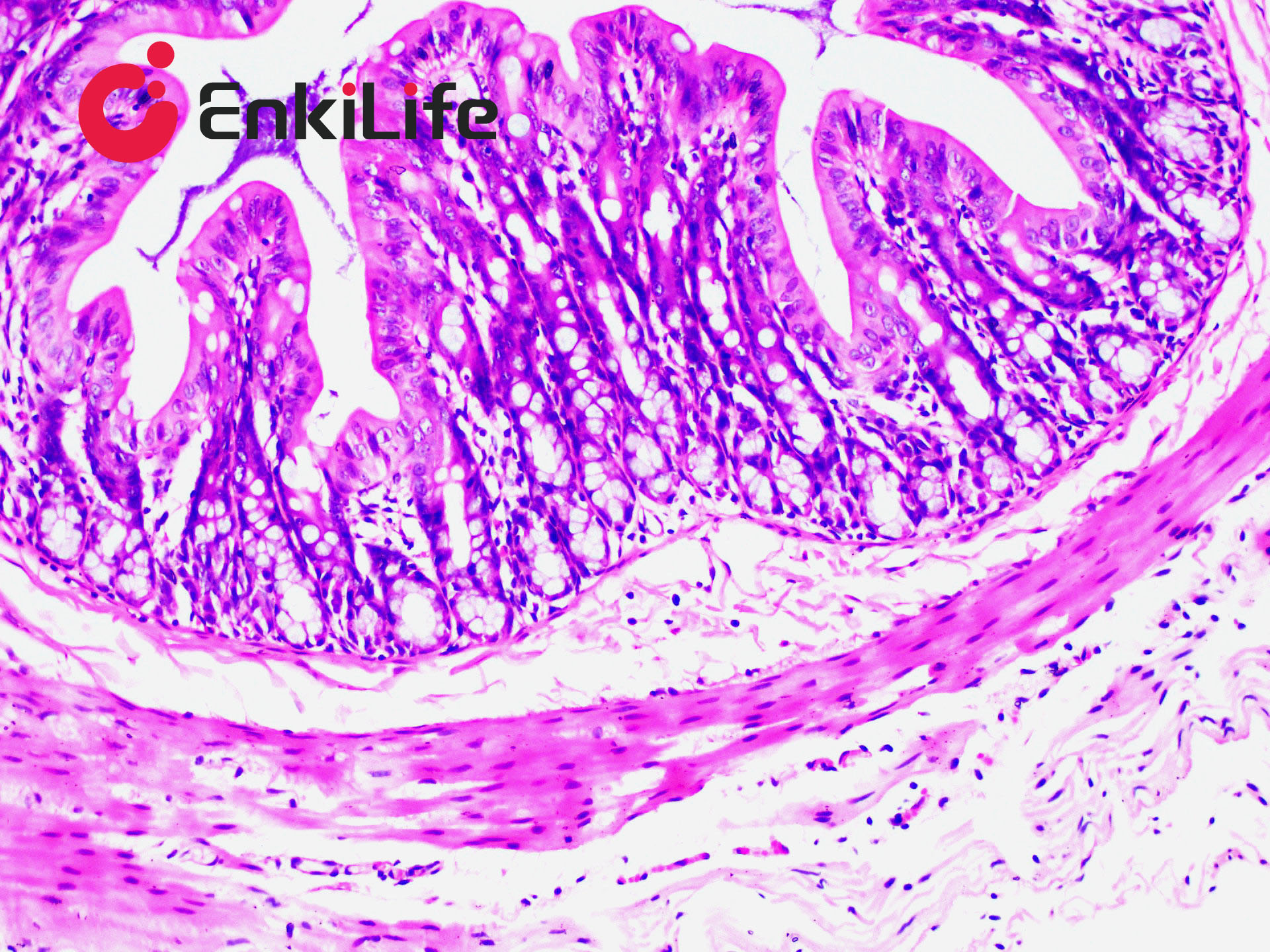
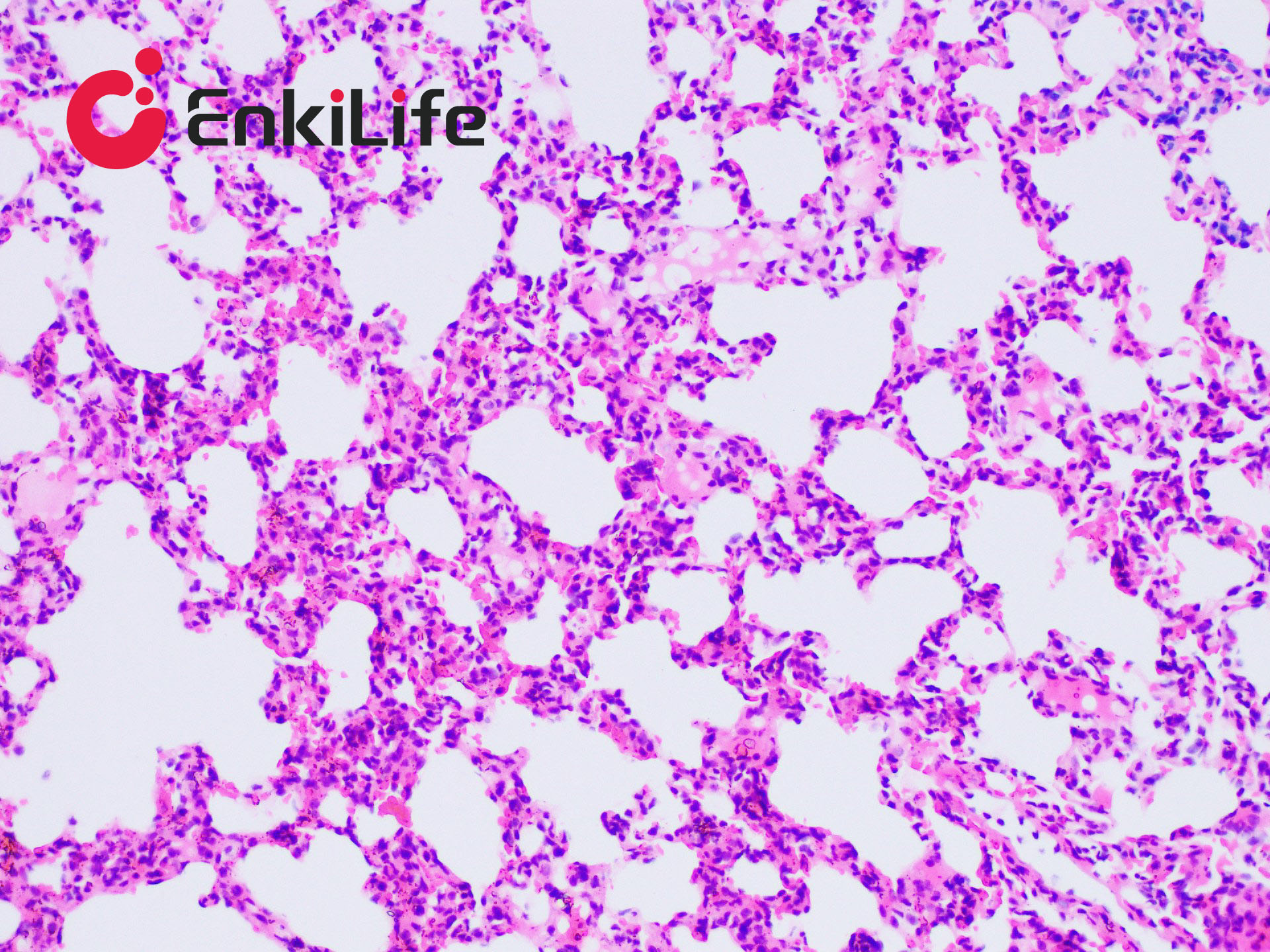
Calcium is abundant in the human body, constituting the skeleton that supports the body and playing essential roles in secretion, transport, muscle contraction, and nerve conduction. Calcium exists in two forms: ionized calcium in the circulation (so-called blood calcium) and bound calcium that is deposited in tissues in combination with proteins, carbonate, or phosphate. Except for bone and teeth, calcium is normally distributed throughout all tissues and cells and usually does not appear in solid form; however, under certain conditions calcium precipitates and deposits in tissues as pathological calcification, mainly calcium phosphate and, to a lesser extent, calcium carbonate. Calcium salts are usually monorefringent, but calcium oxalate is birefringent. With H&E staining calcium appears generally bluish-purple. Many dyes can chelate calcium, including Alizarin Red S, purpurin, and nuclear fast red.
Alizarin Red S, an anthraquinone derivative and the sodium salt of alizarin sulfonate, chelates calcium in calcium carbonate or calcium phosphate to form an orange-red complex. Alizarin Red S often yields more reliable results for small amounts of deposits and is frequently combined with Fast Green or Mayer’s hematoxylin to produce an orange-red precipitate, making it suitable for staining tissues containing scant calcium salts.
Basic Information
Product name | Alizarin Red S Staining Kit, 2%, pH 4.2 |
Sizes | 100 mL |
Storage | RT |
Shipping | RT |
Validity | 12 months |
Notes
1. Staining time must be adjusted according to calcium content; monitor under the microscope and rinse as soon as deposits show a deep orange-red. Over-staining causes diffusion; 5 min is usually sufficient.
2. After Alizarin Red S staining, calcium deposits are birefringent.
3. When Fast Green is used as counterstain, the background appears green; with Mayer’s hematoxylin, nuclei appear blue.
4. This method is particularly useful for identifying and detecting small amounts of calcium, e.g., abnormal calcification in kidney (hypercalciuria).
5. Wear lab coat and disposable gloves for safety.
6. Use the reagent promptly after opening to ensure consistent performance.