

Product Introduction
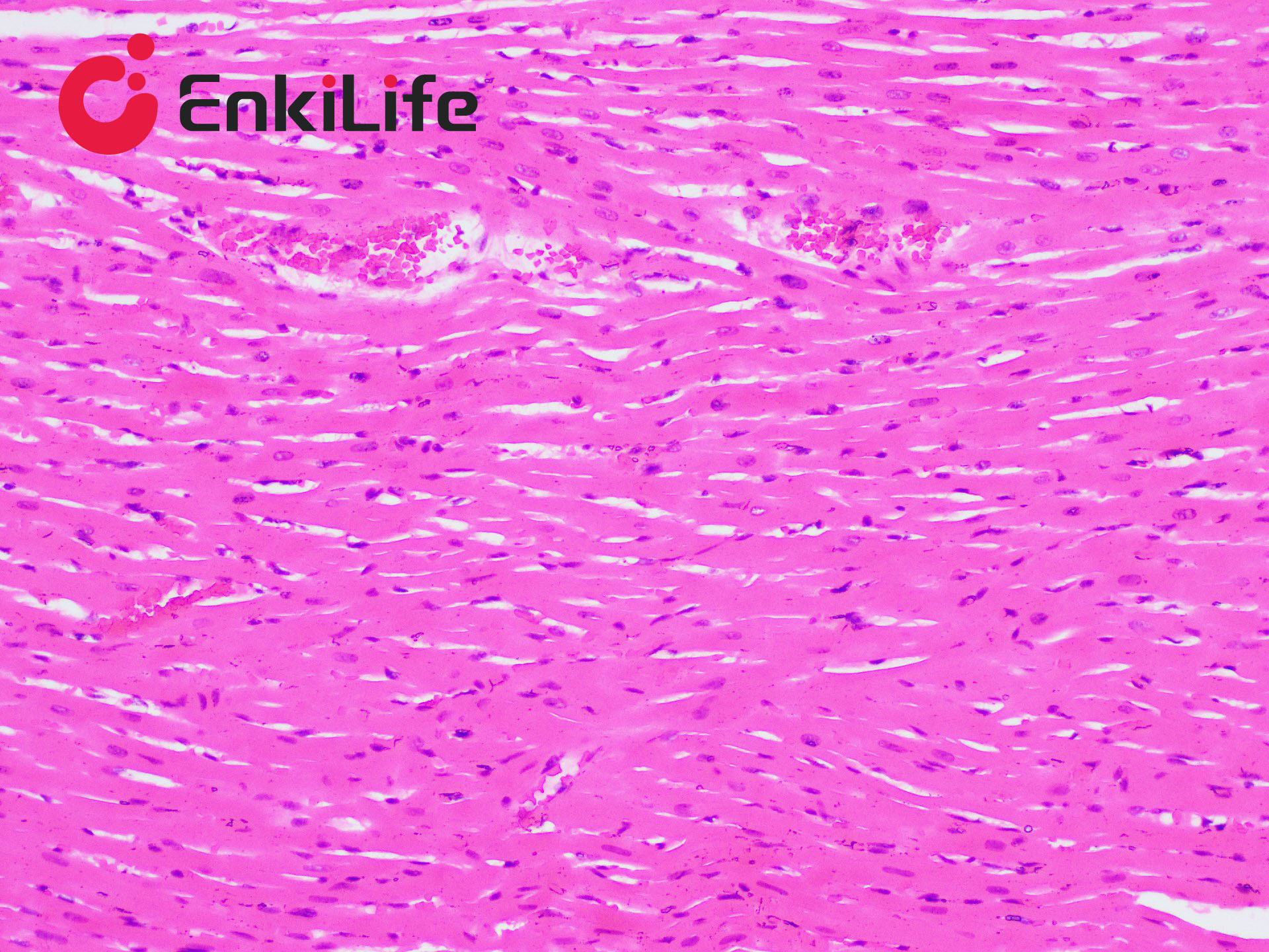
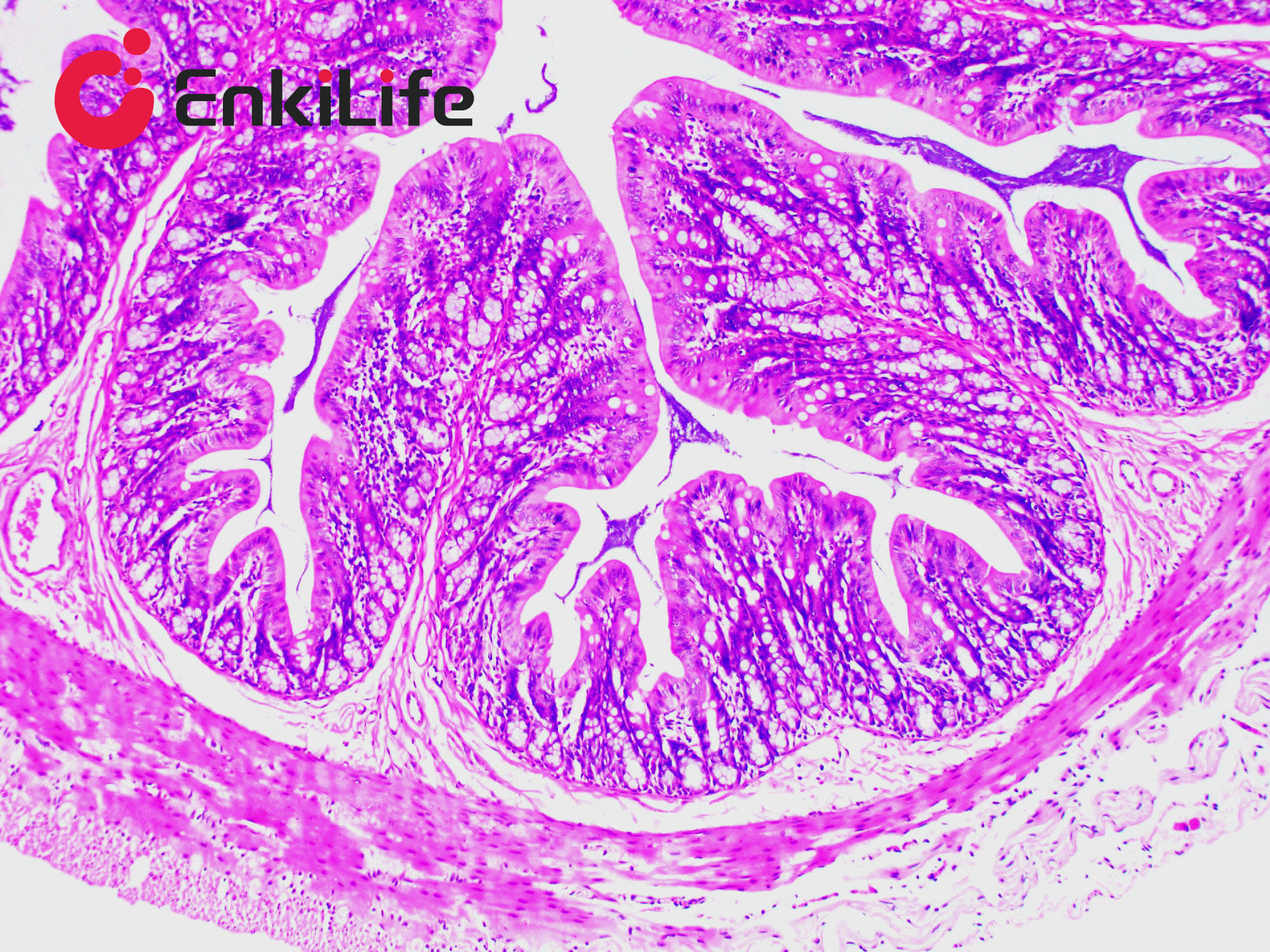
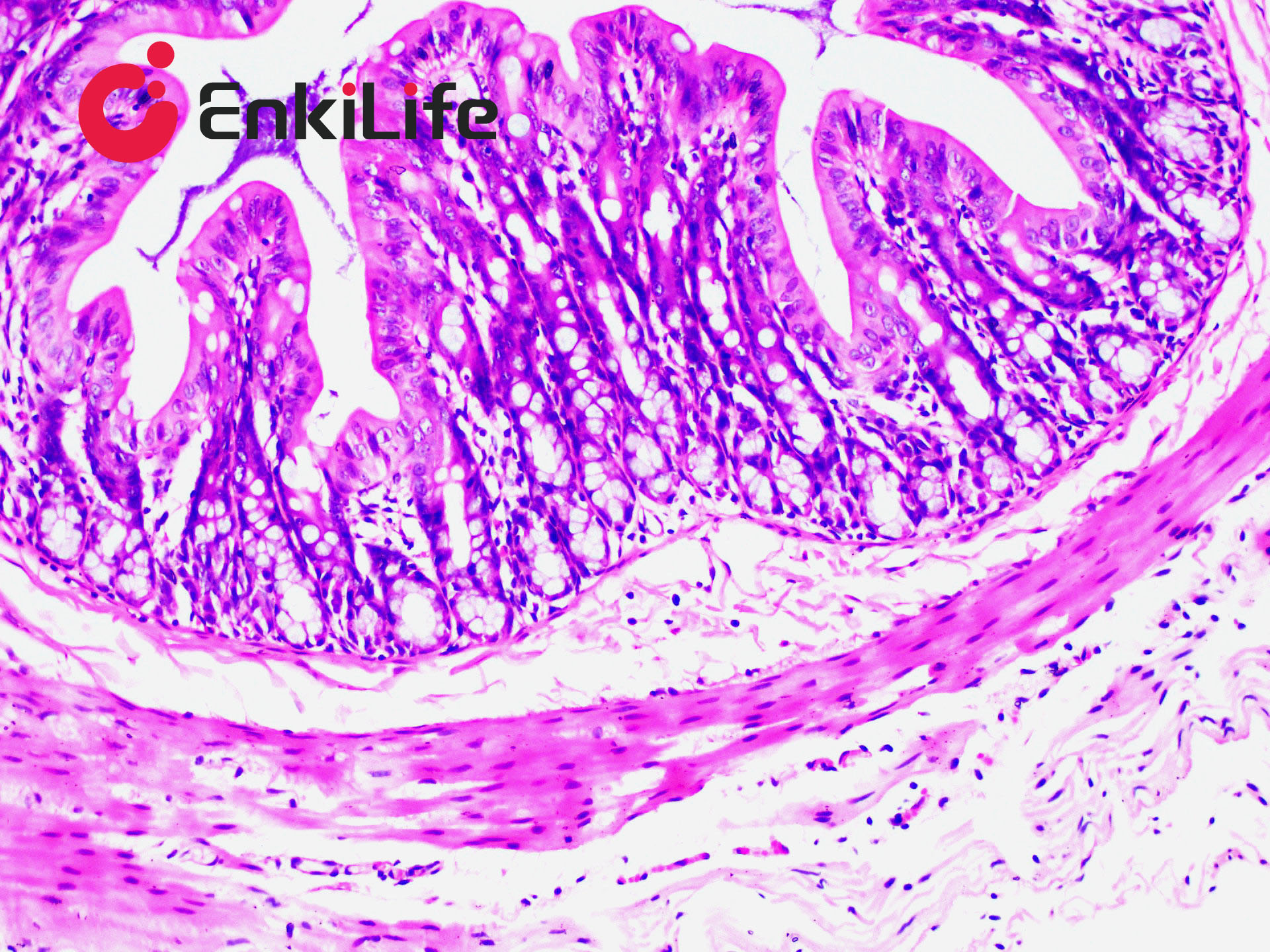
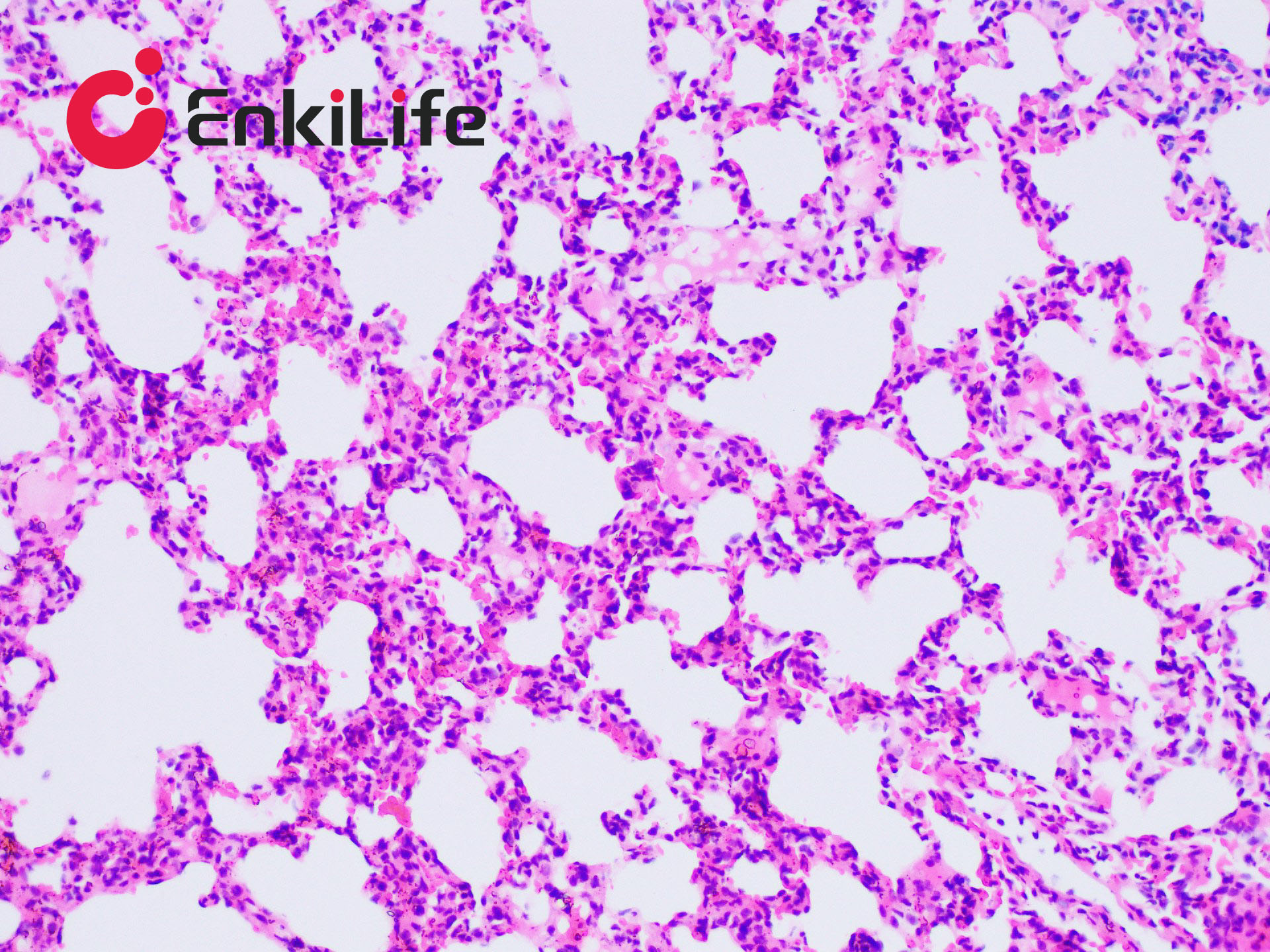
Connective tissue, in a narrow sense, is composed of three types of fibers: collagen fibers, reticular fibers, and elastic fibers. Elastic fibers are mainly distributed in the human arterial walls, alveolar walls, and skin. They appear yellow and are highly refractile when fresh.
Elastic fiber staining can reveal changes in elastic fibers in skin tissues, such as elastic nevus, granuloma annulare, and scleroderma. It is also used to display and assess lesions of the endocardium and arteries, to observe whether certain pathological changes are accompanied by proliferation or destruction of elastic fibers, and to identify tumor components such as elastic fibromas. After elastic fiber staining, elastic fiber balls within the tumor can be clearly observed. Commonly used elastic fiber staining methods include the Gomori aldehyde-fuchsin method, resorcin-fuchsin method, orcein method, Victoria blue method, and iron hematoxylin method.
EnkiLife Elastic Fiber Staining Solution (E.V.G Method) belongs to the iron hematoxylin staining category. The staining principle is based on the fact that elastin and pre-elastin fibers are highly cross-linked via disulfide bonds. After oxidation by iodine, some disulfide bonds are converted into anionic sulfated derivatives, which are strongly basophilic and selectively absorb the basic dye hematoxylin in the staining solution. This product uses the most commonly used Van Gieson (VG) counterstaining method. After staining, thick fibers are deeply stained, while thin fibers are slightly less distinct. Elastic fibers appear black, collagen fibers red, and muscle fibers and erythrocytes yellow, providing a clear contrast.
Basic Information
Product name | Elastic Fiber Staining Kit, E.V.G Method |
Sizes | 50 mL |
Storage | RT, keep away from light |
Shipping | RT |
Validity | 12 months |
Product Components
Components | 3x 50mL | |
Reagent (A): Verhöeff dye solution | A1: Verhöeff Staining Solution A | 30 ml |
A2: Verhöeff Staining Solution B | 12 ml | |
A3: Verhöeff Staining Solution C | 12 ml | |
Before use, mix A1:A2:A3 at a ratio of 5:2:2 to prepare the Verhöeff staining solution. Use within 2–3 h. Do not prepare in advance. | ||
Reagent(B):Verhöeff Differentiating Solution | 50 mL | |
Reagent(C): Van Gieson dye solution 50 mL
| C1: Fuchsin Staining Solution | 5 ml |
C2: Fuchsin Diluent | 45 ml | |
Before use, mix C1:C2 at a ratio of 1:9 to prepare the Van Gieson staining solution. Do not prepare in advance. | ||
Notes
1. Dewax sections thoroughly; if temperature is low, incubate at 60–70 °C in an oven.
2. Replace graded ethanol solutions regularly.
3. Differentiation time with hydrochloric acid ethanol should be adjusted according to section thickness, tissue type and age; ensure adequate washing with tap water after differentiation to remove all acid.
4. Ether-ethanol mixed fixative is prepared by mixing equal volumes of ether and 95% ethanol, adding a small amount of acetic acid, and storing in a sealed container.
5. Keep staining times as short as possible for frozen sections.
6. Common bluing solutions include 0.2–1% ammonia water, Scott’s tap water substitute, or 0.1–1% lithium carbonate solution.
7. For your safety and health, wear a lab coat and disposable gloves during operation.