

Product Introduction
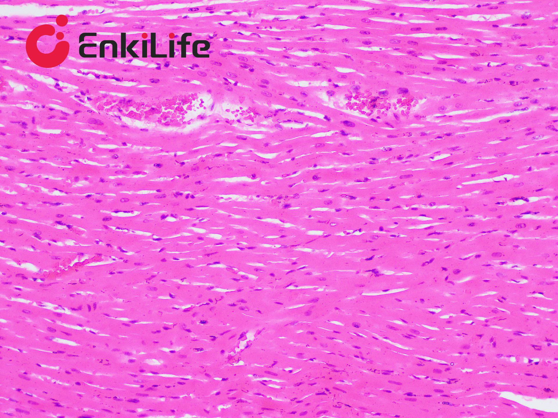
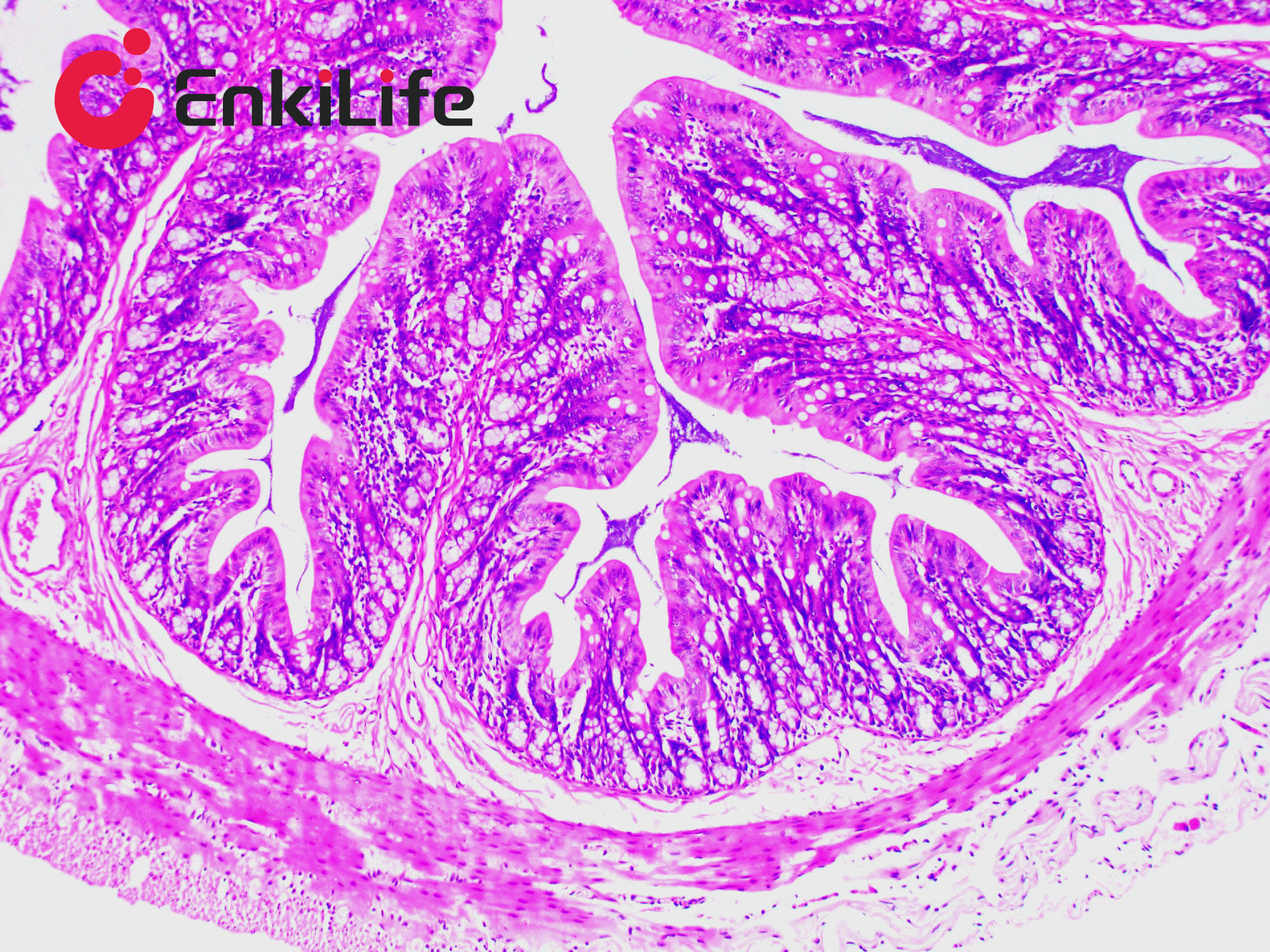
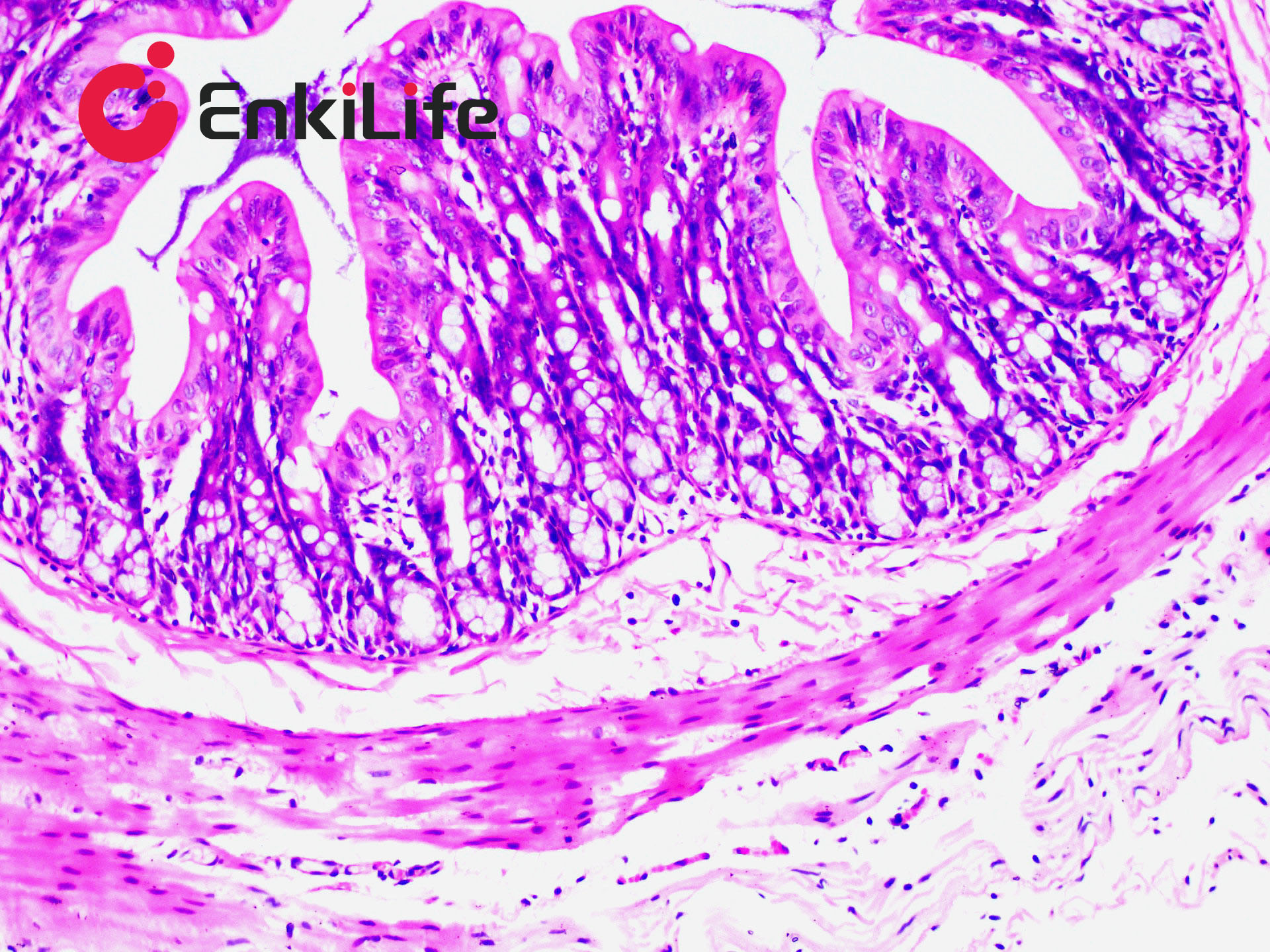
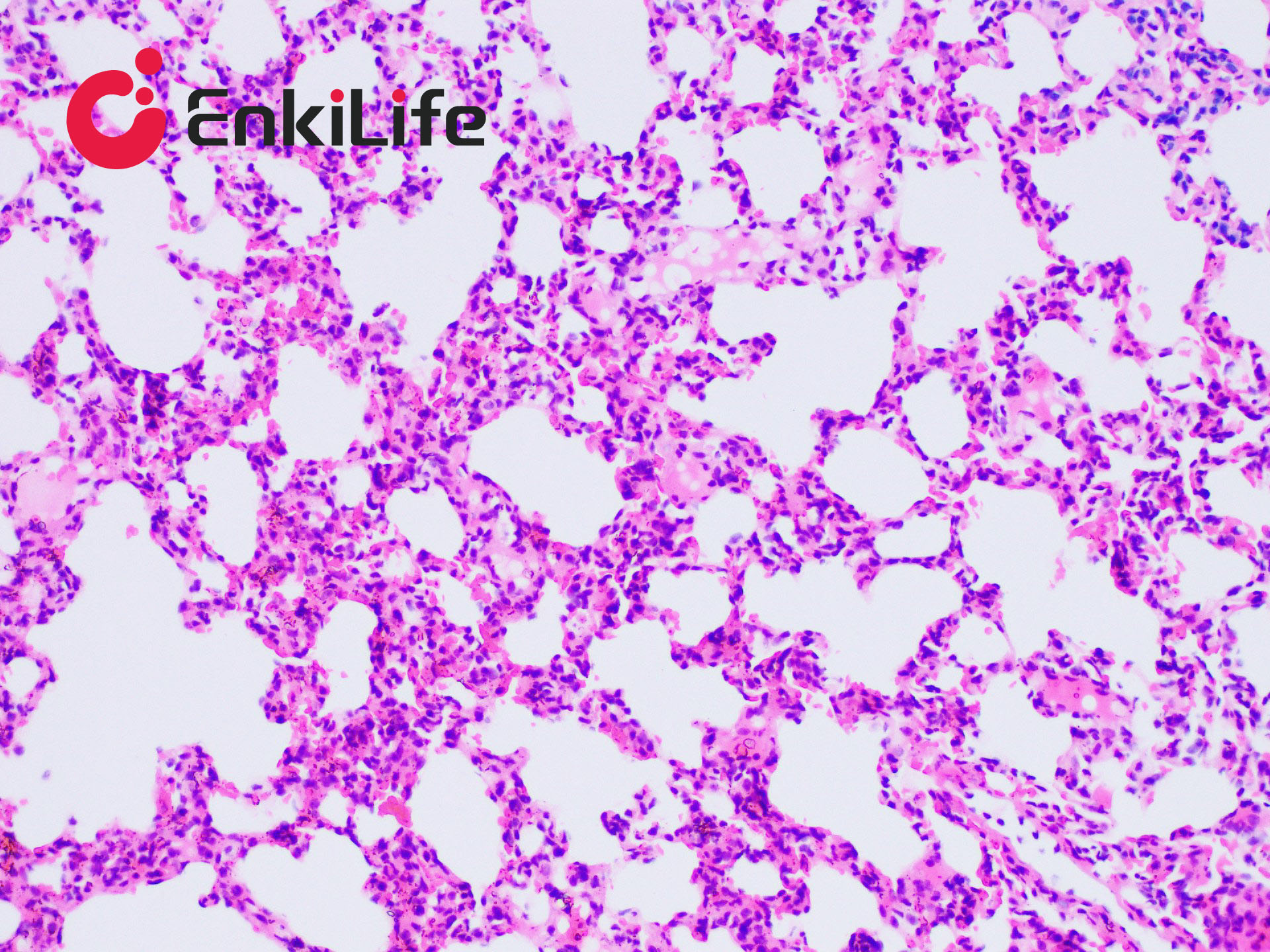
Elastic fibers are primarily distributed in the human arterial walls, alveolar walls, and skin. They appear yellow and are highly refractile when fresh. Commonly used elastic fiber staining methods include the Gomori aldehyde-fuchsin method, resorcin-fuchsin method (Weigert's resorcin-fuchsin), orcein method, Victoria blue method, and iron hematoxylin method. Resorcin-fuchsin staining solution is mainly used for elastic fiber staining and is also known as Weigert's resorcin-fuchsin solution. Elastic fiber staining can reveal changes in elastic fibers in skin tissues, such as elastic nevus, granuloma annulare, and scleroderma. It is also used to display and assess lesions of the endocardium and arteries, to observe whether certain pathological changes are accompanied by proliferation or destruction of elastic fibers, and to identify tumor components such as elastic fibromas. After elastic fiber staining, elastic fiber balls within the tumor can be clearly observed.
EnkiLife Elastic Fiber Staining Solution (Modified Gomori Aldehyde-Fuchsin Method) is based on the principle that mature aldehyde-fuchsin has a strong affinity for specific proteins and sulfated mucopolysaccharides, and binds well with elastic fibers. This staining solution also reveals mast cell granules, lipofuscin, and eosinophilic cells.
Basic Information
Product name | Elastic Fiber Staining Kit, Modified Gomori Aldehyde-Fuchsin Method |
Sizes | 50 mL |
Storage | 2-8 ℃, keep away from light |
Shipping | Shipped with ice pack |
Validity | 6 months |
Product Components
Components | 4x 50mL | |
Reagent (A): Acidic oxidizing solution | A1: Oxidizing Solution | 25 mL |
A2: Acidifying Solution | 25 mL | |
Before use, mix A1 and A2 at a ratio of 1:1 to prepare the acidic oxidizing solution. Do not prepare in advance. | ||
Reagent (B): Acidic Bleaching Solution | 50 mL | |
Reagent (C): Aldehyde-Fuchsin Staining Solution | 50 mL | |
Reagent (D): Orange G Staining Solution | 50 mL | |
Notes
1. This method can stain elastic fibers, pre-elastic fibers, and oxytalan fibers. Slightly thicker sections (7 μm) are recommended.
2. Do not mix the oxidizing and acidifying solutions in advance; prepare fresh before use.
3. Cover the staining dish during aldehyde-fuchsin staining to prevent evaporation.
4. If the aldehyde-fuchsin staining solution has been stored for a long time, its staining capacity may decrease; increase staining time accordingly.
5. When staining pancreatic β-cells, limit staining time to 30 min; for pituitary basophils, limit to 60 min.
6. Orange G staining should be light; excessive staining may obscure the color of elastic fibers.
7. Use reagents promptly after opening to avoid affecting experimental results.
8. For safety and health, wear lab coats and disposable gloves during operation.