

Product Introduction
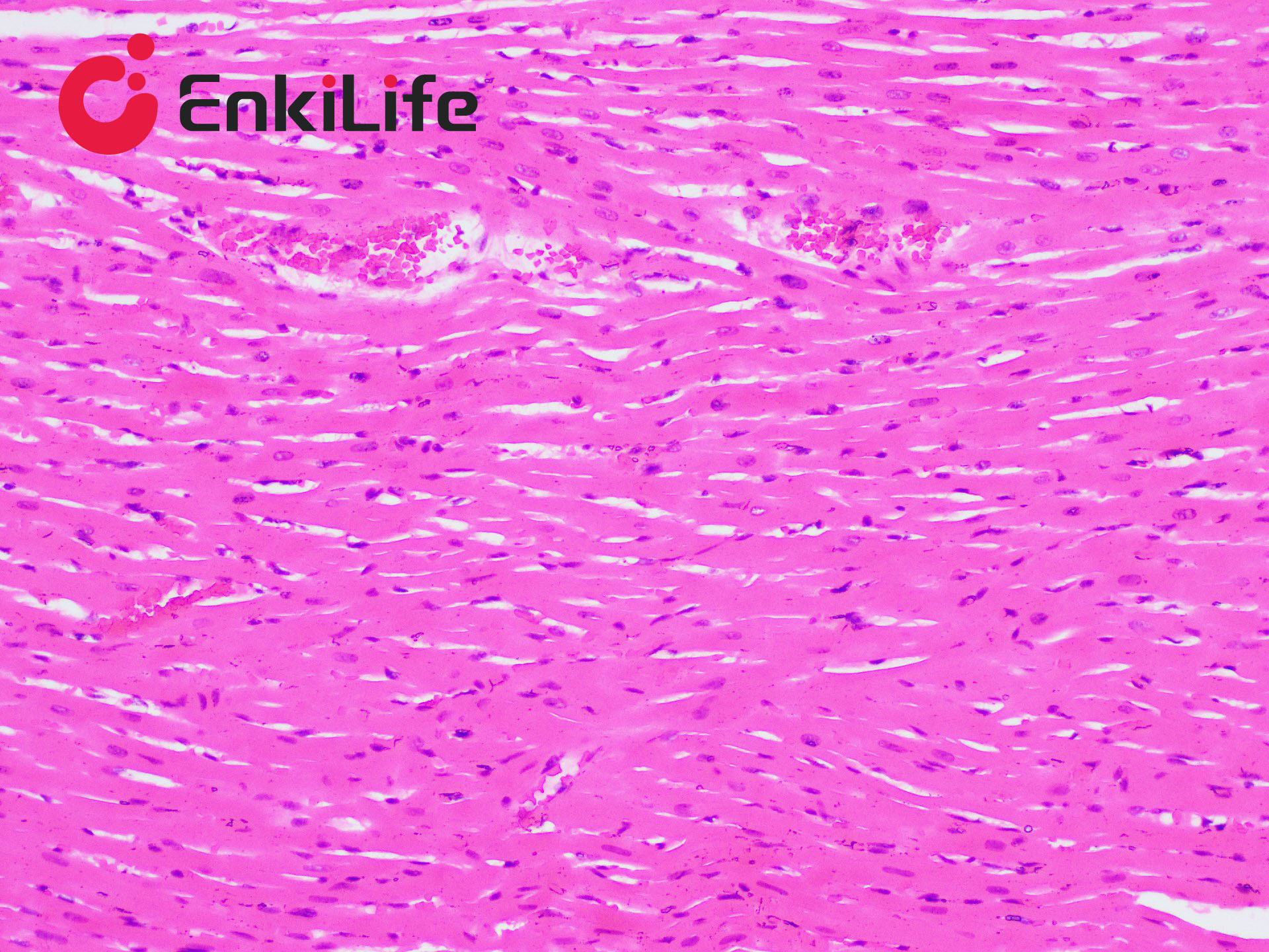
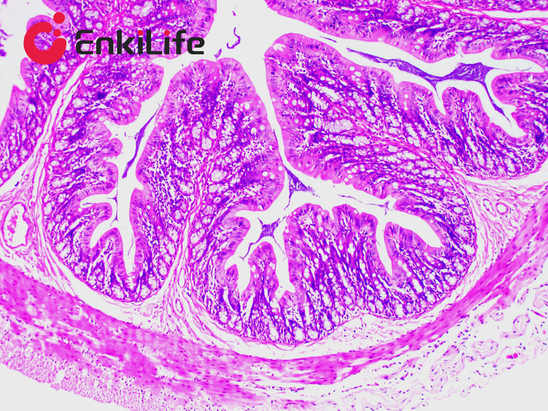
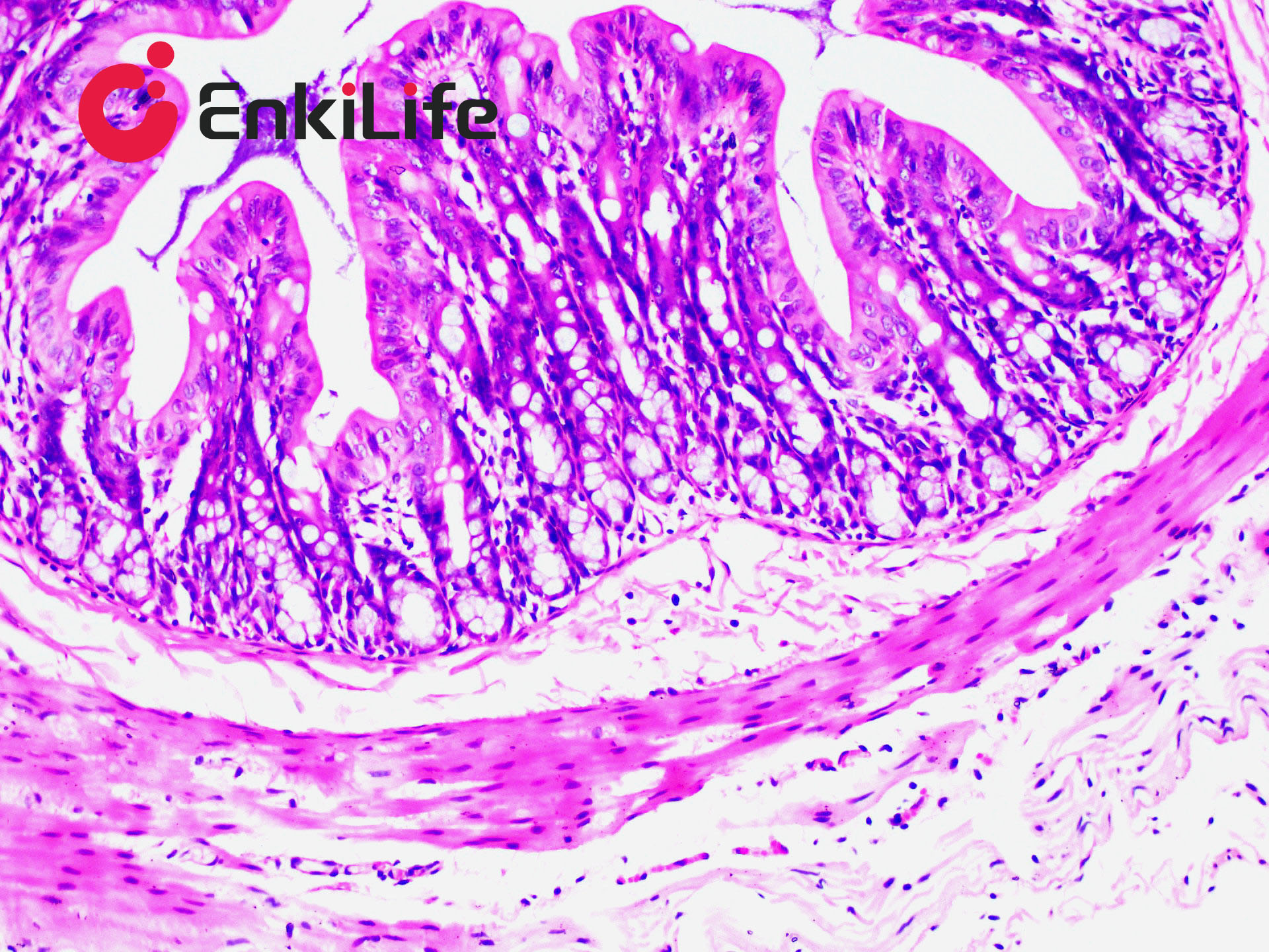
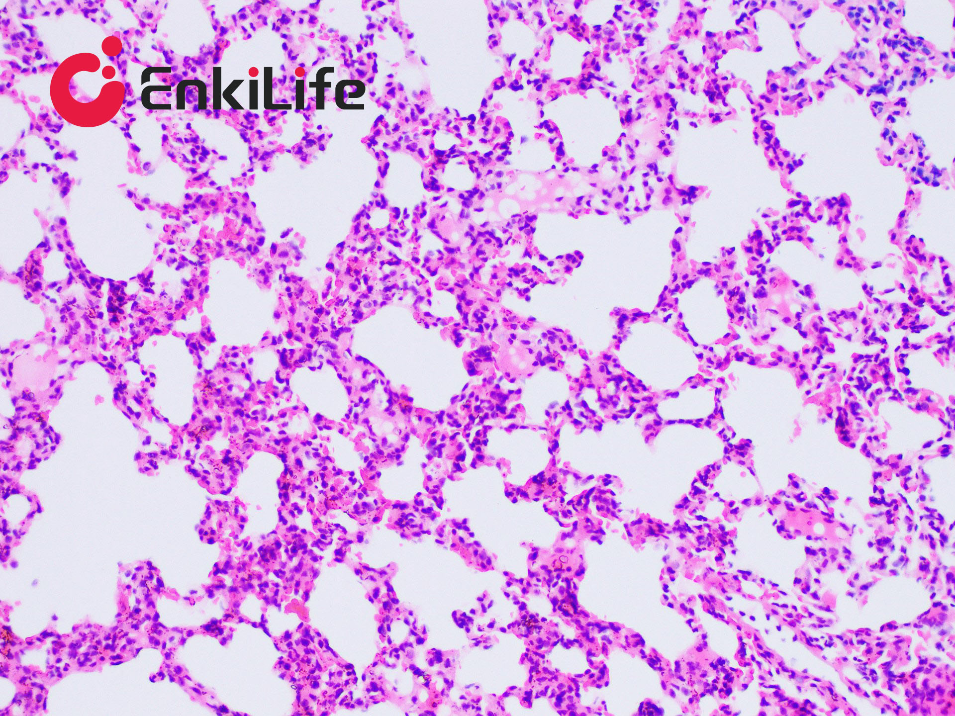
Collagen fibers are the most widely distributed and abundant fibers in connective tissue, found extensively in various organs, especially in the skin, sclera, and tendons. The principle of Van Gieson collagen fiber staining is related to the size of anionic dye molecules and tissue permeability. Molecular size is reflected by molecular weight: smaller molecules penetrate dense, low-permeability tissues more easily, while larger molecules can only enter loose, high-permeability tissues. Phosphomolybdic acid (PA) has the smallest molecular weight, followed by acid fuchsin and ponceau, while light green has the largest. After VG staining, muscle fibers appear yellow, and collagen fibers appear red.
EnkiLife Modified Van Gieson (VG) Staining Solution uses Celestine Blue and Mayer’s Hematoxylin for nuclear staining, resulting in better staining quality and longer storage stability. Ponceau S is used for collagen staining, which is fade-resistant. It is commonly used to distinguish collagen fibers from muscle fibers, to differentiate collagen-derived tumors from muscle-derived tumors, and to observe tissue or organ damage, repair, and fibrosis.
Basic Information
Product name | Modified Van Gieson (VG) Staining Kit |
Sizes | 50 mL |
Storage | 2-8 ℃,keep away from light |
Shipping | Shipped with ice pack |
Validity | 12 months |
Product Components
Components | 4x 50mL | |
Reagent (A): Celestine Blue Staining Solution | 50 mL | |
Reagent (B): Mayer’s Hematoxylin Staining Solution | 50 mL | |
Reagent (C): Acidic Ethanol Differentiation Solution | 50 mL | |
Reagent (D): Improved VG dye solution | D1: Ponceau S Staining Solution | 5 mL |
D2: Phosphomolybdic Acid (PA) Saturated Solution | 45 mL | |
Before use, mix D1 and D2 at a ratio of 1:9 to prepare the modified VG staining solution. Do not prepare in advance. | ||
Notes
1. Acidic ethanol differentiation is typically 1–2 s. After differentiation and rinsing with running water, observe under a microscope. If nuclear staining is too dark, differentiate again for 0.5–1 s. If too light, restain with Celestine Blue and Mayer’s Hematoxylin, then differentiate again with acidic ethanol.
2. Modified VG staining solution consists of D1 and D2. Mix in the required ratio (1:9) before use. For tissues with low collagen content, a 1:7 ratio may be used.
3. After modified VG staining, washing with water or 95% ethanol should be done quickly to avoid washing off Ponceau S and PA.
4. After modified VG staining, you may skip water rinse and directly apply 95% ethanol for differentiation, followed by rapid dehydration with absolute ethanol. This may result in brighter colors, but uneven differentiation may occur. Therefore, a quick water rinse followed by 95% ethanol differentiation is recommended.
5. Use reagents promptly after opening to avoid affecting experimental results.
6. For safety and health, wear lab coats and disposable gloves during operation.