

Product Introduction
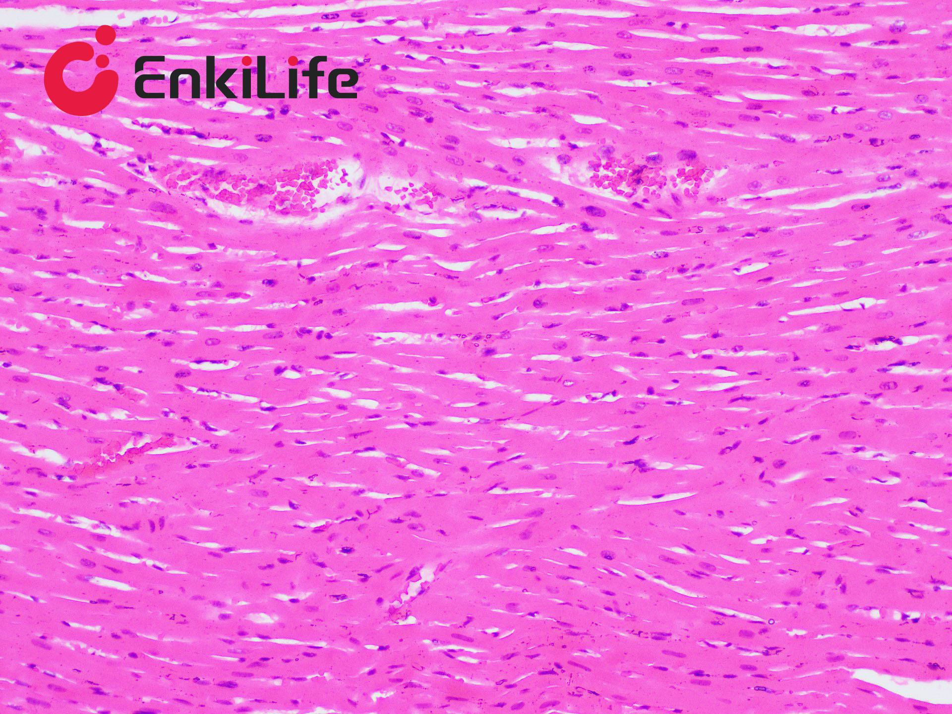
Hemosiderin is a hemoglobin-derived pigment that appears as golden or brownish-yellow granules. Due to its iron content and golden color, it is referred to as hemosiderin. When red blood cells are phagocytosed by macrophages, hemoglobin is broken down into iron-free orange chromogen and iron-containing hemosiderin under the action of lysosomal enzymes. The Prussian blue reaction, also known as hemosiderin staining, produces a blue color after treatment with potassium ferrocyanide and dilute acid. It is commonly observed in phagocytes or interstitial tissues. The staining principle is that potassium ferrocyanide solution, in the presence of dilute hydrochloric acid, dissociates trivalent iron from proteins. The trivalent iron then reacts with potassium ferrocyanide to form an insoluble blue compound—ferric ferrocyanide.
EnkiLife Prussian Blue Staining Solution (Nuclear Fast Red Counterstain Method) is used to visualize various hemorrhagic lesions in local tissues, commonly seen in phagocytes. It effectively distinguishes hemosiderin from other pigments. This staining solution offers good stability, long-term storage capability, minimal precipitation, and a wide range of applications. After the reaction, a red counterstain such as nuclear fast red, eosin, or neutral red may be used. Nuclear fast red is used as the counterstain in this kit, representing the most classic and commonly used method.
Basic Information
Product name | Prussian Blue Staining Kit (Nuclear Fast Red Counterstain Method) |
Sizes | 50 mL |
Storage | RT |
Shipping | RT |
Validity | 12 months |
Product Components
Components | 2x 50mL | |
Reagent (A): Perls Stain | A1: Perls Stain A | 25 mL |
A2: Perls Stain B | 25 mL | |
Mix equal parts of A1 and A2 immediately before use. Do not prepare in advance. | ||
Reagent (B): Nuclear Fast Red Staining Solution | 50 mL | |
Notes
1. Dewax sections thoroughly. Tissue fixation is typically done with 10% neutral formalin. Prolonged fixation with regular formalin may damage tissue. Avoid acidic fixatives; chromate treatment may also interfere with iron preservation.
2. Use clean containers throughout the procedure. Avoid iron-containing metal tools. Use distilled water for rinsing slides and containers, as tap water may contain iron. Adjust staining time with Perls Stain based on sample conditions.
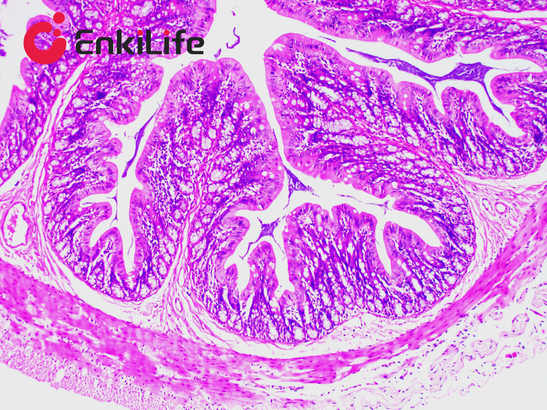
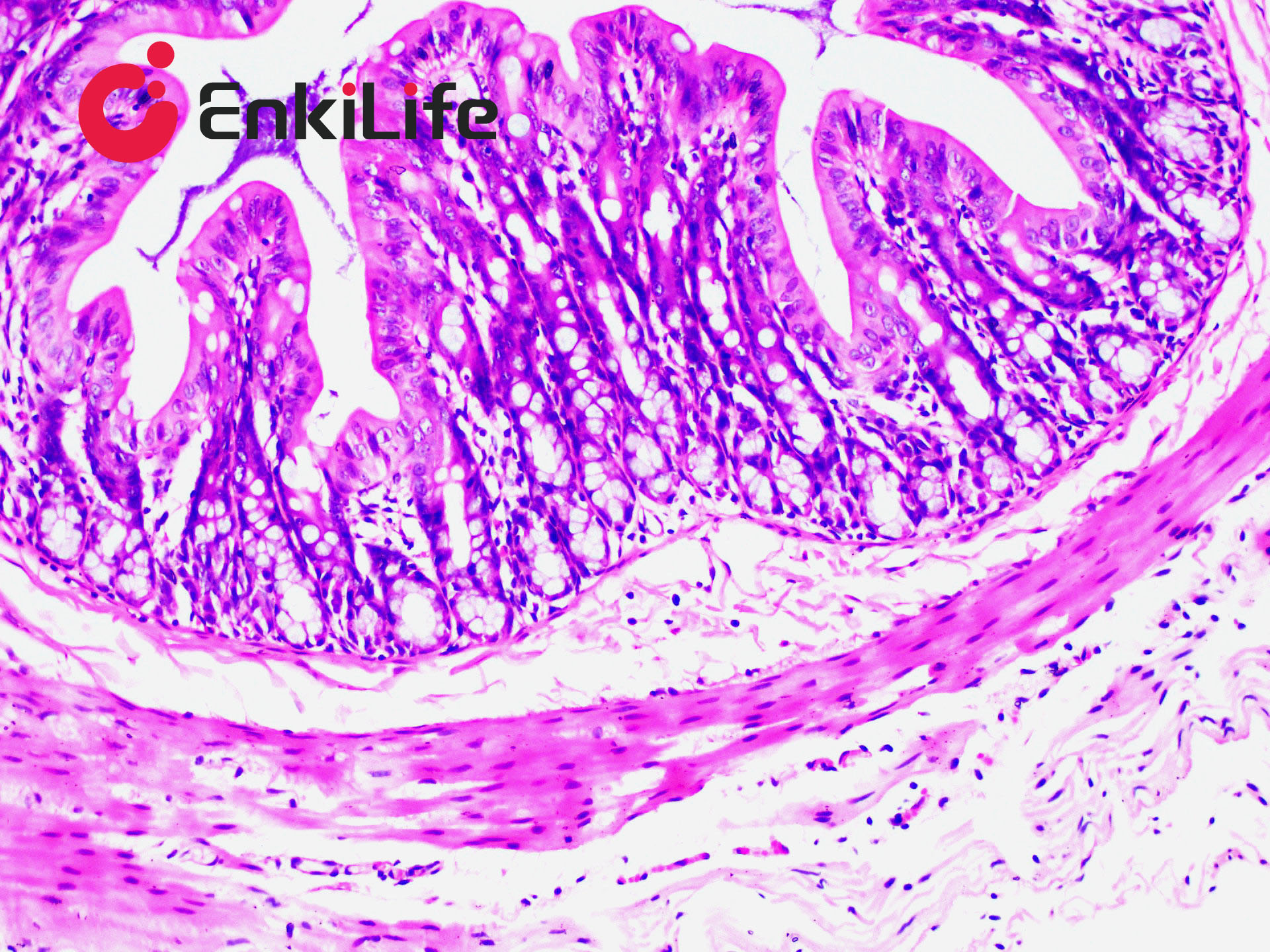
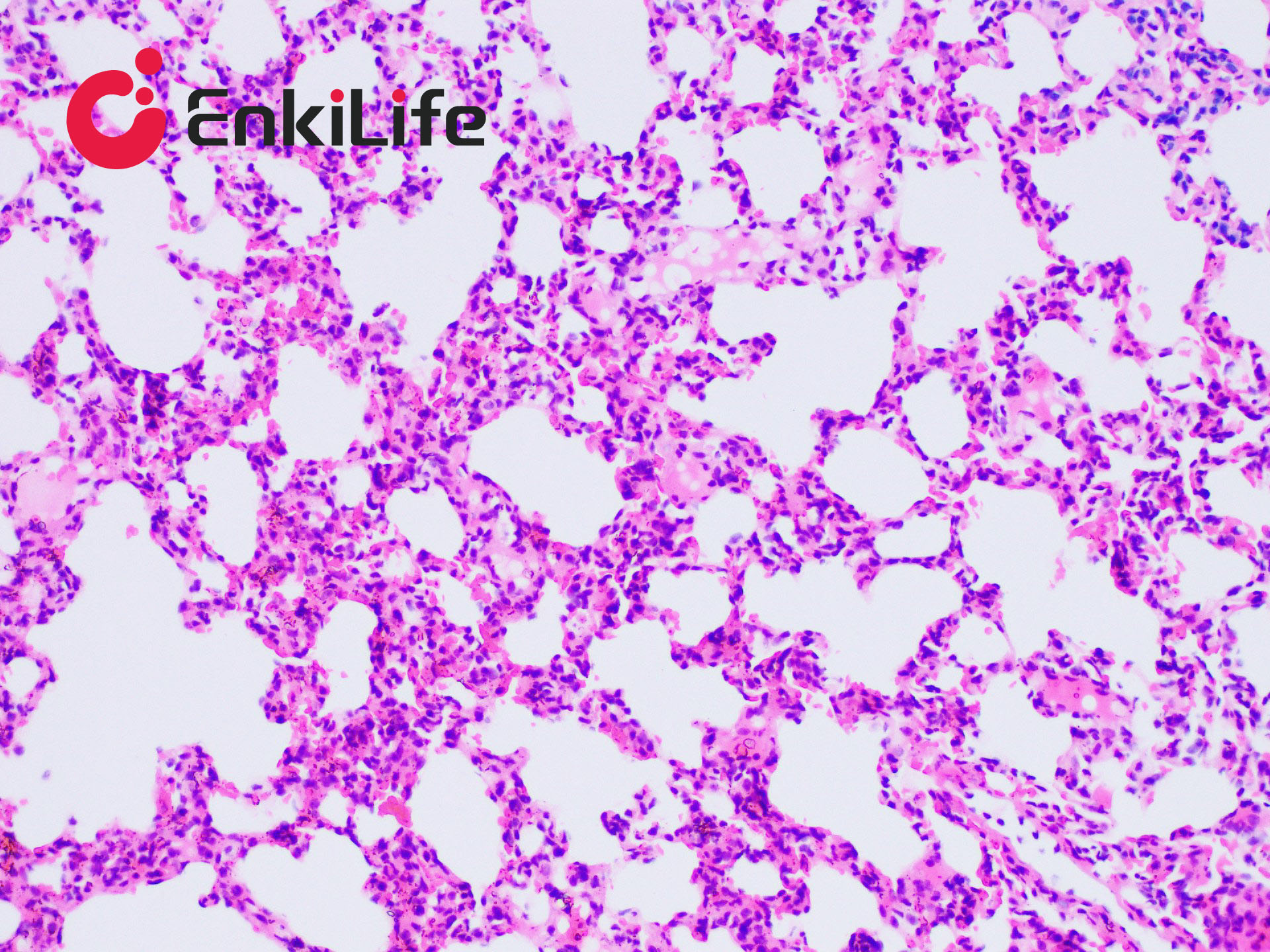
3. Use the same positive control slide for all sections. Choosing an appropriate control is essential. Autopsy lung tissue is a good control, as it contains a significant number of iron-positive macrophages (heart failure cells).
4. For frozen sections and cell staining, optimize experimental conditions based on specific situations.
5. Use reagents promptly after opening to maintain optimal performance.
6. For your safety and health, wear a lab coat and disposable gloves during operation.