

Product Introduction
Collagen fibers are the most widely distributed and abundant fibers in connective tissue, found extensively in various organs, especially in the skin, sclera, and tendons. Type I collagen fibers are mainly found in bone, skin, and tendons; Type II collagen fibers are primarily found in cartilage; Type III collagen fibers are mainly present in embryonic tissues and adult blood vessels and gastrointestinal tract; Type IV collagen fibers are mainly found in the basement membrane.
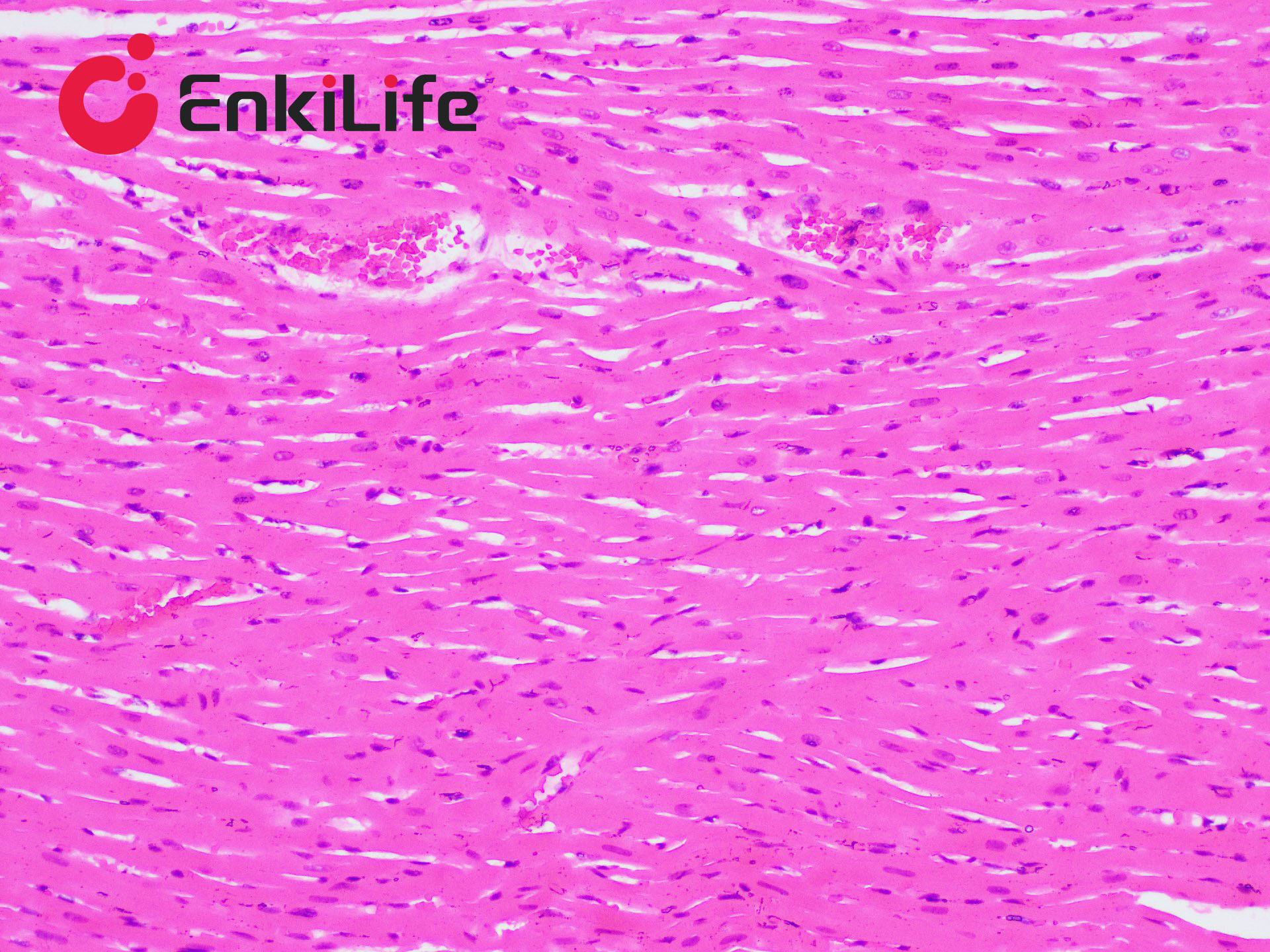
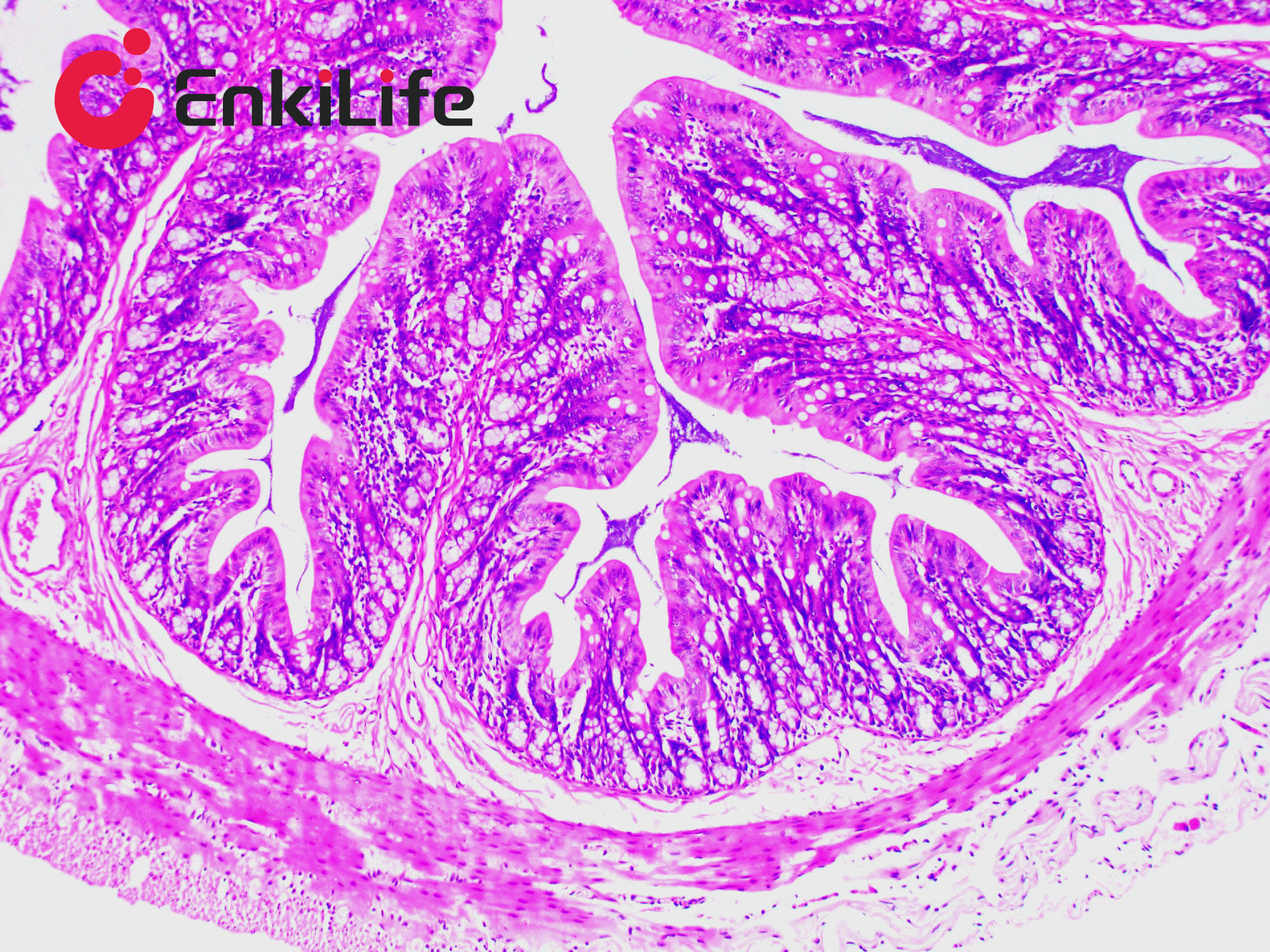
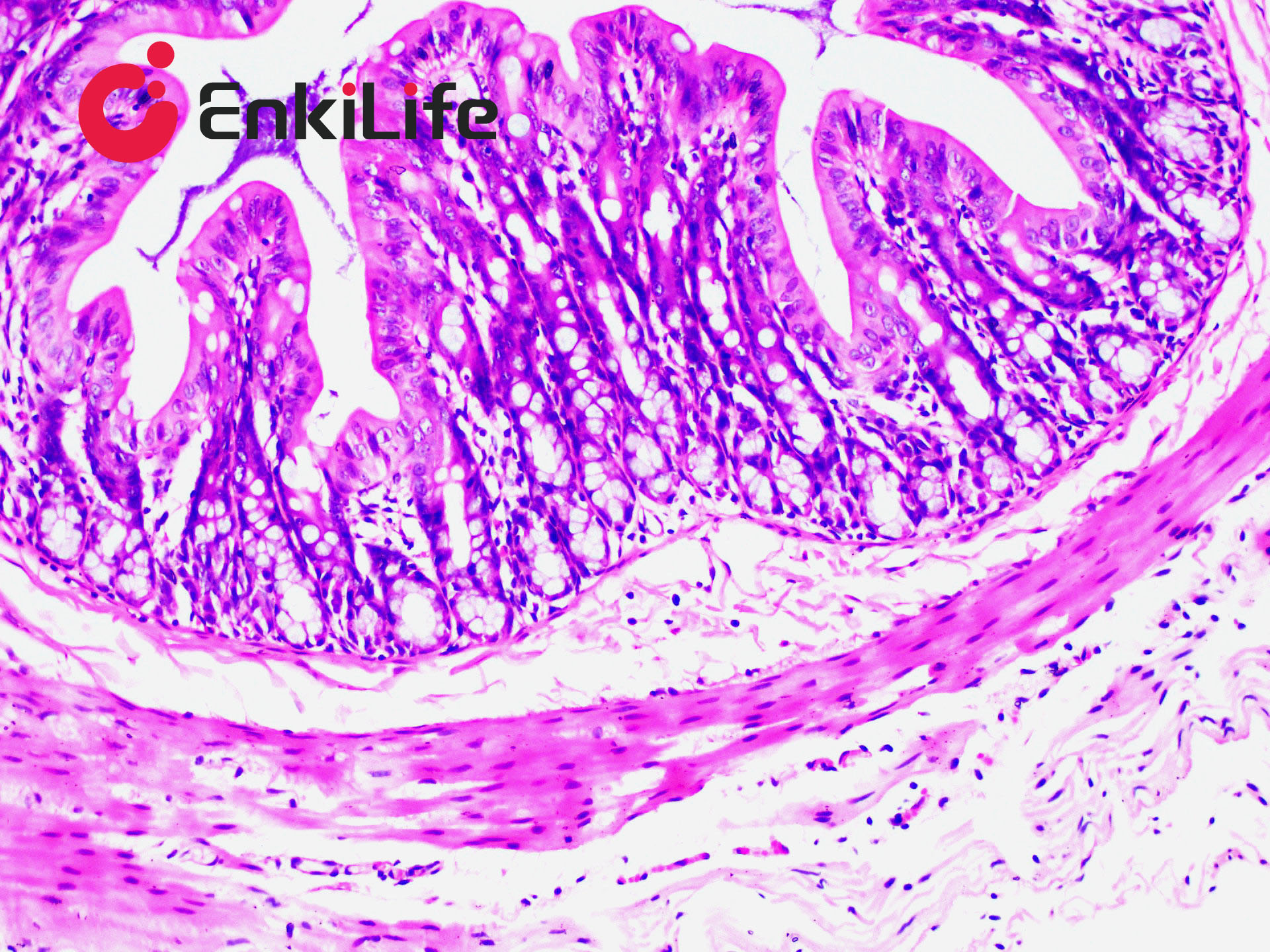
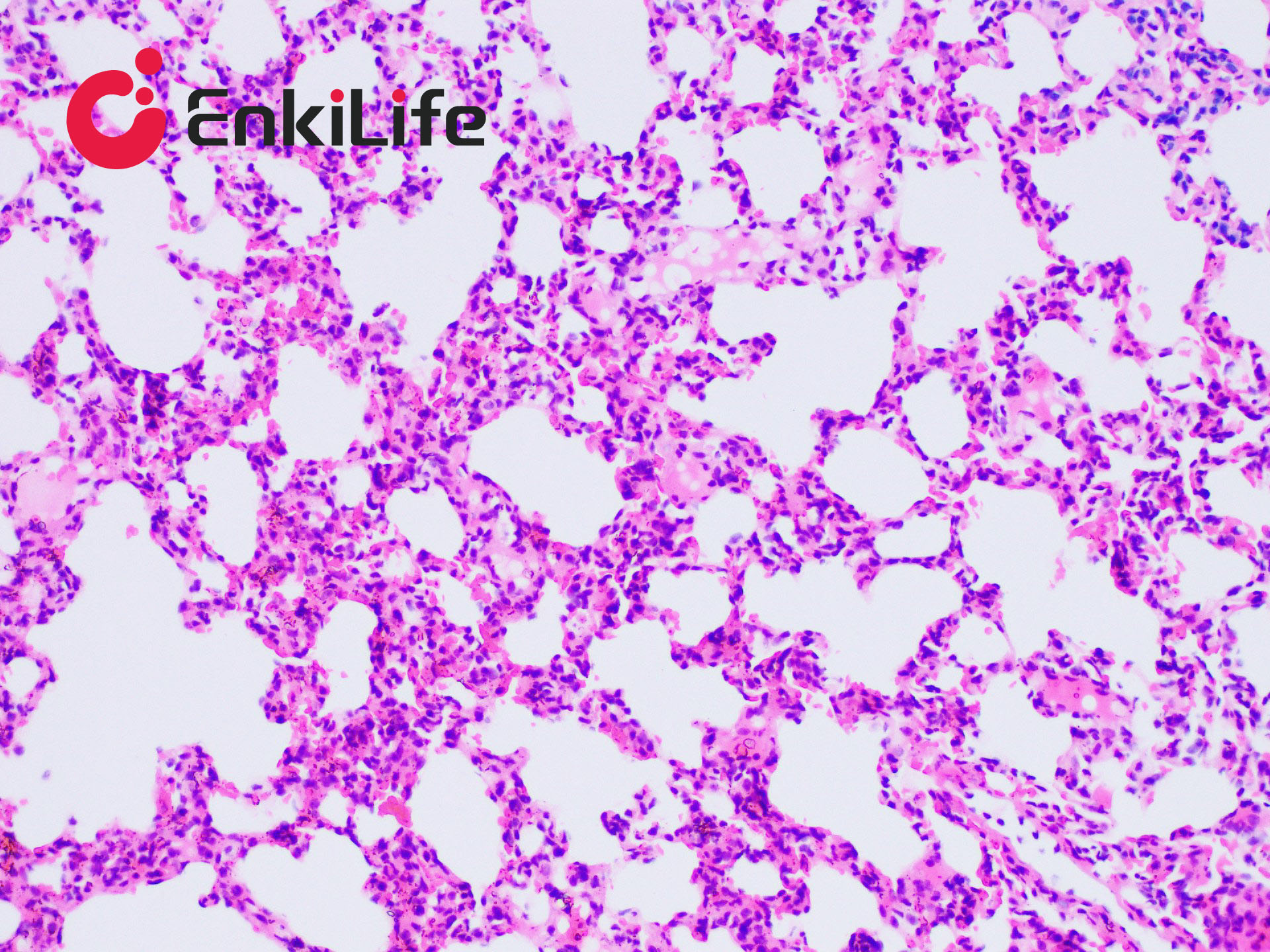
The principle of Van Gieson collagen fiber staining is related to the size of anionic dye molecules and tissue permeability. Molecular size is reflected by molecular weight: smaller molecules penetrate dense, low-permeability tissues more easily, while larger molecules can only enter loose, high-permeability tissues. Phosphomolybdic acid (PA) has the smallest molecular weight, followed by acid fuchsin and ponceau, while light green has the largest. After VG staining, muscle fibers appear yellow, and collagen fibers appear red.
EnkiLife Van Gieson (VG) Staining Solution is commonly used to distinguish collagen fibers from muscle fibers, to differentiate collagen-derived tumors from muscle-derived tumors, and to observe tissue or organ damage, repair, and fibrosis.
Basic Information
Product name | Van Gieson (VG) Staining Kit |
Sizes | 50 mL |
Storage | RT |
Shipping | RT |
Validity | 12 months |
Product Components
Components | 3x 50mL | |
Reagent (A): Weigert hematoxylin staining solution | A1: Weigert Solution A | 25 mL |
A2: Weigert Solution B | 25 mL | |
Before use, mix A1 and A2 in equal parts to prepare Weigert's iron hematoxylin, which appears purple-red. | ||
Reagent (B): Acidic Ethanol Differentiation Solution | 50 mL | |
Reagent (C): VG staining solution | C1: Acid Fuchsin Staining Solution | 5 mL |
C1: Phosphomolybdic Acid (PA) Solution | 45 mL | |
Before use, mix C1 and C2 at a ratio of 1:9 to prepare VG staining solution. Prepare fresh and use immediately. | ||
Notes
1. Prepare Reagent (A) and Reagent (C) fresh before use. Do not prepare in advance; staining capacity is generally lost after 24 h.
2. This staining solution is mainly used for paraffin sections. If using celloidin or LVN (low-viscosity nitrocellulose) sections, rinse celloidin sections with distilled water after VG staining.
3. All reagents mentioned above are irritating; handle with care.
4. Use reagents promptly after opening to avoid affecting experimental results.
5. For safety and health, wear lab coats and disposable gloves during operation.