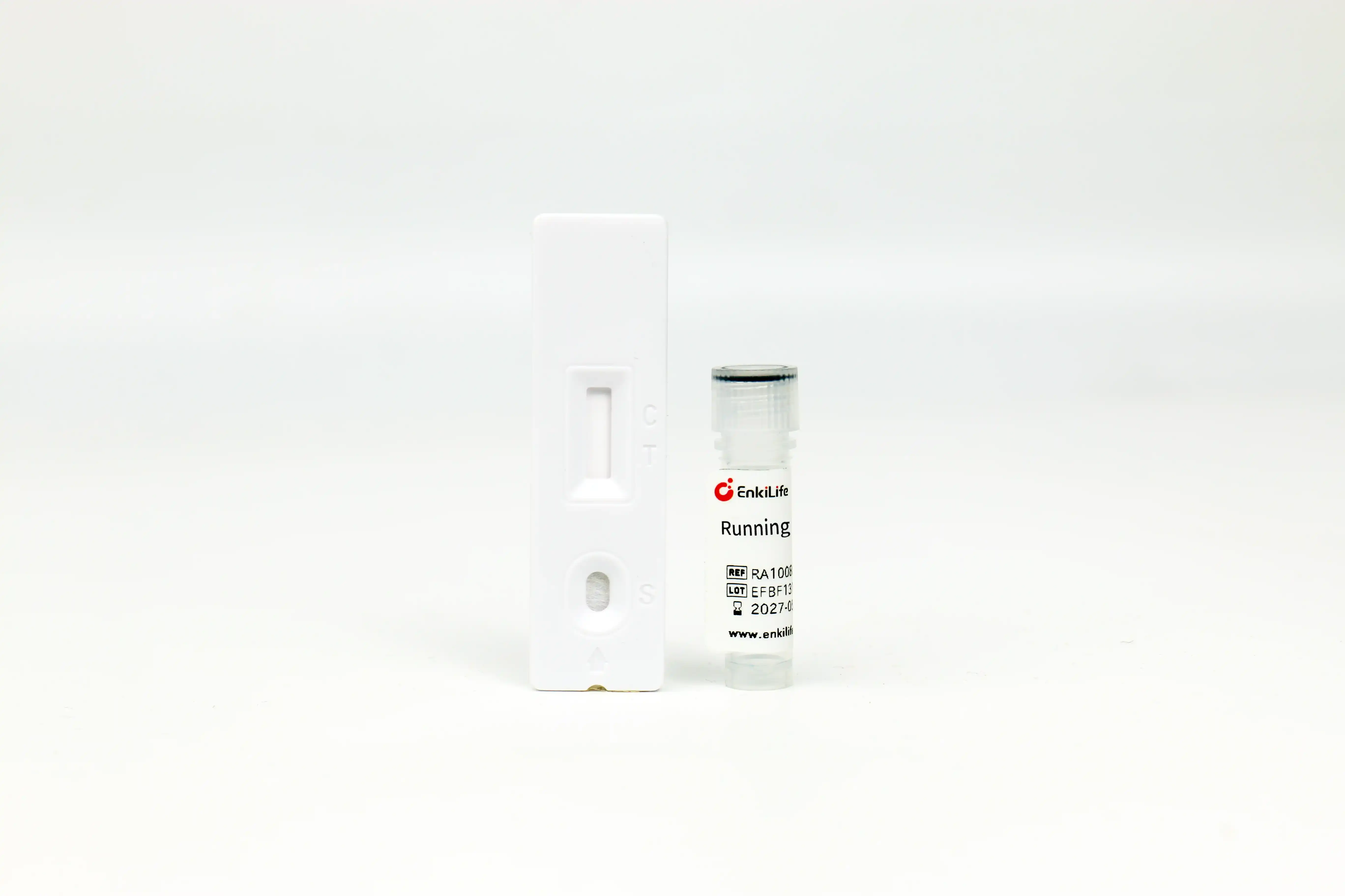



Human IgG(Fc)Rapid Assay Kit
Dedicated for antibody expression detection · Results in 15 min · Latex microsphere labeling technology
Product Overview
Human IgG(Fc)Rapid Assay Kit adopts latex microsphere-labeled immunochromatography based on double-antibody sandwich principle. This kit is specially designed to detect human IgG or recombinant proteins fused with human Fc fragment in samples. During testing, human Fc fragment in specimen binds with labeled antibody to form immune complex, which migrates via capillary action to the test zone (T line) and combines with pre-coated anti-human Fc antibody to generate red bands.
The detection range is 1~30 μg/mL, and the color depth of test line is positively correlated with Fc concentration. The assay shows CV ≤15% for repeatability and relative deviation within ±15% for accuracy, enabling reliable semi-quantitative measurement of antibody expression level.
Core Advantages
Time Comparison with Conventional Methods
This kit cuts antibody detection duration from hours down to only 20 minutes!
Product Specifications
| Catalog No. | Pack Size | Storage Condition | Shelf Life |
|---|---|---|---|
| RA10082 | 10T | Sealed & dry, 2~30℃ | 12 months |
| RA10082 | 50T | Sealed & dry, 2~30℃ | 12 months |
Kit Contents: Individually foil-packed Human IgG(Fc)test cassettes, dropper bottle of running buffer and detailed instruction manual
Wide Application Scenarios
- Quantification of humanized antibody expression: Rapid measurement of human antibody yield in cell culture supernatant
- Screening of recombinant protein clones: Fast selection of high-expression clones producing Fc-fused recombinant proteins
- In-process monitoring during antibody purification: Real-time tracking of antibody content to optimize purification protocols
- Direct testing of cell culture supernatant: Detect supernatant antibody concentration without pre-processing
- Antibody sample quality verification: Confirm antibody integrity and concentration to guarantee subsequent downstream experiments
- Quality control for large-scale manufacturing: Intermediate batch inspection for antibody drug and recombinant protein mass production
Operating Procedure
- Preparation: Read manual carefully; equilibrate refrigerated samples to ambient temperature; verify product lot information matches outer package label
- Take out test strip: Tear aluminum pouch and place cassette on flat bench surface
- Sample dilution: Pipette 80 μL running buffer into empty centrifuge tube, add 20 μL sample to prepare total 100 μL mixture, vortex for 10 seconds
- Sample loading: Transfer 70 μL mixed solution into sample well of test cassette
- Result reading: Incubate at room temperature for 15 min before recording results
Sample Dilution Guide: Dilute known-concentration protein samples to 5 μg/mL; test low-expression samples undiluted; further dilute high-concentration specimens if readings exceed linear range. Adjust dilution factor accordingly when out of valid detection scope.
Low-abundance sample tips: Highly diluted low-content Fc protein may drop below LOD and cause false-negative results. Run undiluted raw sample together with matrix-matched negative control in such case.
Result Interpretation
Both control line (C) and test line (T) develop red bands, confirming human IgG or Fc-tagged recombinant protein exists; darker test line corresponds to higher target concentration.
Only control line (C) shows red band while test line (T) remains blank, meaning no target protein or analyte concentration is below detection limit.
Absent control line (C) invalidates the whole assay regardless of T line status, caused by expired reagent, wrong operation or insufficient sample volume.
Reference Concentration & Color Chart
| Concentration (μg/mL) | Band Color Reference |
|---|---|
| 30 | Deep red band |
| 20 | Red band |
| 10 | Medium red band |
| 5 | Light red band |
| 2 | Faint red band |
| 1 | Very weak red band |
| 0 | No visible band |
Compatibility with Common Lab Buffers
This kit tolerates various routine buffer additives, enabling direct testing of most conventionally prepared protein samples.
| Reagent Name | Max Tolerated Concentration |
|---|---|
| NaCl | 0.5M |
| KCl | 0.5M |
| Urea | 0.4M |
| Triton X-100 | 1% |
| Tween-20 | 1% |
| SDS | 0.2% |
| Reagent Name | Max Tolerated Concentration |
|---|---|
| NP-40 | 1% |
| CHAPS | 1% |
| EDTA | 5mM |
| Glycerol | 10% |
| Imidazole | 0.5M |
| RIPA Lysis Buffer | 100% |
Troubleshooting
| Observation | Possible Causes | Solutions |
|---|---|---|
| No band on control line | Expired test card Moisture damage after opening pouch Incorrect operating steps | Use unexpired new strips Use test card immediately after unsealing Repeat test strictly per manual |
| No visible test line | No target protein in sample Conc. below detection limit Over-diluted specimen | Confirm sample matches kit specificity Reduce dilution or test raw sample Readjust dilution factor and retest |
| Over-dark test line | Excessively high sample concentration Beyond linear detection range | Dilute sample at higher fold and retest |
| High non-specific background | Interfering impurities in sample Abnormal buffer composition | Dilute sample with PBS or running buffer before re-testing |
Precautions
- For research detection of human IgG or human Fc-fused recombinant protein only; discard expired products.
- Single-use consumable; do not reuse test cassettes.
- This is semi-quantitative assay and cannot be used for absolute quantitative measurement.
- Technical defects, misoperation or sample interference may lead to inaccurate results.
- Test results serve only as auxiliary reference; further verification is required for abnormal readings.
- Keep test horizontally stationary during incubation and avoid vibration.
- Do not mix kit components from different production batches.
- Test is invalid once control line fails to develop red band.
- Recommended to use test cassette and buffer right after pouch opening.