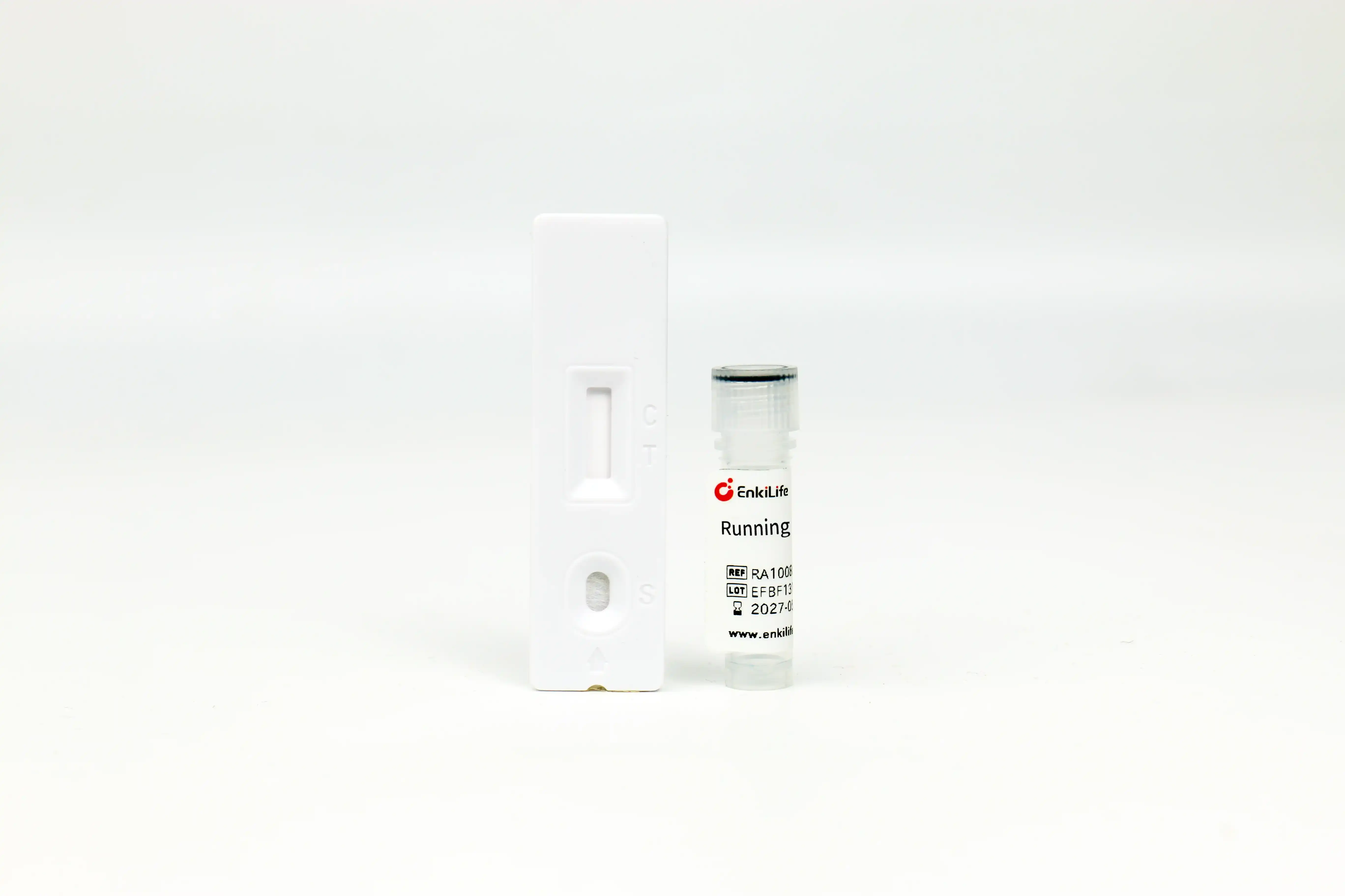



Flag-Tag Rapid Competitive Assay Kit
For Rapid Flag-tagged Protein Detection · Results in 10-15 min · High Specificity via Competitive Assay
Product Overview
Flag-Tag Rapid Competitive Assay Kit adopts colloidal gold lateral flow technology based on competitive immunoassay. Recombinant Flag-Tag protein is immobilized on the test line of chromatography membrane, and anti-secondary antibody is fixed on the control line; colloidal gold-labeled anti-Flag-Tag monoclonal antibody is coated on the conjugate pad.
In the absence of Flag-Tag protein in samples, gold-labeled antibodies migrate freely to the test line and bind with immobilized Flag protein to form distinct purplish-red bands. When Flag-Tag protein exists in samples, target proteins preferentially combine with gold-labeled antibodies to occupy binding sites and block antibody binding to test line antigens. Higher Flag-Tag concentration leads to fainter test line; the test line disappears completely above 10μg/mL. The limit of detection is 1μg/mL, and band intensity is negatively correlated with protein concentration ranging from 1μg/mL to 10μg/mL.
Key Advantages
Time Comparison with Conventional Methods
Cut detection duration from one full day down to only 15 minutes with this product!
Product Information
| Cat.No. | Specification | Storage Condition | Shelf Life |
|---|---|---|---|
| RA10084 | 10 Tests | Dry at Room Temp | 12 Months |
| RA10084 | 100 Tests | Dry at Room Temp | 12 Months |
Components: Individually foil-sealed Flag-Tag test cards, dropper bottle of running buffer and detailed product manual.
Wide Application Fields
- Recombinant clone screening: Rapid identification of high-expression positive transformants to avoid waste of reagents and time on low-expression strains
- Optimization of induction conditions: Compare expression levels under varied induction temperature, duration and inducer concentration quickly
- Monitoring protein purification: Real-time quantification of target protein in loading sample, flow-through and elution fractions
- Eukaryotic transfection efficiency test: Evaluate performance of different transfection reagents and protocols conveniently
- Raw protein QC check: Verify tag integrity of protein samples to guarantee subsequent functional research
- Mass production quality control: Intermediate product testing for recombinant biopharmaceuticals and industrial enzymes.
Operating Procedure
- Preparation: Take test card out from sealed foil bag and lay flat on dry bench; dilute sample appropriately to adjust protein concentration within optimal detection range: 1–10μg/mL
- Sample Loading: Pipette 20μL diluted sample and add slowly into sample well
- Initiate Chromatography: Immediately add 50μL matched running buffer (approx. 2 vertical drops from dropper)
- Result Reading: Read results after 10–15 min at room temperature; readings after 20 min are invalid.
Dilution Guide: Dilute known-concentration samples to ~3μg/mL; dilute bacterial lysate 25-fold initially, mammalian cell supernatant/lysate 5-fold initially. If test line is extremely faint with expected high expression, further dilute 10-fold and retest.
Low-abundance Protein Tip: Undiluted raw sample can be used for low-expression specimens; always run negative control with identical matrix for comparison of band intensity.
Result Interpretation
Both Control line(C) and Test line(T) develop purplish-red bands with equivalent color intensity vs negative control, meaning no Flag-Tag protein or concentration below 1μg/mL LOD.
Control line(C) appears colored, while Test line(T) is absent or obviously paler than negative control, confirming Flag-Tag protein presence; paler T band corresponds to higher protein concentration.
No band on Control line(C) regardless of Test line status; test is invalid due to expired reagent, improper operation or insufficient running buffer.
Compatible Common Reagents
This kit tolerates regular lab buffer components and directly tests routine protein samples without extra pre-treatment.
| Reagent Name | Max Concentration |
|---|---|
| NaCl | 0.25M |
| KCl | 0.25M |
| Urea | 0.4M |
| Triton X-100 | 1% |
| Tween-20 | 1% |
| Reagent Name | Max Concentration |
|---|---|
| NP-40 | 1% |
| CHAPS | 1.0% |
| SDS | 0.20% |
| EDTA | 5mM |
| Glycerol | 10% |
Special Remark: Fully compatible with undiluted 100% RIPA lysis buffer; cell lysate prepared by RIPA can be tested directly without dialysis.
Precautions
- For research use only; prohibited for human clinical diagnosis or therapeutic application.
- Use test strip within 1 hour after opening foil pouch; long-term air exposure causes reagent dampening and failure.
- Appropriate sample dilution is critical for accurate results; select dilution fold based on anticipated protein concentration.
- Negative control is mandatory for competitive assay to compare band intensity for result judgement.
- If unexpected faint test line appears, suspect excessive analyte concentration, dilute further and repeat assay.
- Strictly follow stipulated reading time; premature or delayed reading leads to false conclusion.
- Do not mix test cards and running buffer from different production lots due to minor batch variation.
- Dispose experimental waste per laboratory biosafety regulations.
Troubleshooting
| Phenomenon | Possible Causes | Solutions |
|---|---|---|
| No obvious T line fading vs negative control | No target protein in sample Conc. below detection limit |
Confirm vector matches test card Test undiluted sample with negative control |
| Extremely faint or missing Test line | Sample conc. exceeds upper working range Full competitive inhibition by high antigen |
Dilute sample at higher fold and retest |
| No visible Control line | Misoperation Expired test card Moisture damage Insufficient running buffer |
Retest following manual strictly Replace with valid new strips Store under dry ambient condition Add ~2 drops of buffer |