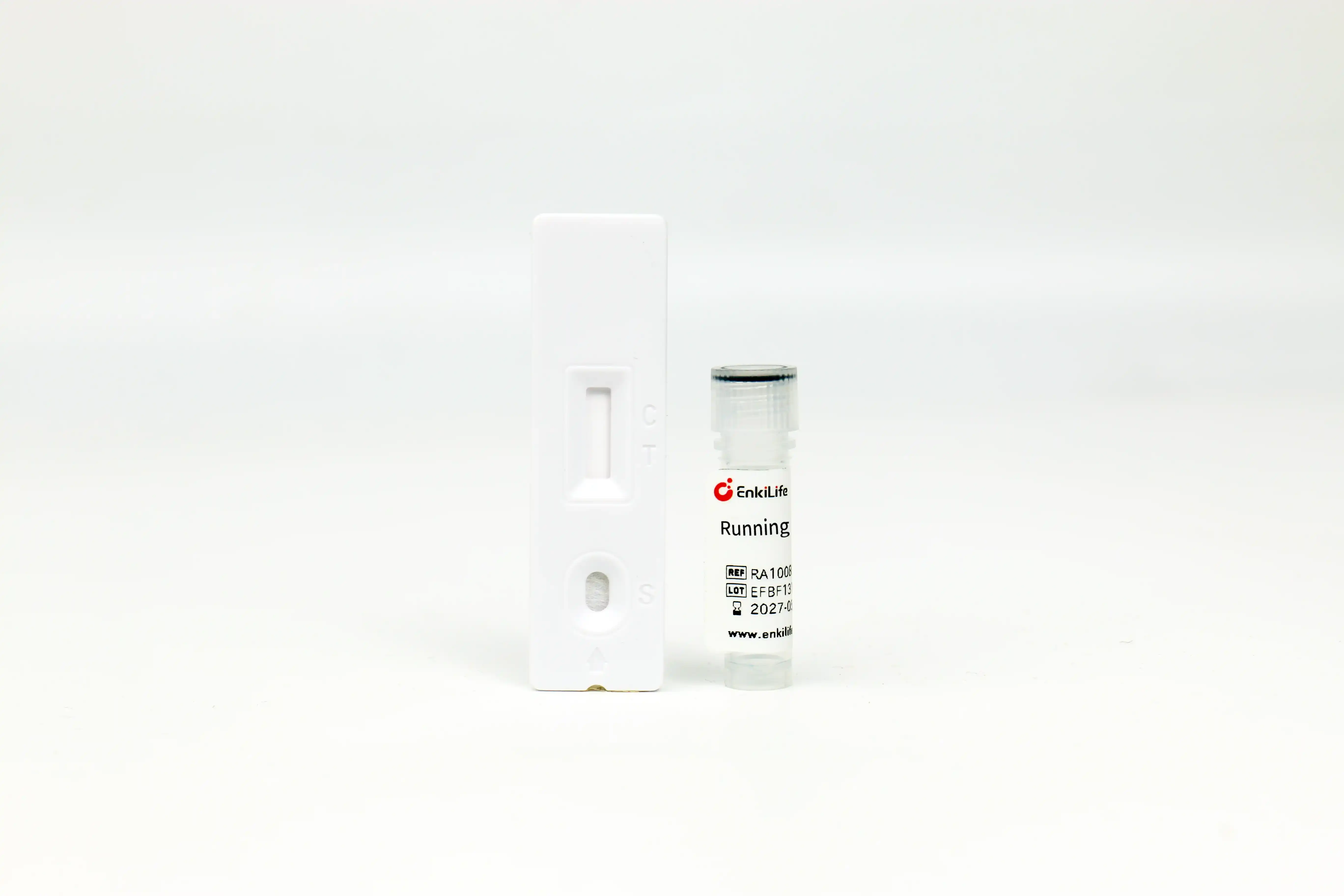



Flag-His-Tag Rapid Assay Kit
For Rapid Protein Expression Detection · Results in 10-15 Minutes · No Equipment Required
Product Overview
The Flag-His-Tag Rapid Assay Kit is developed based on colloidal gold lateral flow technology and the double antibody sandwich immunoassay principle. The chromatography membrane is immobilized with mouse anti-Flag-Tag monoclonal antibody as the test line and goat anti-mouse IgG polyclonal antibody as the control line; the gold conjugate pad is coated with colloidal gold-labeled mouse anti-His-Tag monoclonal antibody.
When a sample containing Flag-His-Tag fusion protein is added to the sample pad, it moves towards the absorbent pad by capillary action. The target protein in the sample first binds to the gold-labeled antibody to form a complex, which is then captured and enriched by the antibody on the test line, forming a visible purplish-red band. This product has a detection sensitivity of 4ng/mL. Within the range of 4ng/mL to 500ng/mL, the color intensity of the test line is positively correlated with the protein concentration; when the protein concentration exceeds 500ng/mL up to 10μg/mL, the test line still maintains high-intensity color development without significant weakening due to antigen excess.
Key Advantages
Time Comparison with Traditional Methods
Using this product reduces protein detection time from hours to just 15 minutes!
Product Information
| Cat. No. | Size | Storage Conditions | Shelf Life |
|---|---|---|---|
| RA10085 | 10 Tests | Room temperature, dry | 12 months |
| RA10085 | 100 Tests | Room temperature, dry | 12 months |
Components: Individually foil-wrapped Flag-His-Tag detection cards, dropper bottle of running buffer, detailed product manual
Wide Range of Applications
- Recombinant protein expression clone screening: Rapidly identify high-expression positive clones among large numbers of transformants, avoiding wasting time and reagents on low-expression clones
- Expression condition optimization: Quickly compare the effects of different induction temperatures, induction times, and inducer concentrations on protein expression levels
- Protein purification process monitoring: Real-time monitoring of target protein content in loading samples, flow-through, eluates, and collected fractions
- Eukaryotic cell transfection efficiency detection: Rapidly evaluate the effects of different transfection reagents and methods on target protein expression
- Protein sample quality verification: Quickly confirm the integrity of tags in protein samples, providing quality assurance for subsequent functional experiments
- Large-scale production quality control: Suitable for rapid detection of intermediate products in the production of recombinant protein drugs, industrial enzymes, etc.
Operating Procedure
- Preparation: Remove the detection card from the sealed foil pouch and place it horizontally on a dry laboratory bench; dilute the sample appropriately according to the expected concentration to ensure the target protein concentration is within the optimal detection range of 4-500ng/mL
- Sample Loading: Use a micropipette to accurately pipette 20μL of the diluted sample and slowly add it to the sample well of the detection card
- Start Chromatography: Immediately add 50μL of the provided running buffer to the sample well (approximately 2 drops from the dropper bottle held vertically)
- Result Interpretation: Observe and record results after 10-15 minutes at room temperature. Results observed after 20 minutes are not valid.
Sample Dilution Guide: For samples with known concentration, dilute directly to approximately 100ng/mL; for cell lysates of unknown concentration (bacterial, mammalian, yeast, or insect cells), a 50-fold dilution is recommended initially. If the initial test shows very weak or no color development on the test line and high protein expression is expected, further dilute the sample 100-fold and retest to exclude the prozone effect caused by high antigen concentration. Dilution factors below 10-fold are not recommended as complex components in the sample may interfere with the detection results.
Result Interpretation
Both the control line (C) and test line (T) appear purplish-red, indicating the presence of Flag-His-Tag fusion protein in the sample. Within the linear range of 4-500ng/mL, the darker the test line color, the higher the protein concentration.
Only the control line (C) appears purplish-red, and the test line (T) shows no obvious color development, indicating that the sample does not contain Flag-His-Tag fusion protein or its concentration is below the detection limit of 4ng/mL.
No purplish-red band appears at the control line (C), regardless of whether the test line (T) develops color, indicating that the test is invalid. Possible causes include reagent failure, improper operation, or insufficient running buffer.
Common Reagent Compatibility
This product has good tolerance to various common laboratory buffer components and can directly detect most routinely prepared protein samples without additional processing.
| Reagent Name | Maximum Concentration |
|---|---|
| NaCl | 1.5M |
| KCl | 1.5M |
| Urea | 0.4M |
| Triton X-100 | 1% |
| Tween-20 | 1% |
| SDS | 0.20% |
| Reagent Name | Maximum Concentration |
|---|---|
| NP-40 | 1% |
| CHAPS | 1.0% |
| EDTA | 7.5mM |
| Glycerol | 10% |
| RIPA Lysis Buffer | 100% |
Special Note: This product is fully compatible with 100% concentration RIPA lysis buffer. Cell samples prepared with RIPA lysis buffer can be directly tested without dialysis or other processing steps.
Troubleshooting
| Symptom | Possible Causes | Solutions |
|---|---|---|
| No visible band at test line | No target protein in sample Sample concentration below detection limit Prozone effect due to excessive antigen concentration | Confirm expression vector matches detection card Reduce dilution factor to 10-fold and retest Dilute sample further and retest |
| Very faint band at test line | Sample concentration near detection limit Prozone effect due to excessive antigen concentration Insufficient chromatography reaction time | Reduce dilution factor and retest Dilute sample further and retest Extend reaction time to 15 minutes before observation |
| No visible band at control line | Incorrect operation steps Detection card expired Detection card damaged by moisture Insufficient running buffer added | Repeat the test strictly following the instructions Replace with a new detection card within validity period Store detection cards in a dry place at room temperature Add sufficient running buffer (approximately 2 drops) |
Precautions
- This product is for scientific research purposes only and is strictly prohibited for human clinical diagnosis or treatment.
- Once removed from the foil pouch, the detection card should be used within 1 hour. Prolonged exposure to air may cause the reagents to become damp and ineffective.
- Proper sample dilution is critical for obtaining accurate detection results. Be sure to select the appropriate dilution factor based on the expected concentration of the sample.
- When the test line shows abnormally weak color development that does not match the expected expression level, the prozone effect caused by high antigen concentration should be highly suspected, and the sample should be further diluted and retested.
- Dilution factors below 10-fold are not recommended, as high concentrations of cell lysate components may interfere with the immune reaction and lead to inaccurate detection results.
- Please read the results strictly according to the time specified in the instructions. Reading too early or too late may lead to incorrect conclusions.
- There may be minor performance differences between products from different production batches. Do not mix detection cards and running buffers from different batches.
- All waste generated during the experiment should be disposed of in accordance with laboratory biosafety management regulations.